

The Impact of Changes in Corneal Back Surface Astigmatism on the Residual Astigmatic Refractive Error following Routine Uncomplicated Phacoemulsification
- PMID: 32774910
- PMCID: PMC7396088
- DOI: 10.1155/2020/7395081
The Impact of Changes in Corneal Back Surface Astigmatism on the Residual Astigmatic Refractive Error following Routine Uncomplicated Phacoemulsification
Abstract
Purpose: To determine the significance of any association between intersessional changes in ocular residual astigmatism (RA) and astigmatism at corneal front (FSA) and back (BSA) surfaces following uneventful routine phacoemulsification.
Methods: Astigmatism was evaluated by autorefractometry and subjective refraction and at both the corneal surfaces with Orbscan II™ (Bausch & Lomb) over central 3 mm and 5 mm optical zones at 1, 2, and 3 months after routine phacoemulsification in 103 patients implanted with monofocal nontoric intraocular lenses (IOLs, one eye/patient). Data were subjected to vector analysis to determine the actual change (Δ) in astigmatism (power and axis) for the refractive and Orbscan II findings.
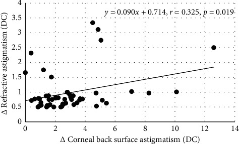
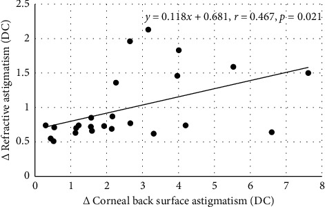
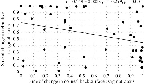
Results: The number of cases that attended where ΔRA was ≥0.50 DC between 1 and 2 months was 52 by autorefractometry and 36 by subjective refraction and between 2 and 3 months was 24 by autorefractometry and 19 by subjective refraction. Vector analysis revealed significant correlations between ΔFSA and ΔRA for data obtained by autorefractometry but not by subjective refraction. At all times, ΔBSA was greater than ΔFSA (p < 0.01). Key findings for ΔBSA values over the central 3 mm zone were between (A) the sine of the axis of ΔRA (y) and sine of the axis of ΔBSA (x) for the data obtained by autorefractometry (between 1 and 2 months, y = 0.749 - 0.303x, r = 0.299, n = 52, p=0.031) and subjective refraction (between 2 and 3 months, y = 0.6614 - 0.4755x, r = 0.474, n = 19, p=0.040) and (B) ΔRA (y) and ΔBSA (x) powers between 2 and 3 months postoperatively for the data obtained by autorefractometry (ΔRA = 0.118 ΔBSA + 0.681 r = 0.467, n = 24, p=0.021) and subjective refraction (ΔRA = 0.072 ΔBSA + 0.545 r = 0.510, n = 19, p=0.026).
Conclusion: Changes in the ocular residual refractive astigmatic error after implanting a monofocal nontoric IOL are associated with changes in astigmatism at the back surface of the cornea within the central optical zone.
Copyright © 2020 Larysa Tutchenko et al.
Conflict of interest statement
The authors declare that they have no conflicts of interest.
Figures
Similar articles
-
The relationship between angle kappa and astigmatism after phacoemulsification with implanting of spherical and aspheric intraocular lens.Indian J Ophthalmol. 2021 Dec;69(12):3503-3510. doi: 10.4103/ijo.IJO_572_21. Indian J Ophthalmol. 2021. PMID: 34826984 Free PMC article.
-
The influence of routine uncomplicated phacoemulsification on the orthogonality of the cornea.Indian J Ophthalmol. 2021 May;69(5):1073-1079. doi: 10.4103/ijo.IJO_1168_20. Indian J Ophthalmol. 2021. PMID: 33913835 Free PMC article.
-
Fluctuations of Anterior Chamber Depth and Astigmatism in Pseudophakic Eyes.Clin Ophthalmol. 2024 Dec 13;18:3739-3752. doi: 10.2147/OPTH.S492253. eCollection 2024. Clin Ophthalmol. 2024. PMID: 39691310 Free PMC article.
-
Posterior corneal astigmatism in refractive lens exchange surgery.Acta Ophthalmol. 2016 May;94(3):295-300. doi: 10.1111/aos.12965. Epub 2016 Jan 30. Acta Ophthalmol. 2016. PMID: 26825986
-
Pseudolentogenic astigmatic effect of multifocal intraocular lenses: non-corneal ocular residual astigmatism (N-CORA) as a new parameter in astigmatic change analysis.Int Ophthalmol. 2017 Aug;37(4):957-964. doi: 10.1007/s10792-016-0359-4. Epub 2016 Sep 24. Int Ophthalmol. 2017. PMID: 27665612
Cited by
-
The Homburg-Adelaide toric IOL nomogram: How to predict corneal power vectors from preoperative IOLMaster 700 keratometry and total corneal power in toric IOL implantation.Acta Ophthalmol. 2025 Feb;103(1):e19-e30. doi: 10.1111/aos.16742. Epub 2024 Jul 16. Acta Ophthalmol. 2025. PMID: 39011876 Free PMC article.
-
Evaluation of keratometric and total corneal astigmatism measurements from optical biometers and anterior segment tomographers and mapping to reconstructed corneal astigmatism vector components.PLoS One. 2025 Jan 8;20(1):e0313574. doi: 10.1371/journal.pone.0313574. eCollection 2025. PLoS One. 2025. PMID: 39775332 Free PMC article.
-
Prediction of corneal power vectors after cataract surgery with toric lens implantation-A vector analysis.PLoS One. 2023 Sep 8;18(9):e0288316. doi: 10.1371/journal.pone.0288316. eCollection 2023. PLoS One. 2023. PMID: 37682881 Free PMC article.
-
Evaluation of Refractive Accuracy of ORA and the Factors Impacting Residual Astigmatism in Patients Implanted with Trifocal IOLs During Cataract Surgery: A Retrospective Observational Study.Clin Ophthalmol. 2022 Aug 10;16:2491-2503. doi: 10.2147/OPTH.S371555. eCollection 2022. Clin Ophthalmol. 2022. PMID: 35974901 Free PMC article.
References
LinkOut - more resources
Full Text Sources
