In vivo Regeneration of Mineralized Bone Tissue in Anisotropic Biomimetic Sponges
- PMID: 32775319
- PMCID: PMC7381345
- DOI: 10.3389/fbioe.2020.00587
In vivo Regeneration of Mineralized Bone Tissue in Anisotropic Biomimetic Sponges
Abstract
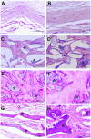
In the last two decades, alginate scaffolds have been variously studied as extracellular matrix analogs for tissue engineering. However, relevant evidence is still lacking concerning their ability to mimic the microenvironment of hierarchical tissues such as bone. Hence, an increasing amount of attention has recently been devoted to the fabrication of macro/microporous sponges with pore anisotropy able to more accurately replicate the cell niche structure as a trigger for bioactive functionalities. This paper presents an in vivo study of alginate sponges with anisotropic microporous domains (MAS) formed by ionic crosslinking in the presence of different fractions (30 or 50% v) of hydroxyapatite (HA). In comparison with unloaded sponges (MAS0), we demonstrated that HA confers peculiar physical and biological properties to the sponge, depending upon the inorganic fraction used, enabling the sponge to bio-mimetically support the regeneration of newly formed bone. Scanning electron microscopy analysis showed a preferential orientation of pores, ascribable to the physical constraints exerted by HA particles during the pore network formation. Energy dispersive spectroscopy (EDS) and X-Ray diffraction (XRD) confirmed a chemical affinity of HA with the native mineral phase of the bone. In vitro studies via WST-1 assay showed good adhesion and proliferation of human Dental Pulp-Mesenchymal Stem Cells (hDP-MSC) that increased in the presence of the bioactive HA signals. Moreover, in vivo studies via micro-CT and histological analyses of a bone model (e.g., a rat calvaria defect) confirmed that the maximum osteogenic response after 90 days was achieved with MAS30, which supported good regeneration of the calvaria defect without any evidence of inflammatory reaction. Hence, all of the results suggested that MAS is a promising scaffold for supporting the regeneration of hard tissues in different body compartments.
Keywords: alginate; anisotropic structure; hard tissues; hydroxyapatite; in vivo models; microporous sponges.
Copyright © 2020 Serrano-Bello, Cruz-Maya, Suaste-Olmos, González-Alva, Altobelli, Ambrosio, Medina, Guarino and Alvarez-Perez.
Figures


Similar articles
-
Synthesis and physicochemical, in vitro and in vivo evaluation of an anisotropic, nanocrystalline hydroxyapatite bisque scaffold with parallel-aligned pores mimicking the microstructure of cortical bone.J Tissue Eng Regen Med. 2015 Dec;9(12):E152-66. doi: 10.1002/term.1729. Epub 2013 Apr 15. J Tissue Eng Regen Med. 2015. PMID: 23585334
-
Fabrication and characterization of novel ethyl cellulose-grafted-poly (ɛ-caprolactone)/alginate nanofibrous/macroporous scaffolds incorporated with nano-hydroxyapatite for bone tissue engineering.J Biomater Appl. 2019 Mar;33(8):1128-1144. doi: 10.1177/0885328218822641. Epub 2019 Jan 16. J Biomater Appl. 2019. PMID: 30651055
-
Biomimetic mineralized hierarchical hybrid scaffolds based on in situ synthesis of nano-hydroxyapatite/chitosan/chondroitin sulfate/hyaluronic acid for bone tissue engineering.Colloids Surf B Biointerfaces. 2017 Sep 1;157:93-100. doi: 10.1016/j.colsurfb.2017.05.059. Epub 2017 May 25. Colloids Surf B Biointerfaces. 2017. PMID: 28578273
-
Scaffolds for bone regeneration made of hydroxyapatite microspheres in a collagen matrix.Mater Sci Eng C Mater Biol Appl. 2016 Jun;63:499-505. doi: 10.1016/j.msec.2016.03.022. Epub 2016 Mar 8. Mater Sci Eng C Mater Biol Appl. 2016. PMID: 27040244
-
High-aspect-ratio nanostructured hydroxyapatite: towards new functionalities for a classical material.Chem Sci. 2023 Dec 1;15(1):55-76. doi: 10.1039/d3sc05344j. eCollection 2023 Dec 20. Chem Sci. 2023. PMID: 38131070 Free PMC article. Review.
Cited by
-
Nanocomposite Methacrylated Silk Fibroin-Based Scaffolds for Bone Tissue Engineering.Biomimetics (Basel). 2024 Apr 6;9(4):218. doi: 10.3390/biomimetics9040218. Biomimetics (Basel). 2024. PMID: 38667229 Free PMC article.
-
3D-Printed Tubular Scaffolds Decorated with Air-Jet-Spun Fibers for Bone Tissue Applications.Bioengineering (Basel). 2022 Apr 27;9(5):189. doi: 10.3390/bioengineering9050189. Bioengineering (Basel). 2022. PMID: 35621467 Free PMC article.
-
Dual nanofiber and graphene reinforcement of 3D printed biomimetic supports for bone tissue repair.RSC Adv. 2024 Oct 15;14(44):32517-32532. doi: 10.1039/d4ra06167e. eCollection 2024 Oct 9. RSC Adv. 2024. PMID: 39411258 Free PMC article.
-
3D Scaffolds Fabrication via Bicomponent Microgels Assembly: Process Optimization and In Vitro Characterization.Micromachines (Basel). 2022 Oct 12;13(10):1726. doi: 10.3390/mi13101726. Micromachines (Basel). 2022. PMID: 36296078 Free PMC article.
-
Evaluating the efficacy of human dental pulp stem cells and scaffold combination for bone regeneration in animal models: a systematic review and meta-analysis.Stem Cell Res Ther. 2023 May 15;14(1):132. doi: 10.1186/s13287-023-03357-w. Stem Cell Res Ther. 2023. PMID: 37189187 Free PMC article.
References
-
- Chatzipetros E., Christopoulos P., Donta C., Tosios K. I., Tsiambas E., Tsiourvas D., et al. (2018). Application of nano-hydroxyapatite/chitosan scaffolds on rat calvarial critical-sized defects: a pilot study. Med. Oral Patol. Oral Cir. Bucal. 23 e625–e632. 10.4317/medoral.22455 - DOI - PMC - PubMed
LinkOut - more resources
Full Text Sources

