. 2020 Jul 9:18:595-606.
doi: 10.1016/j.omtm.2020.07.008. eCollection 2020 Sep 11.

Enhancing Chimeric Antigen Receptor T Cell Anti-tumor Function through Advanced Media Design

Affiliations

Enhancing Chimeric Antigen Receptor T Cell Anti-tumor Function through Advanced Media Design

Saba Ghassemi et al. Mol Ther Methods Clin Dev. .

Abstract

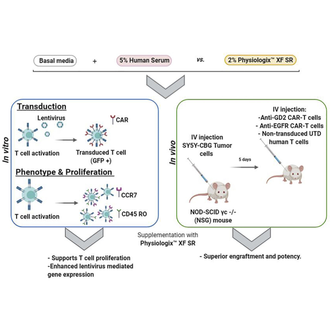
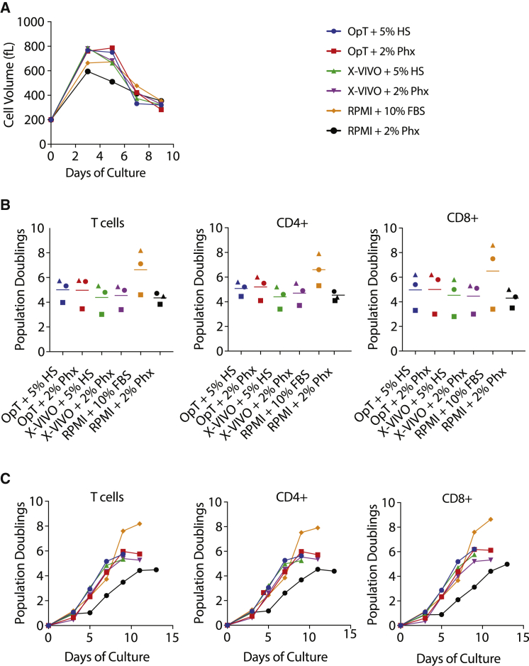
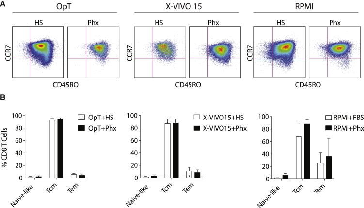
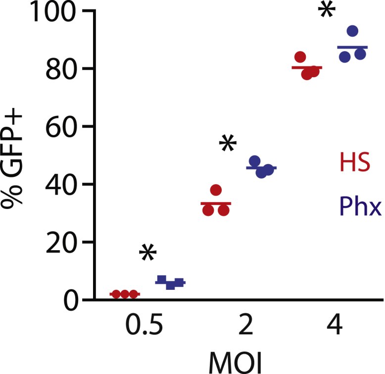
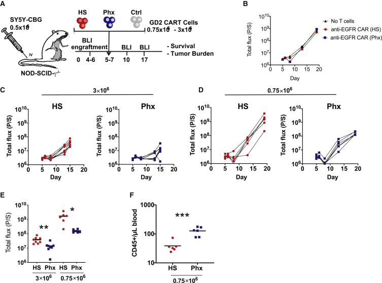
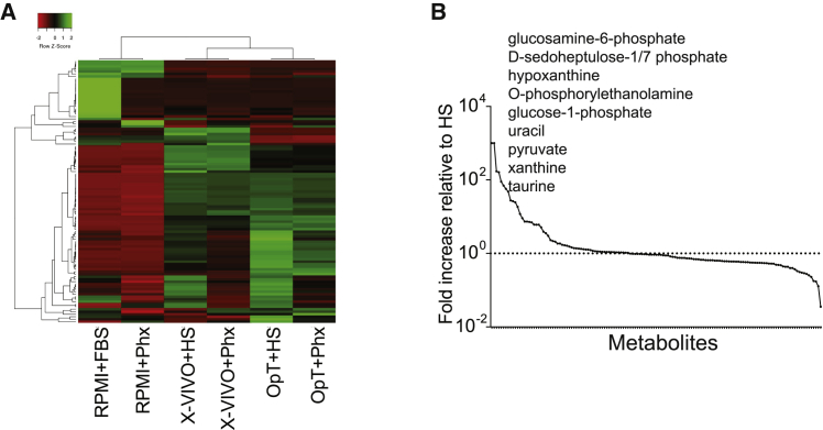
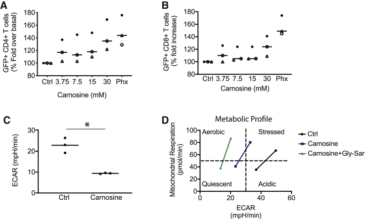
Effective chimeric antigen receptor (CAR)-T cell therapy is dependent on optimal cell culture methods conducive to the activation and expansion of T cells ex vivo, as well as infection with CAR. Media formulations used in CAR-T cell manufacturing have not been optimized for gene delivery, cell expansion, and overall potency. Bioactive components and derivatives that support the generation of functionally-competent T cell progeny with long-lasting persistence are largely undefined. Current media formulations rely on fetal bovine serum (FBS) or human serum (HS), which suffer from a lack of consistency or supply issues. We recognize that components of blood cellular fractions that are absent in serum may have therapeutic value. Here we investigate whether a concentrated growth factor extract, purified from human transfusion grade whole blood fractions, and marketed as PhysiologixTM xeno-free (XF) hGFC (Phx), supports CAR-T cell expansion and function. We show that Phx supports T cell proliferation in clinical and research-grade media. We also show that Phx treatment enhances lentiviral-mediated gene expression across a wide range of multiplicity of infections (MOIs). We compared the ability of anti-GD-2 CAR-T cells expanded ex vivo in medium conditioned with either Phx or HS to clear tumor burden in a human xenograft model of neuroblastoma. We show that T cells expanded in Phx have superior engraftment and potency in vivo, as well as CAR-induced cytolytic activity in vitro. Metabolomic profiling revealed several factors unique to Phx that may have relevance for CAR-T cell preclinical discovery, process development, and manufacturing. In particular, we show that carnosine, a biogenic amine modestly enriched in Phx relative to HS, enhances lentiviral gene delivery in activated T cells. By limiting extracellular acidification, carnosine enhances the metabolic fitness of T cells, shifting their metabolic profile from an acidic, stressed state toward an oxidative, energetic state. These findings are very informative regarding potential derivatives to include in medium customized for gene delivery and overall potency for T cell adoptive immunotherapies.


Figures

None
Graphical abstract
Figure 1
Figure 1
The Concentrated Blood Cell Extract, Physiologix, Supports Primary Human T Cell Expansion in Clinical and Research-Grade Medias (A) A mixed population of T cells were stimulated with anti-CD3/CD28 Dynabeads and expanded in medium conditioned with 5% human serum or 2% Physiologix. The mean cell volume was measured every other day beginning on day 3 until the number of cells in the culture ceased increasing and the mean cell volume is below 350 fL. Representative data from 3 independent experiments are shown. (B) T cells were stimulated as in (A). Cell enumeration was performed every other day beginning on day 3 until the number of cells in the culture ceased increasing and the mean cell volume is below 350 fL. The maximum number of population doublings is plotted with the horizontal bars representing the mean and each symbol representing a separate donor. Data were analyzed by a one-way ANOVA with Neuman-Keuls multiple comparison test. (C) T cells were stimulated as in (A). Cells were enumerated at the indicated time points. Data presented for bulk T cells, as well as CD4+ and CD8+ T cell subsets, are representative of 3 independent experiments with separate donors.
Figure 2
Figure 2
Differentiation Is Similar in T Cells Expanded in Medium Conditioned with Phx versus Human Serum Anti-CD3/CD28 stimulated T cells were expanded over several days in various media conditioned with either 5% human serum or 2% Physiologix. (A) As cells exit their proliferative phase, surface expression of CCR7 and CD45RO were measured by flow cytometry. Representative plots (n = 3) are shown. Cells were pre-gated for live, CD8+ T cells. (B) Relative proportion of naive-like (CCR7+; CD45RO), central memory T cells (Tcm) (CCR7+; CD45RO+), and effector memory T cells (Tem) (CCR7; CD45RO) subsets (after gating on live, CD8+ T cells). Data are mean ± SEM from 3 independent experiments.
Figure 3
Figure 3
Phx Enhances Lentiviral-Mediated Gene Expression A mixed population of T cells were activated with anti-CD3/CD28 Dynabeads and cultured in OpTmizer medium conditioned with either 5% human serum or 2% Physiologix. Following overnight stimulation, previously titered GFP lentiviral supernatant was added at various dilutions. After 3 days, lentiviral infection efficiencies were measured by flow cytometry. The mean number of GFP+ cells ± SEM from 3 independent experiments with separate donors is shown. Data were analyzed by one-way ANOVA (p < 0.05). All groups were compared using a Newman-Keuls multiple comparison test with the difference between the HS and Phx groups statistically significant as indicated.
Figure 4
Figure 4
Investigating the Anti-Tumor Function of Anti-GD2 CAR-T Cells that Were Expanded in Medium Conditioned with Phx In Vivo (A) Schematic of the xenograft model and anti-GD2 CAR-T cell treatment (derived from healthy donors) in NSGs i.v. injected with 0.5 × 106 SY5Y cells. Activated T cells were infected with anti-GD2 CAR lentivirus and expanded in OpTmizer medium conditioned with H.S. or Phx over 9 days. A high (3 × 106) or a low (0.75 × 106) dose of CAR+ T cells were selected to examine the influence of Phx versus HS on CAR-T cell potency. These cells were i.v. injected into NSG mice 5 days after SY5Y injection (n = 6–8 per group). (B–H) Serial quantification of disease burden by luminescence imaging. (B) Tumor size in mice that received no T cells, or T cells expressing an irrelevant CAR engineered to recognize human EGFR. Symbols represent a single mouse each. (C) The effectiveness of 3 × 106 anti-GD2 CAR-T cells expanded in Phx versus anti-GD2 CAR-T cells expanded in HS. (D) The effectiveness of 0.75 × 106 CAR-T cells expanded in Phx versus anti-GD2 CAR-T cells expanded in HS. (E) Quantification of tumor burden by bioluminescence imaging on days 10 (3 × 106) and 14 (0.75 × 106) in mice treated with anti-GD2 CAR-T cells. Values represent mean ± SEM for each group. (F) Blood was collected by retroorbital bleeding 14 days after tumor injection, and absolute peripheral blood CD45+ T cells were enumerated by a TruCount assay.
Figure 5
Figure 5
Metabolomic Assessment of OpTmizer Medium Conditioned with Human Serum Versus Phx (A) Heatmap illustrating the hierarchical relationship between metabolites present in various media conditioned with either human serum or Phx. In this grid, each row represents a unique metabolite and each column corresponds to a unique media formulation. (B) Metabolite abundance in Phx normalized to HS. The levels of metabolites present in both media were normalized to the level of HS. Metabolites were then rearranged in the x axis by the highest to lowest difference between metabolite levels derived from both supplementation methods. The dotted line represents the levels present in HS, while the solid line represents Phx.
Figure 6
Figure 6
Carnosine Enhances GFP+ Lentivirus Expression and Improves the Metabolic Properties of T Cells (A and B) Freshly isolated T cells were activated in cell culture medium containing human serum and supplemented with varying levels of carnosine. As a positive control, T cells were activated in cell culture medium containing 2% Phx. After 24 h, T cells were infected with GFP lentivirus. The proportion of live, GFP+, T cells in the CD4+ (A) and CD8+ (B) compartment are shown. T cell subsets were enumerated using bead-based flow cytometry, as described in the Materials and Methods. Values are expressed as a percentage of OpT+HS. Mean ± SEM from 3 independent experiments with separate donors are shown. (C) T cells were activated with Dynabeads. After overnight stimulation, the cells were switched to a bicarbonate-free XF assay medium containing 30 mM carnosine. Metabolic parameters were measured by extracellular flux assay. Steady state extracellular acidification rates (ECARs) are shown. Values are mean ± SEM from 3 independent experiments with separate donors. ∗p < 0.01 for control versus carnosine as analyzed by an unpaired Student t test. (D) T cells were activated with Dynabeads for 1–3 days. The cells were switched to a bicarbonate-free XF assay medium containing 10 mM carnosine and 30 mM glycylsarcosine (Gly-Sar). Representative data from two independent experiments with separate donors are shown.

References

    1. June C.H., O’Connor R.S., Kawalekar O.U., Ghassemi S., Milone M.C. CAR T cell immunotherapy for human cancer. Science. 2018;359:1361–1365. - PubMed
    1. O’Sullivan D., Sanin D.E., Pearce E.J., Pearce E.L. Metabolic interventions in the immune response to cancer. Nat. Rev. Immunol. 2019;19:324–335. - PubMed
    1. Bieback K., Fernandez-Muñoz B., Pati S., Schäfer R. Gaps in the knowledge of human platelet lysate as a cell culture supplement for cell therapy: a joint publication from the AABB and the International Society for Cell & Gene Therapy. Cytotherapy. 2019;21:911–924. - PubMed
    1. Carson B.P., Patel B., Amigo-Benavent M., Pauk M., Kumar Gujulla S., Murphy S.M., Kiely P.A., Jakeman P.M. Regulation of muscle protein synthesis in an in vitro cell model using ex vivo human serum. Exp. Physiol. 2018;103:783–789. - PubMed
    1. Cantor J.R., Abu-Remaileh M., Kanarek N., Freinkman E., Gao X., Louissaint A., Lewis C.A., Sabatini D.M. Physiologic Medium Rewires Cellular Metabolism and Reveals Uric Acid as an Endogenous Inhibitor of UMP Synthase. Cell. 2017;169:258–272. - PMC - PubMed

LinkOut - more resources