

Removal of Large Primary Vaginal Calculus Using the Nephroscope and Endoscopic Ultrasonic Lithotrite: A Case Report
- PMID: 32775688
- PMCID: PMC7383409
- DOI: 10.1089/cren.2019.0099
Removal of Large Primary Vaginal Calculus Using the Nephroscope and Endoscopic Ultrasonic Lithotrite: A Case Report
Abstract
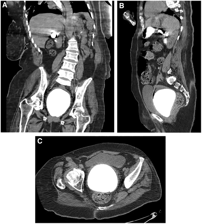
Background: Vaginal calculi are rare and can grow quite large if they remain undetected. Vaginal stones are caused by the pooling of urine in the vagina and can be classified as either primary or secondary, depending on the absence or presence, respectively, of a nidus. Primary stones without any urethrovaginal or vesicovaginal fistula are even more uncommon but appear to be more commonly reported in incontinent women with significant physical disabilities. Case Presentation: We present a case of an ∼11 cm primary vaginal stone in a 61-year-old woman with cerebral palsy. This was removed using a nephroscope and an endoscopic ultrasonic lithotrite through the vaginal introitus with subsequent analysis demonstrating a struvite stone composition. Conclusion: This case is unique not only for the large size of the calculi but also for our less invasive approach, using a nephroscope and endoscopic ultrasonic lithotrite to fragment and remove the stone. We hope that this report will assist other providers in the timely and accurate diagnosis and treatment of future vaginal stone patients.
Keywords: instrumentation; nephroscope; urolithiasis; vaginal stone.
Copyright 2020, Mary Ann Liebert, Inc., publishers.
Conflict of interest statement
No competing financial interests exist.
Figures


References
-
- Ikeda Y, Oda K, Matsuzawa N, Shimizu K. Primary vaginal calculus in a middle-aged woman with mental and physical disabilities. Int Urogynecol J 2013;24:1229–1231 - PubMed
-
- Lin CJ, Hsu HH, Chen CP, Wu CH, Chen HY. Huge primary vaginal stone in a recumbent woman. Taiwan J Obstet Gynecol 2005;44:80–82
-
- Jaspers JW, Kuppens SM, van Zundert AA, de Wildt MJ. Vaginal stones in a 5-year-old girl: A novel approach of removal. J Pediatr Adolesc Gynecol 2010;23:e23–e25 - PubMed
Publication types
LinkOut - more resources
Full Text Sources