Case Reports
. 2020 Jul 11:19:100806.
doi: 10.1016/j.ajoc.2020.100806. eCollection 2020 Sep.

Orbital myeloid sarcoma (chloroma): Report of 2 cases and literature review

Affiliations
Case Reports

Orbital myeloid sarcoma (chloroma): Report of 2 cases and literature review

Mohammad A AlSemari et al. Am J Ophthalmol Case Rep. .

Abstract

Purpose: Myeloid sarcoma (MS) of the orbit is an uncommon condition in occurring in children, generally coupled to myeloproliferative neoplasms.

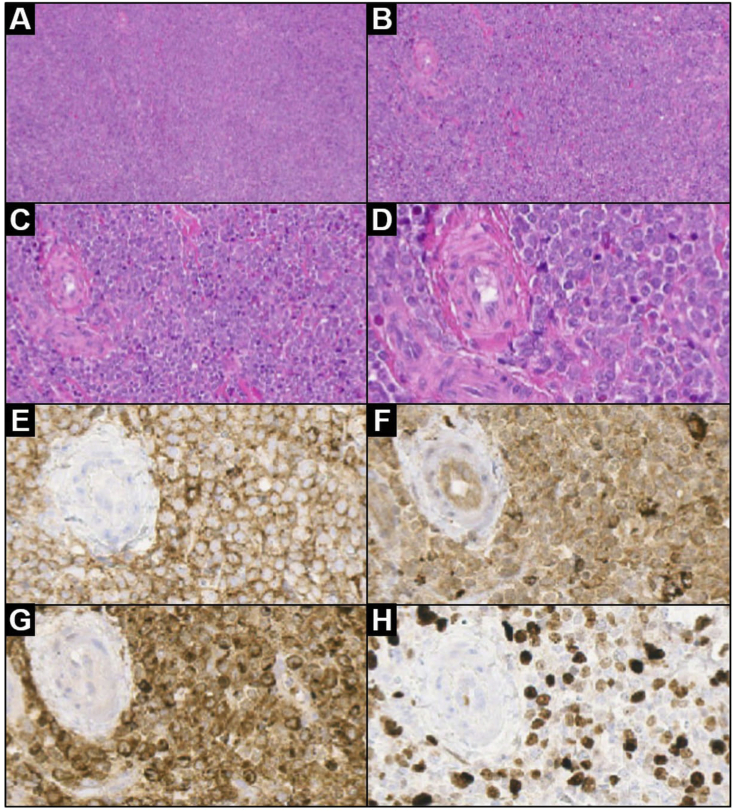
Observations: We describe two rare cases of orbital MS in young boys with aggressive local symptoms but without evidence of acute myeloid leukemia (AML), both patients underwent orbitotomy for gross-tumor resection and biopsy. At follow up, there was no evidence of recurrence nor evolution of the myeloproliferative neoplasms clinically and by radiological and laboratory work-up. We also provide a detailed description of the magnetic resonance imaging presentation, with an extensive pathological analysis correlation.

Conclusions and importance: A comprehensive revision of the literature on isolated orbital MS was carried out with particular emphasis on clues for differential diagnosis and treatment options, stressing the need to consider MS even in the absence of sign and symptoms of an underlying myeloproliferative disorders.

Keywords: Acute myeloid leukemia; Magnetic resonance imaging; Myeloid sarcoma; Orbital pathology; Pediatric tumor.


Conflict of interest statement

The following authors have no financial disclosures (MP, MA, CR, HA, AM, SE, RM, MM, AE, LR, RC, DS).

Figures

Fig. 1
Fig. 1
Axial (a) and coronal (b–d) T2w images showing expansive extra-conal orbital lesion arising along the external border of superior and lateral rectus muscles and superior oblique muscle. The lesion presents mild hyperintensity with inhomogeneous core, and slightly restricted water diffusion on DWI (c) indicating hypercellularity. Extra-ocular muscles and optic nerve are displaced (white dotted line); marked left eye proptosis is also present. The lesion does not spear lacrimal gland that seems to be infiltrated (not shown); bony erosion and invasion of the lateral wall of the orbit are clearly visible (white arrow).
Fig. 2
Fig. 2
Axial pre (a) and post-contrast (b) T1w showing lesion vivid enhancement and inner un-enhancing necrotic area (black arrow); the great wing of the sphenoid seems to be infiltrated (white arrow). Multi-planar reconstruction on coronal plane shows lesion extent from the lacrimal fossa of the frontal bone (c) (black arrowhead) to the orbital apex (d) (white arrowhead).
Fig. 3
Fig. 3
Low and high magnification lesion histology after surgery: (a) low magnification showing a dense, diffuse infiltrate (H&E 4×); (b) the tumor consists of a cohesive proliferation of small/medium-sized cells (H&E 10×); (c) a discrete number of eosinophils are admixed with tumor cells (H&E 20×); (d) neoplastic cells show large oval nuclei, prominent nucleoli and poor cytoplasm (H&E 40×). Lesion immunophenotype: (e–g) neoplastic cells diffusely expressed CD45RO/LCA (anti-CD45RO/LCA, 40×), CD68 (anti-CD68, 40×) and myeloperoxidase (anti-MPO, 40×); (h) Ki-67 proliferative index is positive in about 50% of neoplastic cells (anti-Ki-67, 40×). Legend: H&E = hematoxylin and eosin stain.
Fig. 4
Fig. 4
Axial CT scan orbit (a) and sagittal T1 fat suppressed post contrast (b,c) and axial DWI (D) images showing solid lobulated extra-conal orbital lesion arising superiorly along the orbital roof and nasal infiltrating the recti muscles and superior oblique muscle. The lesion presents marked restricted water diffusion on DWI (c) indicating high degree of cellularity. The lesion abutting the lacrimal gland with no definite line of separation.
Fig. 5
Fig. 5
Low and high magnification lesion histology after surgery: (a) high magnification showing a highly vascular soft tissue of poorly differentiated malignant cells infiltrating the adipose tissue with eosinophils (H&E 400×); (b) low magnification showing a dense, diffuse infiltrate (H&E 100×); (c) CD34 expression in neoplastic cells (CD34, ×100) (d) neoplastic cells diffusely expressed CD45RO/LCA (anti-CD45RO/LCA, 100×). Legend: H&E = hematoxylin and eosin stain.

References

    1. Guermazi A., Feger C., Rousselot P. Granulocytic sarcoma (chloroma): imaging findings in adults and children. AJR. 2002;178(February):319–325. - PubMed
    1. Lee B., Fatterpekar G.M., Kim W., Som P.M. Granulocytic sarcoma of the temporal bone. AJNR. 2002;23(October):1497–1499. - PMC - PubMed
    1. Maka E., Lukáts O., Tóth J., Fekete S. Orbital tumour as initial manifestation of acute myeloid Leukemia : granulocytic sarcoma: case report. Pathol Oncol Res. 2008;14:209–211. doi: 10.1007/s12253-008-9028-x. - DOI - PubMed
    1. Thakur B., Varma K., Misra V., Chauhan S. Granulocytic sarcoma presenting as an orbital mass: report of two cases. J Clin Diagn Res. 2013;7(8):1704–1705. doi: 10.7860/JCDR/2013/5582.3260. - DOI - PMC - PubMed
    1. Lee J.Y., Chung H., Cho H. Clinical characteristics and treatment outcomes of isolated myeloid sarcoma without bone marrow involvement: a single-institution experience. Blood Res. 2017;52(3):184–192. - PMC - PubMed

Publication types

LinkOut - more resources