. 2020 Aug 28;40(8):BSR20201807.
doi: 10.1042/BSR20201807.

Tanshinone IIA induces ferroptosis in gastric cancer cells through p53-mediated SLC7A11 down-regulation

Affiliations

Tanshinone IIA induces ferroptosis in gastric cancer cells through p53-mediated SLC7A11 down-regulation

Zhenhua Guan et al. Biosci Rep. .

Abstract

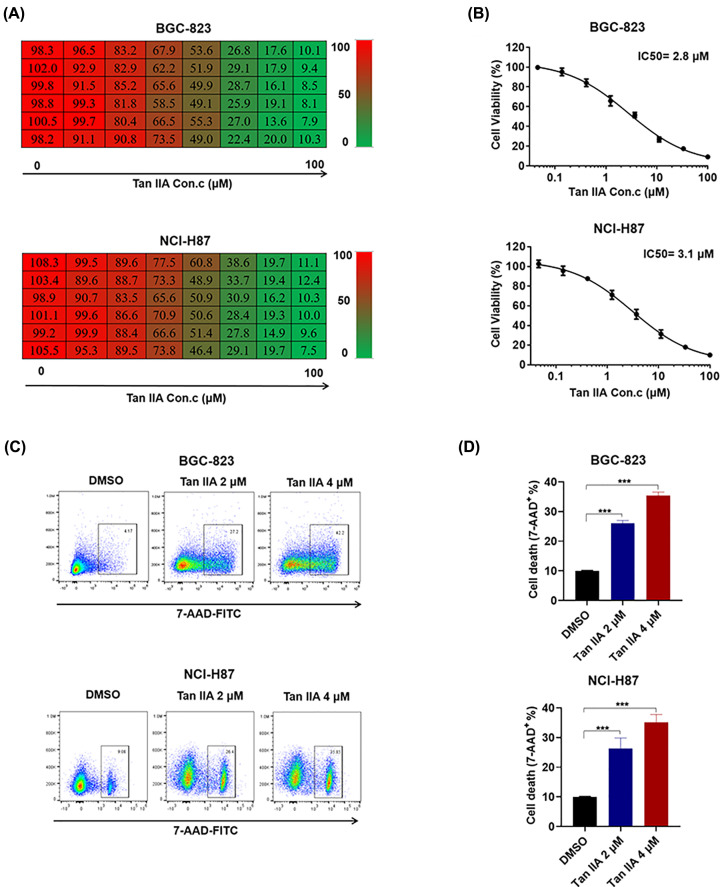
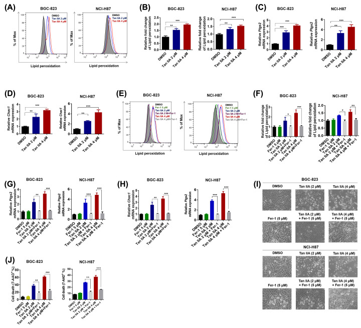
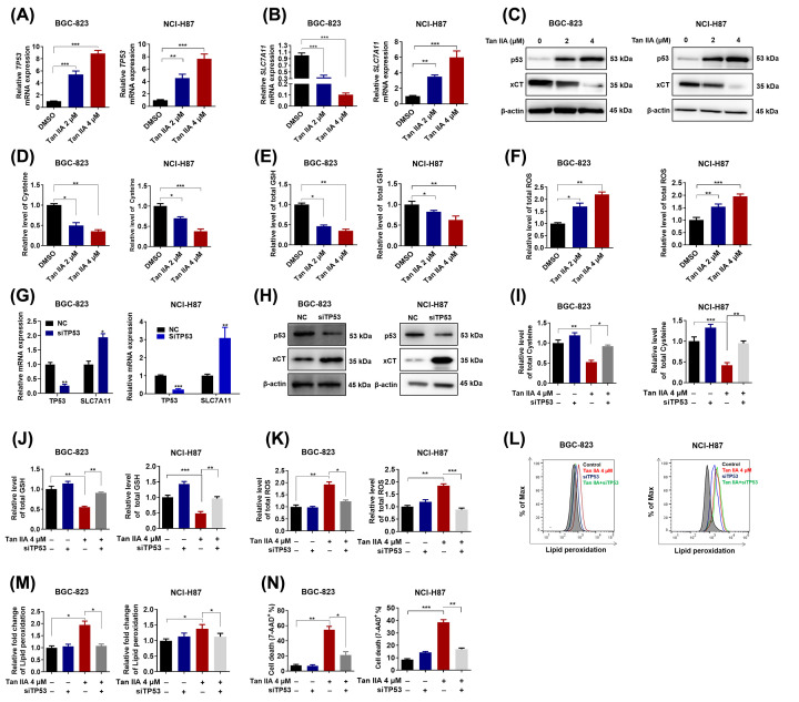
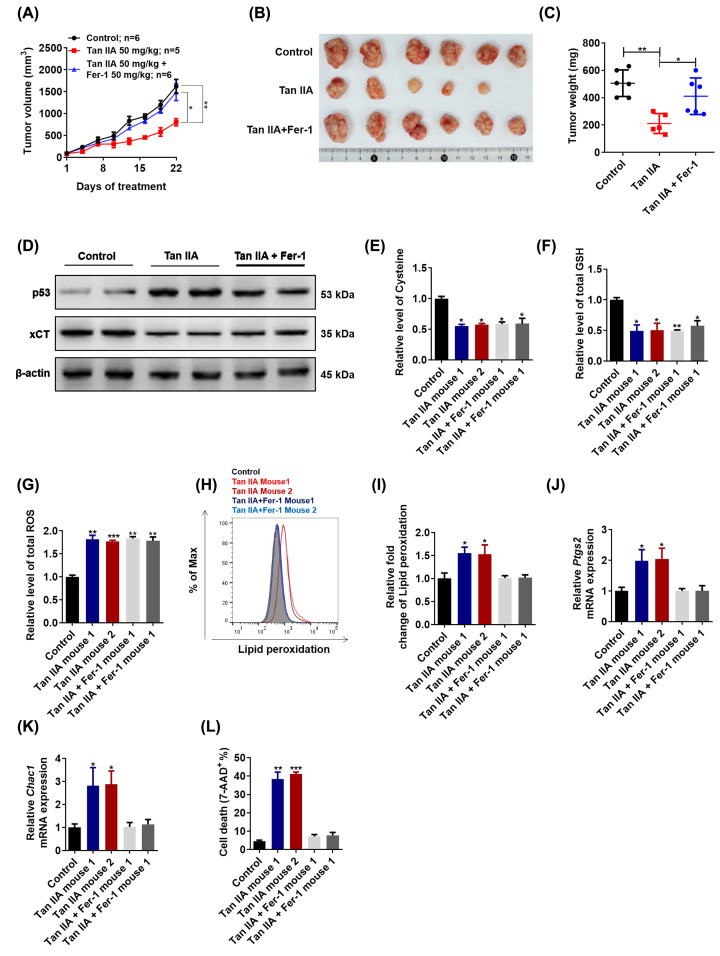
Gastric cancer represents a malignant type of cancer worldwide. Tanshinone IIA (Tan IIA), a pharmacologically active component isolated from the rhizome of the Chinese herb Salvia miltiorrhiza Bunge (Danshen), has been reported to possess an anti-cancer effect in gastric cancer. However, its mechanisms are still not fully understood. In the present study, we found that Tan IIA induced ferroptosis in BGC-823 and NCI-H87 gastric cancer cells. Tan IIA increased lipid peroxidation and up-regulated Ptgs2 and Chac1 expression, two markers of ferroptosis. Ferrostatin-1 (Fer-1), an inhibitor of lipid peroxidation, inhibited Tan IIA caused-lipid peroxidation and Ptgs2 and Chac1 expression. In addition, Tan IIA also up-regulated p53 expression and down-regulated xCT expression. Tan IIA caused decreased intracellular glutathione (GSH) level and cysteine level and increased intracellular reactive oxygen species (ROS) level. p53 knockdown attenuated Tan IIA-induced lipid peroxidation and ferroptosis. Tan IIA also induced lipid peroxidation and ferroptosis in BGC-823 xenograft model, and the anti-cancer effect of Tan IIA was attenuated by Fer-1 in vivo. Therefore, Tan IIA could suppress the proliferation of gastric cancer via inducing p53 upregulation-mediated ferroptosis. Our study have identified a novel mechanism of Tan IIA against gastric cancer, and might provide a critical insight into the application of Tan IIA in gastric cancer intervention.

Keywords: Ferroptosis; Gastric cancer; ROS; Tanshinone IIA; p53; xCT.


Conflict of interest statement

The authors declare that there are no competing interests associated with the manuscript.

Figures

Figure 1
Figure 1. Tan IIA inhibits cell growth and induces cell death in BGC-823 and NCI-H87 gastric cancer cells
(A) BGC-823 and NCI-H87 cells were treated with serially diluted (1:3) concentrations of Tan IIA with the highest concentration of 10 μM for 72 h. Cell viability was detected by MTT. (B) IC50 of Tan IIA on BGC-823 and NCI-H87 cell growth. (C) BGC-823 and NCI-H87 cells were treated with 2 and 4 μM Tan IIA for 72 h, cells were stained with 7-AAD and cell death was assessed by flow cytometry. (D) Histogram statistics of cell death in (C). Data are shown as the mean ± SD (n=3). ***P<0.001 vs control group.
Figure 2
Figure 2. Tan IIA induces ferroptosis in BGC-823 and NCI-H87 gastric cancer cells
(A) BGC-823 and™ NCI-H87 cells were treated with 2 and 4 μM Tan IIA for 72 h, cells were stained with BODIPY™ 581/591 C11 dye and lipid peroxidation was detected by flow cytometry. (A) Histogram statistics of cell death in (B). (C,D) BGC-823 cells were treated with Tan IIA as mentioned above. The expression of two marker genes of ferroptosis, Ptgs2 and Chac1 was detected by RT-qPCR. (E) BGC-823 and NCI-H87 cells were treated with 2 and 4 μM Tan IIA or Tan IIA combined with 5 μM Fer-1 for 72 h, cells were stained with BODIPY™ 581/591 C11 dye and lipid peroxidation was detected by flow cytometry. (F) Histogram statistics of cell death in (E). (G,H) BGC-823 and NCI-H87 cells were treated as mentioned in (E). The expression of two marker genes of ferroptosis, Ptgs2 and Chac1 was detected by RT-qPCR. (I) Representative pictures of BGC-823 and NCI-H87 cells post Tan IIA or Tan IIA combined with 5 μM Fer-1 treatment. (J) BGC-823 and NCI-H87 cells were treated as mentioned in (E), cells were stained with 7-AAD and cell death was assessed by flow cytometry. Data are shown as the mean ± SD (n=3). *P<0.05 vs control group; **P<0.01 vs control group; ***P<0.001 vs control group.
Figure 3
Figure 3. p53 mediates Tan IIA-induced ferroptosis in BGC-823 and NCI-H87 gastric cancer cells
(A,B) BGC-823 and NCI-H87 cells were treated with 2 and 4 μM Tan IIA for 72 h, the mRNA expression of TP53 and its target gene SLC7A11 was detected by RT-qPCR. (C) BGC-823 and NCI-H87 cells were treated with Tan IIA as mentioned above. The protein expression of p53 and xCT was detected by Western Blot. (DF) BGC-823 and NCI-H87 cells were treated with Tan IIA as mentioned above. Intracellular cysteine level, GSH level and ROS level were measured. (G,H) BGC-823 and NCI-H87 cells were transfected with 20 μM TP53 siRNA and negative control (NC) sequence using Lipofectamine 3000 for 4 h, after 24 h more culture with fresh RPMI-1640 medium with 10% FBS, cells were harvested. The knockdown efficiency of TP53 and the expression of its target gene SLC7A11 in both BGC-823 and NCI-H87 gastric cancer cells was detected by RT-qPCR and Western Blot. (IK) BGC-823 and NCI-H87 cells were pretreated with TP53 siRNA, then the cells were treated with Tan IIA for 72 h, intracellular cysteine level, GSH level and ROS level were measured. (L–N) BGC-823 and NCI-H87 cells were treated with TP53 siRNA and Tan IIA as mentioned above. Then lipid peroxidation and cell death was detected by flow cytometry. Data are shown as the mean ± SD (n=3). *P<0.05 vs control group; **P<0.01 vs control group; ***P<0.001 vs control group.
Figure 4
Figure 4. Tan IIA suppresses tumor growth and induces ferroptosis in vivo
(A) Mice were treated with Tan IIA (50 mg/kg), Fer-1 (50 mg/kg) and Tan IIA (50 mg/kg) combined with Fer-1 (50 mg/kg) for 3 weeks, tumor volume was measured every 3 days (n=5–6). (B, C) After treatment with Tan IIA (50 mg/kg), Fer-1 (50 mg/kg) and Tan IIA (50 mg/kg) combined with Fer-1 (50 mg/kg) for 3 weeks, tumors in these groups were removed, tumor pictures were captured (B) and tumors were weighed (C). (D) A total of 20 mg tumor tissue in two mice of each group was isolated. The expression of p53 and xCT was detected by Western Blot and RT-qPCR. (EG) A total of 20 mg tumor tissue in two mice of each group was isolated, intracellular cysteine level, GSH level and ROS level were measured. (H, I) A total of 20 mg tumor tissue in two mice of each group was isolated and digested into single cell. Then the cells of each group were stained with BODIPY™ 581/591 C11 and lipid peroxidation was analyzed by flow cytometry. (J, K) A total of 20 mg tumor tissue in two mice of each group was isolated and total RNA sample was extracted. The expression of two marker genes, Ptgs2 and Chac1 was detected by RT-qPCR. (L) A total of 20 mg tumor tissue in two mice of each group was isolated and digested into single cell. Then the cells of each group were stained with 7-AAD and cell death was analyzed by flow cytometry. Data are shown as the mean ± SD (n=3). *P<0.05 vs control group; **P<0.01 vs control group; ***P<0.001 vs control group.

References

    1. Torre L.A., Bray F., Siegel R.L.et al. . (2015) Global cancer statistics. CA Cancer J. Clin. 65, 87–108 10.3322/caac.21262 - DOI - PubMed
    1. Cavatorta O., Scida S., Miraglia C.et al. . (2018) Epidemiology of gastric cancer and risk factors. Acta Biomed. 89, 82–87 - PMC - PubMed
    1. Laterza L., Scaldaferri F. and Gasbarrini A. (2019) Risk factors for gastric cancer: is it time to discard PPIs? Gut 68, 176–177 10.1136/gutjnl-2017-315621 - DOI - PubMed
    1. Venerito M., Vasapolli R., Rokkas T.et al. . (2018) Gastric cancer: epidemiology, prevention, and therapy. Helicobacter 23, e12518 10.1111/hel.12518 - DOI - PubMed
    1. Bebber C.M., Muller F., Prieto C.L.et al. . (2020) Ferroptosis in cancer cell biology. Cancers (Basel) 12, 10.3390/cancers12010164 - DOI - PMC - PubMed

Publication types

MeSH terms