Review
. 2020 Aug 10;7(1):27.
doi: 10.1186/s40580-020-00237-4.

Graphene impregnated electrospun nanofiber sensing materials: a comprehensive overview on bridging laboratory set-up to industry

Affiliations
Review

Graphene impregnated electrospun nanofiber sensing materials: a comprehensive overview on bridging laboratory set-up to industry

Adel Mohammed Al-Dhahebi et al. Nano Converg. .

Abstract

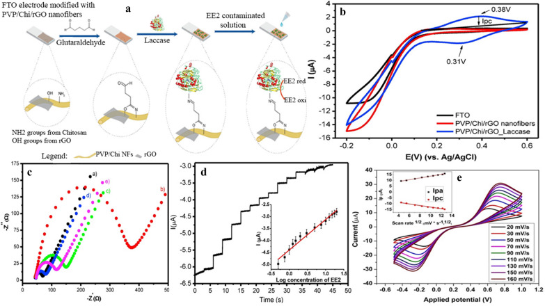
Owing to the unique structural characteristics as well as outstanding physio-chemical and electrical properties, graphene enables significant enhancement with the performance of electrospun nanofibers, leading to the generation of promising applications in electrospun-mediated sensor technologies. Electrospinning is a simple, cost-effective, and versatile technique relying on electrostatic repulsion between the surface charges to continuously synthesize various scalable assemblies from a wide array of raw materials with diameters down to few nanometers. Recently, electrospun nanocomposites have emerged as promising substrates with a great potential for constructing nanoscale biosensors due to their exceptional functional characteristics such as complex pore structures, high surface area, high catalytic and electron transfer, controllable surface conformation and modification, superior electric conductivity and unique mat structure. This review comprehends graphene-based nanomaterials (GNMs) (graphene, graphene oxide (GO), reduced GO and graphene quantum dots) impregnated electrospun polymer composites for the electro-device developments, which bridges the laboratory set-up to the industry. Different techniques in the base polymers (pre-processing methods) and surface modification methods (post-processing methods) to impregnate GNMs within electrospun polymer nanofibers are critically discussed. The performance and the usage as the electrochemical biosensors for the detection of wide range analytes are further elaborated. This overview catches a great interest and inspires various new opportunities across a wide range of disciplines and designs of miniaturized point-of-care devices.

Keywords: Electrochemical biosensors; Electrospinning; Electrospun nanofibers; Graphene; Graphene oxide; Graphene quantum dots; Nanocomposites; Reduced graphene oxide.


Conflict of interest statement

The authors declare that they have no known competing financial interests that could have appeared to influence the work reported in this paper.

Figures

Fig. 1
Fig. 1
a Major fabrication methods of graphene: Top-down and bottom-up fabrication methods. Principal top-down methods include liquid-phase exfoliation and micromechanical cleavage of graphite. An additional method involves the exfoliation of initially oxidized graphite, leading to GO, which is chemically and/or thermally reduced to graphene. The bottom-up fabrication of graphene is usually performed by epitaxial growth on SiC or chemical vapour deposition, typically on Cu using small molecules, such as methane, as precursors. Reproduced with permission from [174] Copyright 2017 Nature Publishing Group. b SEM (a, c, e) images and TEM images (b, d, f) of nanofibers (a, b), nanofibers-rGO-5 (c, d), and nanofibers-rGO-10 (e, f) with different magnifications Reproduced with permission from [173] Copyright 2019 Wiley
Fig. 2
Fig. 2
(1): Pristine and GO-SnO2 NTs preparation and gas sensor mechanism and (2) SEM images of (a) as-prepared Sn + poly (vinyl pyrrolidone) (PVP) nanofibers (b, c) pristine SnO2, and (d, e) GO incorporate SnO2 NTs, (f) Histogram of GO-SnO2 NT diameters Reproduced with permission from [181] Copyright 2019 Elsevier
Fig. 3
Fig. 3
a Technological flow chart of the patterned STNNE. b FESEM image of the networked nanofibers. c FESEM image of the intersections of the nanofibers. d Optical photographs of the stretchable and transparent networked nanofibers film. Dispersion of PU/rGO/AgNPs in nanofibers. e Raman spectra of PU/GO/AgNPs nanofiber and PU/rGO/AgNPs nanofiber samples with a GO:AgNPs loading ratio of 1:1.25. f TEM images of nanofibers with diameters of ~ 290, ~ 484, and ~ 933 nm. g Schematics of the functional groups on GO, chemical structure of polyurethane, and negative surface charges of AgNPs. GO nanosheets can be hydrogen-bonded to the PU matrix by the functional moieties of the carboxyl and hydroxyl groups. h Optical transmittance-sheet resistance of the networked nanofibers for different types of nanofibers: rGO-coated PU, PU/rGO, PU/AgNPs, and PU/rGO/AgNPs nanofibers with that of copper nanowires, PEDOT: PSS/Zonyl/DMSO and graphene. i Stress–strain curves of PU/rGO and PU/rGO/AgNPs nanofibers. Evaluation of STNNEs under stretching conditions. j Resistance change (ΔR/R0) versus elongation of the PU/rGO and PU/rGO/AgNPs nanofiber electrodes on PDMS substrates. k Resistance change (ΔR/R0) versus low strain under tensile and compressive bending of STNNEs Reproduced with permission from [153] Copyright 2019 Royal Society of Chemistry
Fig. 4
Fig. 4
Dimensionally stable anodes (DSC) (a) and thermogravimetric analysis (TGA) (b) curves of pure PS matrix and the TRG/PS nanocomposites. Reproduced with permission from [158] Copyright 2018 Elsevier. c TGA curves of electrospun PVA mats mixed with GO. Reproduced with permission from [184] Copyright 2019 American Scientific Publishers. d TGA curves of rGO, rGO and AM-rGO. e TGA curves electrospun PMMA/PANI/AM-rGO, PMMA/PANI/rGO and PMMA/PANI nanofibers. As shown in e, the thermal degradation temperature of PMMA/PANI/Am-rGO nanofibers increased to ~ 441 °C, a magnitude higher than that of the PMMA/PANI samples at ~ 348 °C. Both d, e are reproduced with permission from [163] Copyright 2017 MDPI
Fig. 5
Fig. 5
a Schematic presentation of electrospinning for producing PVA/GQD onto GCE for electrochemical biosensing and catalyzing of H2O2, b the possible detection mechanism, c Zeta potentials of GQDs, PVA, and PVA/GQD nanofibrous membranes at varied pH, d CVs of GCEs modified with PVA and PVA/GQD nanofibrous membranes, sensitivity of the biosensor at different potentials (inset), e CVs of the PVA/GQD nanofibrous membranes modified GCE 0.1 M PBS with different addition of H2O2 (Reproduced with permission from [202], Copywrite 2015 Royal Society of Chemistry)
Fig. 6
Fig. 6
a Schematic representation of the fabrication of EE2 electrochemical biosensor. b Cyclic voltammetry measurements using a PBS buffer solution (pH 7.4) and scan rate of 100 mV s−1 for PTO, PVP/Chi/rGO ES NFs and PVP/Chi/rGO ES NFs coated with Laccase enzyme. c Nyquist plots of EIS for (a) FTO, (b) PVP nanofibers, (c) PVP/Chi nanofibers, (d) PVP/Chi/rGO nanofibers and (e) PVP/Chi/rGO nanofibers coated with Laccase in a 5 mmol L-1[FeCN)63-/4- solution with 0.1 mol L−1 KCl. d Amperometric response upon successive additions of EE2 ethanol solution recorded at PVP/Chi/rGO_Laccase coated electrode in a phosphate buffer solution pH 7.0 in concentrations ranging from 0.25 to 20 pmol L−1 at a fixed potential of − 0.3 V. The inset shows the calibration curve with the respective linear fit. ad reproduced from with permission from [162] Copyright 2018 Elsevier. (E) Schematic of cyclic voltammetry shown the electrochemical behaviour of BSA/BH/PNF/GCE in presence of [Fe(CN)6]3−/4− at different scan rates (20–160 mV/s). It can be revealed that, the increase in the peak to peak voltage difference is also an indication of the progressive immobilization and the anodic peak shifts towards the higher potential value whereas the cathodic peaks shift towards lower potential value with the increase in the scan rate Reproduced with permission from [129] Copyright 2019 Wiley

References

    1. Krishnan SK, Singh E, Singh P, Meyyappan M, Nalwa HS. A review on graphene-based nanocomposites for electrochemical and fluorescent biosensors. RSC Adv. 2019;9:8778–8781. doi: 10.1039/c8ra09577a. - DOI - PMC - PubMed
    1. Wongkaew N, Simsek M, Griesche C, Baeumner AJ. Functional nanomaterials and nanostructures enhancing electrochemical biosensors and lab-on-a-chip performances: recent progress, applications, and future perspective. Chem. Rev. 2019;119:120–194. doi: 10.1021/acs.chemrev.8b00172. - DOI - PubMed
    1. Asif MH, Razaq A, Akbar N, Danielsson B, Sultana I. Facile synthesis of multisegment Au/Ni/Au nanowire for high performance electrochemical glucose sensor. Mater. Res. Express. 2019;6:95028.
    1. Asmatulu R, Veisi Z, Uddin MN, Mahapatro A. Highly sensitive and reliable electrospun polyaniline nanofiber based biosensor as a robust platform for COX-2 enzyme detections. Fibers Polym. 2019;20:966–974. doi: 10.1007/s12221-019-1096-x. - DOI
    1. Niu Y, Xie H, Luo G, Weng W, Ruan C, Li G, Sun W. Electrochemical performance of myoglobin based on TiO2-doped carbon nanofiber decorated electrode and its applications in biosensing. RSC Adv. 2019;9:4480–4487. doi: 10.1039/c8ra07910b. - DOI - PMC - PubMed

Grants and funding

LinkOut - more resources