

UV biomarker genes for classification and risk stratification of cutaneous actinic keratoses and squamous cell carcinoma subtypes
- PMID: 32776588
- PMCID: PMC8323755
- DOI: 10.1096/fj.202001412R
UV biomarker genes for classification and risk stratification of cutaneous actinic keratoses and squamous cell carcinoma subtypes
Abstract
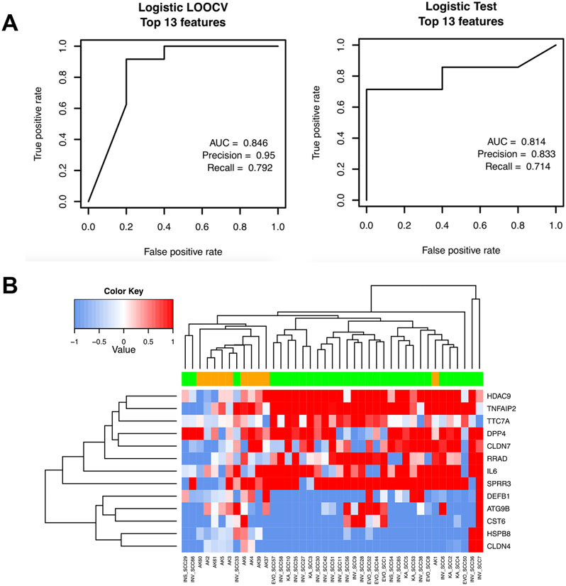
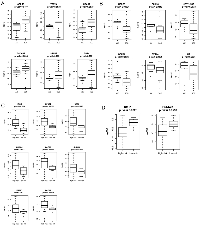
Currently, there is no sensitive molecular test for identifying transformation-prone actinic keratoses (AKs) and aggressive squamous cell carcinoma (SCC) subtypes. Biomarker-based molecular testing represents a promising tool for risk stratifying these lesions. We evaluated the utility of a panel of ultraviolet (UV) radiation-biomarker genes in distinguishing between benign and transformation-prone AKs and SCCs. The expression of the UV-biomarker genes in 31 SCC and normal skin (NS) pairs and 10 AK/NS pairs was quantified using the NanoString nCounter system. Biomarker testing models were built using logistic regression models with leave-one-out cross validation in the training set. The best model to classify AKs versus SCCs (area under curve (AUC) 0.814, precision score 0.833, recall 0.714) was constructed using a top-ranked set of 13 UV-biomarker genes. Another model based on a 15-gene panel was developed to differentiate histologically concerning from less concerning SCCs (AUC 1, precision score 1, recall 0.714). Finally, 12 of the UV-biomarker genes were differentially expressed between AKs and SCCs, while 10 genes were uniquely expressed in the more concerning SCCs. UV-biomarker gene subsets demonstrate dynamic utility as molecular tools to classify and risk stratify AK and SCC lesions, which will complement histopathologic diagnosis to guide treatment of high-risk patients.
Keywords: actinic keratosis; squamous cell carcinoma; ultraviolet radiation.
© 2020 Federation of American Societies for Experimental Biology.
Conflict of interest statement
Figures

Similar articles
-
Molecular profiling of cutaneous squamous cell carcinomas and actinic keratoses from organ transplant recipients.BMC Cancer. 2013 Feb 5;13:58. doi: 10.1186/1471-2407-13-58. BMC Cancer. 2013. PMID: 23379751 Free PMC article.
-
Somatic mutations in kinetochore gene KNSTRN are associated with basal proliferating actinic keratoses and cutaneous squamous cell carcinoma.J Eur Acad Dermatol Venereol. 2019 Aug;33(8):1535-1540. doi: 10.1111/jdv.15615. Epub 2019 May 8. J Eur Acad Dermatol Venereol. 2019. PMID: 30972880
-
Gene expression patterns of normal human skin, actinic keratosis, and squamous cell carcinoma: a spectrum of disease progression.Arch Dermatol. 2010 Mar;146(3):288-93. doi: 10.1001/archdermatol.2009.378. Arch Dermatol. 2010. PMID: 20231500
-
Actinic keratosis and squamous cell carcinoma: clinical and pathological features.G Ital Dermatol Venereol. 2015 Aug;150(4):379-84. Epub 2015 Jun 23. G Ital Dermatol Venereol. 2015. PMID: 26099352 Review.
-
Evaluation of the Use of Capecitabine for the Treatment and Prevention of Actinic Keratoses, Squamous Cell Carcinoma, and Basal Cell Carcinoma: A Systematic Review.JAMA Dermatol. 2020 Oct 1;156(10):1117-1124. doi: 10.1001/jamadermatol.2020.2327. JAMA Dermatol. 2020. PMID: 32639538
Cited by
-
Innovation in Actinic Keratosis Assessment: Artificial Intelligence-Based Approach to LC-OCT PRO Score Evaluation.Cancers (Basel). 2023 Sep 7;15(18):4457. doi: 10.3390/cancers15184457. Cancers (Basel). 2023. PMID: 37760425 Free PMC article.
-
Loss of CELF2 promotes skin tumorigenesis and increases drug resistance.Int J Dermatol. 2025 Jan;64(1):101-110. doi: 10.1111/ijd.17295. Epub 2024 Jun 17. Int J Dermatol. 2025. PMID: 38887832 Free PMC article.
-
Transcriptomic Study on Human Skin Samples: Identification of Two Subclasses of Actinic Keratoses.Int J Mol Sci. 2023 Mar 21;24(6):5937. doi: 10.3390/ijms24065937. Int J Mol Sci. 2023. PMID: 36983009 Free PMC article.
-
UVREK: Development and Analysis of an Expression Profile Knowledgebase of Biomolecules Induced by Ultraviolet Radiation Exposure.ACS Omega. 2025 Jan 8;10(2):1927-1942. doi: 10.1021/acsomega.4c06708. eCollection 2025 Jan 21. ACS Omega. 2025. PMID: 39866619 Free PMC article.
References
-
- Karia PS, Han J, and Schmults CD (2013) Cutaneous squamous cell carcinoma: estimated incidence of disease, nodal metastasis, and deaths from disease in the United States, 2012. Journal of the American Academy of Dermatology 68, 957–966 - PubMed
-
- Rogers HW, Weinstock MA, Feldman SR, and Coldiron BM (2015) Incidence Estimate of Nonmelanoma Skin Cancer (Keratinocyte Carcinomas) in the U.S. Population, 2012. JAMA dermatology 151, 1081–1086 - PubMed
-
- Karia PS, Jambusaria-Pahlajani A, Harrington DP, Murphy GF, Qureshi AA, and Schmults CD (2014) Evaluation of American Joint Committee on Cancer, International Union Against Cancer, and Brigham and Women's Hospital tumor staging for cutaneous squamous cell carcinoma. Journal of clinical oncology : official journal of the American Society of Clinical Oncology 32, 327–334 - PMC - PubMed
-
- Schmults CD, Karia PS, Carter JB, Han J, and Qureshi AA (2013) Factors predictive of recurrence and death from cutaneous squamous cell carcinoma: a 10-year, single-institution cohort study. JAMA dermatology 149, 541–547 - PubMed
-
- Criscione VD, Weinstock MA, Naylor MF, Luque C, Eide MJ, and Bingham SF (2009) Actinic keratoses: Natural history and risk of malignant transformation in the Veterans Affairs Topical Tretinoin Chemoprevention Trial. Cancer 115, 2523–2530 - PubMed
Publication types
MeSH terms
Substances
Grants and funding
LinkOut - more resources
Full Text Sources
Medical
Research Materials
Miscellaneous
