Review
. 2020 Sep;30(5):555-561.
doi: 10.1111/jon.12770. Epub 2020 Aug 18.

COVID-19 as a Blood Clotting Disorder Masquerading as a Respiratory Illness: A Cerebrovascular Perspective and Therapeutic Implications for Stroke Thrombectomy

Affiliations
Review

COVID-19 as a Blood Clotting Disorder Masquerading as a Respiratory Illness: A Cerebrovascular Perspective and Therapeutic Implications for Stroke Thrombectomy

Vallabh Janardhan et al. J Neuroimaging. 2020 Sep.

Abstract

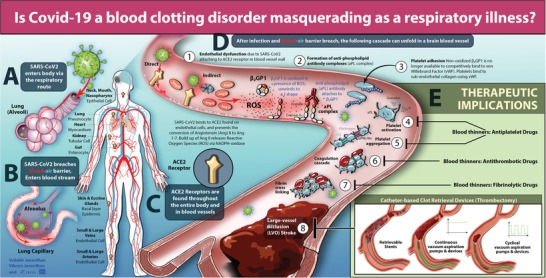
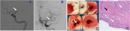
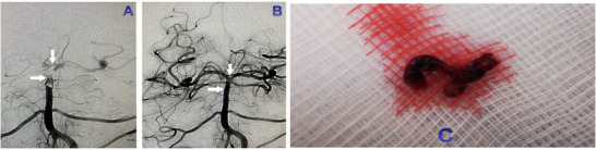
Severe acute respiratory syndrome coronavirus 2 (SARS-CoV-2) as the name suggests was initially thought to only cause a respiratory illness. However, several reports have been published of patients with ischemic strokes in the setting of coronavirus disease 2019 (COVID-19). The mechanisms of how SARS-CoV-2 results in blood clots and large vessel strokes need to be defined as it has therapeutic implications. SARS-CoV-2 enters the blood stream by breaching the blood-air barrier via the lung capillary adjacent to the alveolus, and then attaches to the angiotensin-converting enzyme II receptors on the endothelial cells. Once SARS-CoV-2 enters the blood stream, a cascade of events (Steps 1-8) unfolds including accumulation of angiotensin II, reactive oxygen species, endothelial dysfunction, oxidation of beta 2 glycoprotein 1, formation of antiphospholipid antibody complexes promoting platelet aggregation, coagulation cascade, and formation of cross-linked fibrin blood clots, leading to pulmonary emboli (PE) and large vessel strokes seen on angiographic imaging studies. There is emerging evidence for COVID-19 being a blood clotting disorder and SARS-CoV-2 using the respiratory route to enter the blood stream. As the blood-air barrier is breached, varying degrees of collateral damage occur. Although antiviral and immune therapies are studied, the role of blood thinners in the prevention, and management of blood clots in Covid-19 need evaluation. In addition to ventilators and blood thinners, continuous aspiration and clot retrieval devices (approved in Europe, cleared in the United States) or cyclical aspiration devices (approved in Europe) need to be considered for the emergent management of life-threatening clots including PE and large vessel strokes.

Keywords: Antiphospholipid antibodies; COVID-19; blood clots; stroke; thrombectomy.


Figures

Fig 1
Fig 1
Coronavirus disease 2019 (COVID‐19), blood clots, and stroke—mechanisms and therapeutic implications: In Panel A, severe acute respiratory syndrome coronavirus 2 (SARS‐CoV‐2) enters the body predominantly via the respiratory route. In Panel B, SARS‐CoV‐2 breaches the blood‐air barrier, and enters the blood stream via the lung capillary that is adjacent to the alveolus. In Panel C, once SARS‐CoV‐2 enters the blood stream, the spike protein (key) can attach to angiotensin‐converting enzyme 2 (ACE2) receptors (lock) across the body including endothelial cells in neck or brain blood vessels. Mechanisms of blood clots and stroke in COVID‐19: In Panel D, a cascade of events unfolds resulting in blood clots and strokes. In Step 1, endothelial dysfunction can occur either directly by viral entry into the endothelial cell or indirectly with the accumulation of Angiotensin (Ang) II as SARS‐CoV‐2 attaches to ACE2 and is not able to convert Ang II to Ang 1,7. Ang II can result in reactive oxygen species (ROS), oxidative stress, and endothelial dysfunction. In Step 2, oxidation of beta 2 glycoprotein 1 (β2GP1) occurs due to endothelial dysfunction and ROS, and results in the formation of antiphospholipid (aPL) antibody complexes. In Step 3, platelet adhesion occurs and platelets attach to the subendothelial collagen using von Willebrand Factor (vWF). This happens because the non‐oxidized β2GP1 is no longer available to competitively bind vWF. In Step 4, platelet activation occurs due to platelets binding to vWF resulting in granule release (α and dense) and the presence of aPL complexes further promotes platelet activation. In Steps 5‐7, platelet aggregation using vWF or fibrinogen (released by α granules), formation of thrombus via the coagulation cascade, and subsequent cross‐linking of fibrin strands to stabilize the clot occur. In Step 8, a pulmonary embolism (PE) and/or a large vessel occlusion stroke occurs. Therapeutic implications: In Panel E, several blood thinners like antiplatelet drugs can impact Steps 4 and 5 (eg, oral aspirin, oral clopidogrel, etc), antithrombotic drugs can impact Step 6 (antithrombin III binding agent, eg, parenteral heparin, vitamin K antagonist, eg, oral warfarin, newer direct Xa or thrombin inhibitors, etc), and fibrinolytic drugs can impact Step 7 (eg, intravenous tissue plasminogen activator or tenecteplase, etc). Several catheter‐based devices (approved in Europe, and cleared to market in the United States) can impact Step 8 (eg, clot retrieval devices that try to use clot integration, continuous vacuum aspiration pumps, and devices that try to use uniform negative suction pressure to ingest clots) or newer devices (approved in Europe) can impact Step 8 (eg, cyclical vacuum aspiration pumps and devices that use pulsating negative suction pressure to improve complete clot ingestion and reduce clot fragmentation), help remove blood clots, and treat PE and large vessel strokes.
Fig 2
Fig 2
An illustrative example of a cerebral angiogram (pretreatment, see Panel A; posttreatment, see Panel B) in a young stroke patient (47‐year‐old gentleman) with coronavirus disease 2019 who had three (3) recurrent reocclusions of the proximal segment of the left middle cerebral artery (see white arrow in Panel A) despite thrombectomy (using a combination of a stent retriever and continuous uniform aspiration) and finally on the fourth pass had partial revascularization (see white arrow in Panel B) due to an extensive clot burden (see white arrow in Panel C) suggestive of a prothrombotic state with refractory platelet‐fibrin‐rich clots on histopathology (Fig 2D). In Panel D, predominantly there are fibrin‐rich clots (shown as the bluish‐pink areas, see white arrows in Panel D), with interspersed cells, for example, leucocytes (see blue dots around the white arrows in Panel D), and small amounts of red blood cells (shown as the pinkish‐red areas, see black arrow in Panel D). (Courtesy: Dr Andrew R. Xavier, Michigan, USA has given permission to reproduce the figure).
Fig 3
Fig 3
An illustrative example of a cerebral angiogram (pretreatment, see Panel A; posttreatment, see Panel B) in an elderly stroke patient (82‐year‐old lady) with a basilar artery thrombus extending from the mid‐basilar artery proximal to the origin of the bilateral superior cerebellar arteries (see horizontal white arrow in Panels A‐B) to the top of the basilar artery near the bifurcation of the bilateral posterior cerebral arteries (see vertical white arrow in Panels A‐B). Patient with the basilar artery thrombus was treated with one of the newer vacuum suction devices to remove blood clots (CLEARTM Aspiration System, approved in the European Union recently). Cyclical Aspiration was performed with complete vessel reopening (TICI 3) on the first pass (Fig 3B) and successful removal of the basilar artery thrombus (Fig 3C). (Courtesy: Dr Vladimir Kalousek, Zagreb, Croatia, has given permission to reproduce the figure).

References

    1. Zhou P, Yang X, Wang X, et al. A pneumonia outbreak associated with a new coronavirus of probable bat origin. Nature 2020;579:270‐3. - PMC - PubMed
    1. Zhang Y, Xiao M, Zhang S, et al. Coagulopathy and antiphospholipid antibodies in patients with COVID‐19. N Engl J Med 2020;382:e38. - PMC - PubMed
    1. Oxley TJ, Mocco J, Majidi S, et al. Large‐vessel stroke as a presenting feature of COVID‐19 in the young. N Engl J Med 2020;382:e60. - PMC - PubMed
    1. Janardhan V, Wolf PA, Kase CS, et al. Anticardiolipin antibodies and risk of ischemic stroke and transient ischemic attack: the Framingham Cohort and Offspring Study. Stroke 2004;35:736‐41. - PubMed
    1. Hoffman M, Kleine‐Weber H, Schroeder S, et al. SARS‐CoV‐2 cell entry depends on ACE2 and TMPRSS2 and is blocked by a clinically proven protease inhibitor. Cell 2020;181:271‐80. - PMC - PubMed

Publication types