. 2020 Aug 10;18(8):e3000826.
doi: 10.1371/journal.pbio.3000826. eCollection 2020 Aug.

CaMKII controls neuromodulation via neuropeptide gene expression and axonal targeting of neuropeptide vesicles

Affiliations

CaMKII controls neuromodulation via neuropeptide gene expression and axonal targeting of neuropeptide vesicles

Alessandro Moro et al. PLoS Biol. .

Abstract

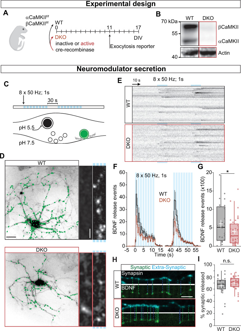
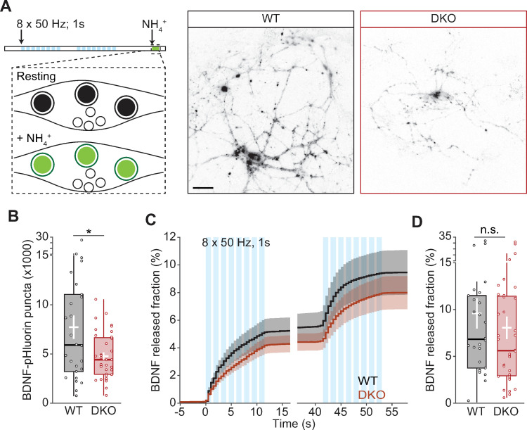
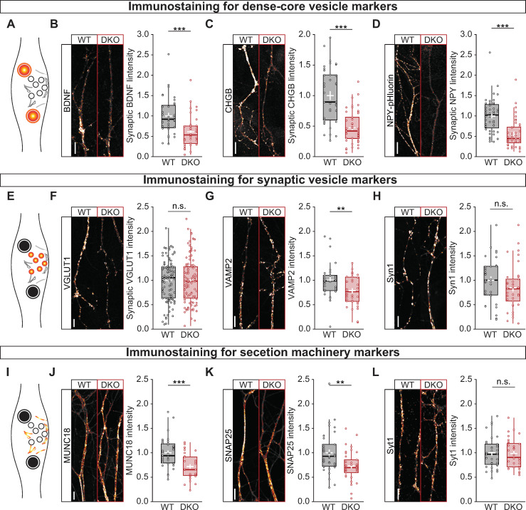
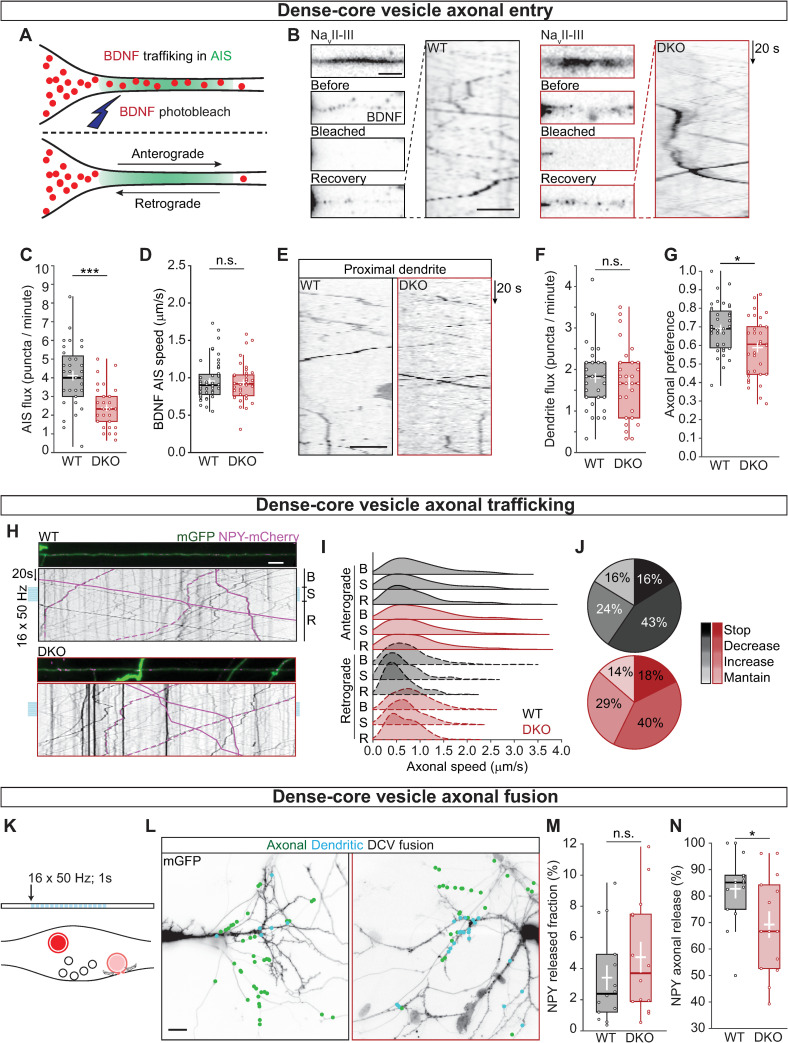
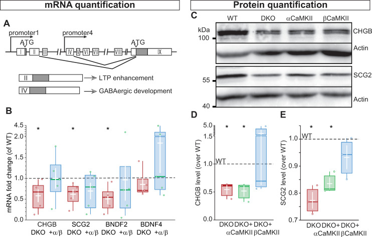
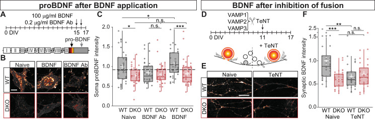
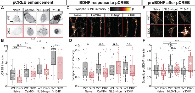
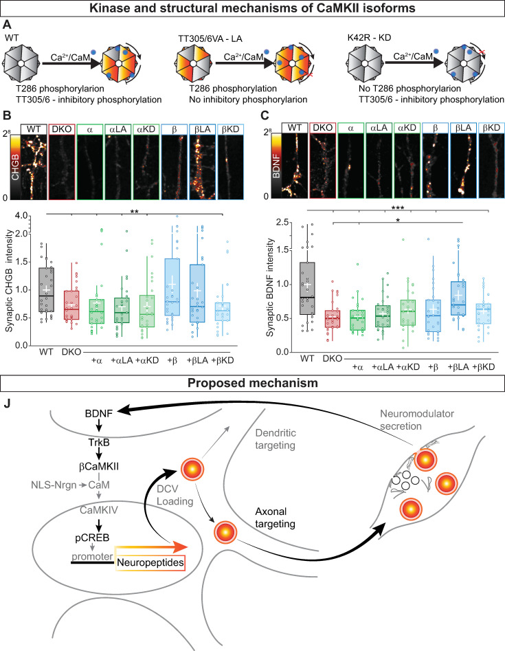
Ca2+/calmodulin-dependent kinase II (CaMKII) regulates synaptic plasticity in multiple ways, supposedly including the secretion of neuromodulators like brain-derived neurotrophic factor (BDNF). Here, we show that neuromodulator secretion is indeed reduced in mouse α- and βCaMKII-deficient (αβCaMKII double-knockout [DKO]) hippocampal neurons. However, this was not due to reduced secretion efficiency or neuromodulator vesicle transport but to 40% reduced neuromodulator levels at synapses and 50% reduced delivery of new neuromodulator vesicles to axons. αβCaMKII depletion drastically reduced neuromodulator expression. Blocking BDNF secretion or BDNF scavenging in wild-type neurons produced a similar reduction. Reduced neuromodulator expression in αβCaMKII DKO neurons was restored by active βCaMKII but not inactive βCaMKII or αCaMKII, and by CaMKII downstream effectors that promote cAMP-response element binding protein (CREB) phosphorylation. These data indicate that CaMKII regulates neuromodulation in a feedback loop coupling neuromodulator secretion to βCaMKII- and CREB-dependent neuromodulator expression and axonal targeting, but CaMKIIs are dispensable for the secretion process itself.


Conflict of interest statement

The authors have declared that no competing interests exist.

Figures

Fig 1
Fig 1. CaMKII regulates neuromodulation.
(A) Schematic representation of the experimental design. Neuronal cultures from floxed αβCaMKII P1 pups were infected at DIV0 with Cre-recombinase to generate DKO neurons and with inactive Cre as control. Cultures were infected at DIV11 with BDNF-pHluorin as exocytosis reporter, and live-cell imaging was performed at DIV17. (B) Typical Western blot showing the complete KO for both α and βCaMKII. (C) Schematic representation of DCV fusion assay. DCVs are labeled with BDNF-pHluorin, and neurons are stimulated with 2 trains of 8 bursts of 50 APs at 50 Hz (light blue bars). (D) Representative neurons expressing CFP driven by CaMKII promoter superimposed with BDNF-pHluorin events (green dots). Insert shows a neurite at resting state (left) and upon stimulation (marked by light blue bars on the right). (E) Kymographs of BDNF-pHluorin fusion events. Individual DCV fusion events, black dots in the images, were quantified semi-automatically. (F) Histogram of the average number of DCV fusion events per cell during stimulation (light blue bars). (G) Total number of BDNF-pHluorin release events per cell. (H) Typical example of BDNF-pHluorin fusion events at synapses visualized by Synapsin-mCherry (dashed lines) and at extrasynaptic sites. (I) Percentage of BDNF-pHluorin fusion events at synapses. Traces show mean ± SEM (shaded area); boxplots with 95% CI whiskers, the white cross represents mean ± SEM, and the central bar is median. Columns and dots represent individual litters and neurons, respectively. The presented data can be found in S1 Data; original Western blot can be found in S1 Raw images. *p = 0.057. Scale bar = 25 μm (D); 5 μm (D insert); 10 μm (I). AP, action potential; BDNF, brain-derived neurotrophic factor; CaMKII, Ca2+/calmodulin-dependent kinase II; CFP, cyan fluorescence protein; CI, confidence interval; DCV, dense-core vesicle; DIV, day in vitro; DKO, double-knockout; WT, wild type.
Fig 2
Fig 2. CaMKII regulates DCV content but not their fusion efficiency.
(A) Schematic representation of DCV fusion assay. DCVs are labeled with BDNF-pHluorin, and neurons are stimulated with 2 trains of 8 bursts of 50 APs at 50 Hz (light blue bars). At the end of the stimulation, the bath application of NH4Cl dequenches all BDNF-pHluorin-containing vesicles. Right, representative neurons with BDNF-pHluorin-labeled vesicles dequenched. (B) Total number of DCVs per cell quantified upon NH4+ superfusion. (C) Cumulative fraction of DCV fusion events during stimulation. The fraction is calculated as the number of DCVs fusing per frame divided by the total number of BDNF-pHluorin puncta per neuron. (D) Fraction of BDNF-pHluorin-labeled DCV fusing during stimulation. Traces show mean ± SEM (shaded area); boxplots with 95% CI whiskers, the white cross represents mean ± SEM, and the central bar is median. Columns and dots represent individual litters and neurons, respectively. The presented data can be found in S1 Data. *p < 0.05. Scale bar = 25 μm (A). AP, action potential; BDNF, brain-derived neurotrophic factor; CaMKII, Ca2+/calmodulin-dependent kinase II; CI, confidence interval; DCV, dense-core vesicle; DIV, day in vitro; DKO, double-knockout; WT, wild type.
Fig 3
Fig 3. CaMKII controls the abundance of DCVs, not SVs or secretory machinery.
(A) Schematic representation of the immunostaining of DCV markers, in color. (B) Left, typical images of neurites of WT (left) and DKO (right) immunostained for BDNF. Right, quantification of BDNF intensity at VGLUT1 labeled synapses in WT and DKO neurons. (C) Left, typical images of neurites of WT (left) and DKO (right) immunostained for CHGB. Right, quantification of CHGB intensity at VGLUT1 labeled synapses in WT and DKO neurons. (D) Left, typical images of neurites of WT (left) and DKO (right) immunostained for the exogenous DCV marker NPY-pHluorin. Right, quantification of NPY-pHluorin intensity at VGLUT1 labeled synapses in WT and DKO neurons. (E) Schematic representation of the immunostaining of SV markers, in color. (F) Left, typical images of neurites of WT (left) and DKO (right) immunostained for VGLUT1. Right, quantification of VGLUT1 intensity in WT and DKO neurons. (G) Left, typical images of neurites of WT (left) and DKO (right) immunostained for VAMP2. Right, quantification of VAMP2 intensity at VGLUT1-labeled synapses in WT and DKO neurons. (H) Left, typical images of neurites of WT (left) and DKO (right) immunostained for Syn1. Right, quantification of Syn1 intensity at VGLUT1 labeled synapses in WT and DKO neurons. (I) Schematic representation of the immunostaining of exocytic proteins, in color. (J) Left, typical images of neurites of WT (left) and DKO (right) immunostained MUNC18. Right, quantification of MUNC18 intensity at VGLUT1-labeled synapses in WT and DKO neurons. (K) Left, typical images of neurites of WT (left) and DKO (right) immunostained for SNAP25. Right, quantification of SNAP25 intensity at VGLUT1 labeled synapses in WT and DKO neurons. (L) Left, typical images of neurites of WT (left) and DKO (right) immunostained for Syt1. Right, quantification of Syt1 intensity at VGLUT1-labeled synapses in WT and DKO neurons. Boxplots with 95% CI whiskers, white cross shows mean ± SEM, central bar is median. Columns and dots represent individual litters and neurons, respectively. The presented data can be found in S1 Data. *p < 0.05, **p < 0.01, ***p < 0.001. Scale bar = 5 μm. CaMKII, Ca2+/calmodulin-dependent kinase II; CI, confidence interval; DCV, dense-core vesicle; DKO, double-knockout; MUNC18, mammalian uncoordinated 18; NPY, neuropeptide Y; n. s., not significant; SNAP25, synaptosomal-associated protein 25; SV, synaptic vesicle; Syn1, synapsin 1; VAMP2, vesicle-associated membrane protein 2; VGLUT1, vesicular glutamate transporter 1; WT.
Fig 4
Fig 4. CaMKII regulates the number of DCVs entering the axon but not their trafficking properties.
(A) Schematic representation of AIS targeting; NavII-III BFP was used to visualize the region of the AIS. BDNF-mCherry vesicles were bleached at the AIS to allow quantification of new vesicles entering the axon. (B) Left, typical example of AIS (top) with BDNF-mCherry-labeled DCVs before bleaching, after bleaching, and at the end of the recovery (top to bottom). Right, kymograph showing DCVs in the AIS after bleaching. (C) Quantification of DCV flux at the AIS calculated as the number of BDNF-mCherry positive puncta that enter the NavII-III BFP area per minute in anterograde (from the soma to the axon) direction. (D) Quantification of BDNF-mCherry speed at the AIS in the anterograde direction. (E) Kymograph showing DCVs in proximal dendrites after bleaching. (F) Quantification of DCV flux in the proximal dendrites in the anterograde direction. (G) Axonal preference of DCV targeting, calculated as BDNF-mCherry-positive puncta entering in the NavII-III BFP area in anterograde direction divided by the total number of BDNF-mCherry positive puncta exiting the soma. (H) Typical examples of axons labeled with mGFP showing NPY-mCherry labeled DCVs; kymographs represent axonal trafficking of NPY-mCherry in anterograde (solid lines) and retrograde (dashed lines) direction. Neurons were stimulated with 16 trains of 50 APs at 50 Hz (light blue bars). DCV speed was calculated before stimulation (B), during stimulation (S), and during recovery (R). (I) Ridgelines representing the density function of NPY-mCherry puncta speed along the axon, showing the characteristic differences in speed for anterograde and retrograde (filled and dashed density respectively) and the lower speed during stimulation. (J) Pie charts for WT (black) and DKO neurons (red) with the fraction of vesicles that stopped during stimulation (Stop), decreased their speed (Decrease), increased their speed (Increase), and maintained the same average speed (Maintain). (K) Schematic representation of DCV fusion assay. DCVs are labeled with NPY-mCherry, neurons are stimulated with 16 trains of 50 APs at 50 Hz (light blue bars). Individual DCV fusion events were quantified manually as puncta that suddenly lost their fluorescence signal. (L) Typical neurons labeled with mGFP overlaid with NPY-mCherry-labeled DCV fusion events (green dots). Axons were identified by the lack of spines. (M) Quantification of NPY-mCherry release fraction upon stimulation. (N) Quantification of the percentage of NPY-mCherry fusion events at axons. Boxplots with 95% CI whiskers, white cross shows mean ± SEM, and central bar is median. Columns and dots represent individual litters and neurons, respectively. The presented data can be found in S1 Data. *p < 0.05, **p < 0.01, ***p < 0.001. Scale bar = 5 μm (B for still frames), 10 μm (B kymograph and E), 25 μm (H), 25 μm (L). AIS, axon initial segment; AP, action potential; BDNF, brain-derived neurotrophic factor; BFP, blue fluorescence protein; CaMKII, Ca2+/calmodulin-dependent kinase II; CI, confidence interval; DCV, dense-core vesicle; DKO, double-knockout; mGFP, membrane-bound green fluorescence protein; NavII-III,voltage-gated sodium channel intracellular domain; NPY, neuropeptide Y; n. s., not significant; WT, wild type.
Fig 5
Fig 5. CaMKII regulates neuropeptide expression.
(A) Representation of BDNF genomic locus, exons are shown as white boxes, the gray box represents the conserved exon shared between the different transcripts. (B) Quantification of mRNA fold change, expressed as ΔCt values normalized to WT, showing the reduction of mRNA levels for CHGB, SCG2, and BDNF transcript 2 in DKO neurons compared with DKO neurons rescued with α and βCaMKII. BDNF transcript 4 is not affected by the lack of CaMKII. (C) Western blot showing the DCV cargo proteins CHGB (top) and SCG2 (bottom) in WT, DKO, and DKO-expressing αCaMKII or βCaMKII neurons. Actin was used as loading control. (D) Quantification of CHGB levels normalized to WT. (E) Quantification of SCG2 levels normalized to WT. Boxplots with 95% CI whiskers, white cross shows mean ± SEM, and central bar is the median. Columns and dots represent individual litters and neurons, respectively. The presented data can be found in S1 Data; original Western blot can be found in S1 Raw images. ATG, transcription starting codon; BDNF, brain-derived neurotrophic factor; CaMKII, Ca2+/calmodulin-dependent kinase II; CHGB, chromogranin B; CI, confidence interval; DCV, dense-core vesicle; DKO, double-knockout; LTP, long-term potentiation; SCG2, secretogranin2; WT, wild type.
Fig 6
Fig 6. Secreted BDNF is sufficient to increase BDNF levels in the presence of CaMKII.
(A) Schematic representation of the experimental design, 0.2 μg/ml of BDNF Ab or 100 ng/ml of recombinant BDNF were added every 24 hours for 2 days before fixing the neurons and immunostained for proBDNF. (B) Typical examples of proBDNF in the soma from untreated neurons (Naive), treated with BDNF Ab or BDNF. (C) Quantification of somatic proBDNF intensity normalized to WT (dash line). (D) Schematic representation of the experimental design, lentiviral particles containing TeNT, which is responsible to cleaved VAMP1-3 (hence, inhibiting SV and DCV fusion), were added 6 DIVs before fixing the neurons and immunostained for BDNF. (E) Typical examples of BDNF in synapses from untreated (Naive) cells or treated with TeNT. (F) Quantification of synaptic BDNF intensity normalized to WT (dash line). Boxplots with 95% CI whiskers, white cross shows mean ± SEM. Columns and dots represents individual litters and neurons, respectively. The presented data can be found in S1 Data. *p < 0.05, **p < 0.01, ***p < 0.001. Scale bar = 20 μm (B-E). BDNF, brain-derived neurotrophic factor; BDNF-Ab, BDNF antibody; CaMKII, Ca2+/calmodulin-dependent kinase II; CI, confidence interval; DCV, dense-core vesicle; DIV, day in vitro; DKO, double-knockout; SV, synaptic vesicle; TeNT, tetanus neurotoxin; VAMP, vesicle-associated membrane protein; WT, wild type.
Fig 7
Fig 7. CaMKII is required for BDNF-induced BDNF production via pCREB.
(A) Typical example of nuclei immunostained for pCREB at Ser-133. (B) Quantification of pCREB signal intensity normalized to WT (dashed line). (C) Typical examples of BDNF in synapses from untreated (Naive) cells, or treated with CaMKIV, NLS-Nrgn, or CREB-Y134F. (D) Quantification of synaptic BDNF intensity normalized to WT. (E) Typical examples of proBDNF in the soma from untreated neurons (Naive), treated NLS-Nrgn or CREB-Y134F. (F) Quantification of somatic proBDNF intensity normalized to WT (dash line). Boxplots with 95% CI whiskers, white cross shows mean ± SEM. Columns and dots represents 0individual litters and neurons, respectively. The presented data can be found in S1 Data. *p < 0.05, **p < 0.01, ***p < 0.001. Scale bar = 20 μm (A-E) and 10 μm (C). BDNF, brain-derived neurotrophic factor; CaMKII, Ca2+/calmodulin-dependent kinase II; CaMKIV, Ca2+/calmodulin-dependent kinase IV; CREB, cAMP-response element binding protein; CREB-Y134F, phosphorylation of CREB with Tyr-to-Phe substitution at position 134; DKO, double-knockout; NLS-Nrgn, nuclear-localized neurogranin; n.s., not significant; pCREB, CREB phosphorylation; WT, wild type.
Fig 8
Fig 8. CaMKII activity controls neuropeptide levels.
β (A) Schematic representation of WT αCaMKII (left) with Ca/CaM-induced autophosphorylation of T286 triggering the activity of the kinase. The TT305/306VA mutation (middle) inhibits TT305/6 inhibitory phosphorylation leading to an LA form of the kinase. The K42R mutation (right) does not allow T286 autophosphorylation producing a KD CaMKII. For βCaMKII, the mutations are TT306/307VA and K43R for the LA and KD, respectively. (B) Top, typical neurites immunostained for the endogenous DCV marker CHGB; bottom, quantification of synaptic CHGB intensity levels at VGLUT1 positive synapses for DKO and DKO rescued with the indicated constructs normalized to WT. (C) Top, typical neurites immunostained for endogenous BDNF; bottom, quantification of synaptic BDNF intensity levels at VGLUT1 positive synapses for DKO and DKO rescued with the indicated constructs normalized to WT. (D) Schematic representation of the mechanism for CaMKII- and BDNF-dependent gene expression. BDNF binds to TrkB receptors; BDNF-TrkB binding leads to activation of βCaMKII that triggers a downstream pathway leading to Ca/CaM to enter into the nucleus and activating CaMKIV, which in turn phosphorylate CREB at S133. Neuropeptides and neuromodulator are then loaded into DCVs and targeted to dendrites and synapses, where they will fuse with the plasma membrane releasing their content. This mechanism will create a positive feedback loop to regulate neuromodulators content. Boxplots with 95% CI whiskers, white cross shows mean ± SEM. Columns and dots represents individual litters and neurons, respectively. The presented data can be found in S1 Data. *p < 0.05, **p < 0.01, ***p < 0.001. Scale bar = 10 μm (B and D). Ca/CaM, Ca2+/Calmodulin; CaMKII, Ca2+/calmodulin-dependent kinase II; CaMKIV, Ca2+/calmodulin-dependent kinase IV; CHGB, chromogranin B; CI, confidence interval; CREB, cAMP-response element binding protein; DCV, dense-core vesicle; DKO, double-knockout; KD, kinase-dead; LA, long-active; NLS-Nrgn, nuclear-localized neurogranin; SV, synaptic vesicle; Syn1, synapsin1; TrKB, tyrosine receptor kinase B; VAMP2, vesicle-associated membrane protein 2; VGLUT1, vesicular glutamate transporter 1; WT, wild type.

Similar articles

Cited by

References

    1. Poo MM. Neurotrophins as synaptic modulators. Nat Rev Neurosci. 2001;2(1):24–32. 10.1038/35049004 - DOI - PubMed
    1. Dean C, Liu H, Dunning FM, Chang PY, Jackson MB, Chapman ER. Synaptotagmin-IV modulates synaptic function and long-term potentiation by regulating BDNF release. Nat Neurosci. 2009;12(6):767–76. 10.1038/nn.2315 - DOI - PMC - PubMed
    1. Korte M, Carroll P, Wolf E, Brem G, Thoenen H, Bonhoeffer T. Hippocampal long-term potentiation is impaired in mice lacking brain-derived neurotrophic factor. Proc Natl Acad Sci U S A. 1995;92(19):8856–60. 10.1073/pnas.92.19.8856 - DOI - PMC - PubMed
    1. Linnarsson S, Bjorklund A, Ernfors P. Learning deficit in BDNF mutant mice. Eur J Neurosci. 1997;9(12):2581–7. 10.1111/j.1460-9568.1997.tb01687.x - DOI - PubMed
    1. Hartmann M, Heumann R, Lessmann V. Synaptic secretion of BDNF after high-frequency stimulation of glutamatergic synapses. EMBO J. 2001;20(21):5887–97. 10.1093/emboj/20.21.5887 - DOI - PMC - PubMed

Publication types

MeSH terms

Substances

LinkOut - more resources