. 2020 Aug 10;15(8):e0236781.
doi: 10.1371/journal.pone.0236781. eCollection 2020.

Bach1 promotes muscle regeneration through repressing Smad-mediated inhibition of myoblast differentiation

Affiliations

Bach1 promotes muscle regeneration through repressing Smad-mediated inhibition of myoblast differentiation

Katsushi Suzuki et al. PLoS One. .

Abstract

It has been reported that Bach1-deficient mice show reduced tissue injuries in diverse disease models due to increased expression of heme oxygenase-1 (HO-1)that possesses an antioxidant function. In contrast, we found that Bach1 deficiency in mice exacerbated skeletal muscle injury induced by cardiotoxin. Inhibition of Bach1 expression in C2C12 myoblast cells using RNA interference resulted in reduced proliferation, myotube formation, and myogenin expression compared with control cells. While the expression of HO-1 was increased by Bach1 silencing in C2C12 cells, the reduced myotube formation was not rescued by HO-1 inhibition. Up-regulations of Smad2, Smad3 and FoxO1, known inhibitors of muscle cell differentiation, were observed in Bach1-deficient mice and Bach1-silenced C2C12 cells. Therefore, Bach1 may promote regeneration of muscle by increasing proliferation and differentiation of myoblasts.


Conflict of interest statement

The authors have declared that no competing interests exist.

Figures

Fig 1
Fig 1. Bach1 is dispensable for muscle growth.
(A-C) Box plots showing body weight (A), muscle weight (B) and length of tibia (C) of uninjured Bach1-deficient and WT mice.(n = 5).
Fig 2
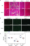
Fig 2. Muscle regeneration is impaired in Bach1-deficient mice.
(A) HE staining of tibialis anterior muscle sections from Bach1-deficient and WT mice 3, 7, or 21 days after muscle injury. Uninjured samples are also shown. Mononuclear cells infiltrated on day 3 (arrow heads). (B) Immunofluorescence staining with anti-Laminin antibody (green) of tibialis anterior muscle sections of Bach1-deficient and WT. Nuclei were stained with DAPI (blue). (C) CSA of injured muscle was calculated using myofibers with central nuclei. (n = 5–8; * p< 0.05).
Fig 3
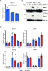
Fig 3. Bach1 protein increases after skeletal muscle injury.
(A) Expression level of Bach1 mRNA measured by RT-qPCR in muscle of WT mice at indicated time points after muscle injury (n = 3; * p< 0.05). (B) Western blotting of muscle of Bach1-deficient and WT mice as above with antibodies against Bach1, or GAPDH as an internal control. (C) Expression of Myod1 (MyoD), Myf5, Myog (Myogenin) and Myf6 (MRF4) mRNA measured by RT-qPCR in muscle of Bach1-deficient and WT mice as above. (n = 3).
Fig 4
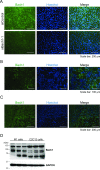
Fig 4. Bach1 protein increases in regenerated muscle fibers.
(A) Immunofluorescence staining with anti-Bach1 antibody (green) of C2C12 cells transfected with Bach1 siRNA (siBach1-1) or control siRNA. Nuclei were stained with Hoechst (blue). (n = 3). (B) Immunofluorescence staining with anti-Bach1 antibody (green) of tibialis anterior muscle sections from WT mice 3 days after muscle injury. Nuclei were stained with Hoechst (blue) (n = 3). (C) Immunofluorescence staining with anti-Bach1 antibody (green) of tibialis anterior muscle sections from WT mice 7 days after muscle injury. Nuclei were stained with Hoechst (blue) (n = 6). (D) Western blotting of M1 cells and C2C12 cells 6 days after inducing differentiation.
Fig 5
Fig 5. Bach1 is necessary for proliferation of C2C12 cells.
(A) Experimental scheme of the proliferation analysis. Cells were cultured in 12-well plate. (B) Cell numbers of C2C12 cells transfected with Bach1 siRNA (siBach1-1 and siBach1-2) or control siRNA. (n = 3). (C) Proliferation rates of C2C12 cells with Bach1 silencing or control C2C12 cells at indicated periods. (n = 3; * p< 0.05).
Fig 6
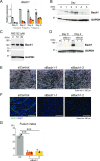
Fig 6. Bach1 is required for myoblasts differentiation of C2C12 cells.
(A) Expression levels of Bach1 mRNA were measured by RT-qPCR in C2C12 cells transfected with indicated siRNA before (day 0) and after inducing differentiation (day 2, 4 and 6). (n = 3). (B) Western blotting of C2C12 cells before (day 0) and after inducing differentiation(day 1–5). (C) Western blotting of MG132-treated and control C2C12 cells. (D) Western blotting of Bach1 in indicated C2C12 cells before (day 0) and after inducing differentiation (day 2). (E) Morphology of C2C12 cells 5 days after inducing differentiation. (F) Immunohistochemical staining of MHC and nuclei (DAPI) of Bach1-silenced and control C2C12 cells 6 days after inducing differentiation. (G) Fusion index after 6 days. (n = 3; *** p < 0.001).
Fig 7
Fig 7. Bach1 regulates Mhc and MRFs mRNA expression.
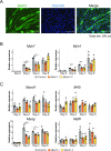
(A) Immunohistochemical staining of Bach1 and nuclei (Hoechst) of C2C12 cells 6 days after inducing differentiation. (B, C) Expression levels of Myh7 (MHC Ⅰ/b) and Myh1 (MHC Ⅱd/x)mRNA (B) and Myogenic regulatory factors (MRFs: Myod1 (MyoD), Myf5, Myog (myogenin) and Myf6 (Mrf4))mRNA (C) measured by RT-qPCR of Bach1-silenced and control C2C12 cells before (day 0) and after inducing differentiation (day 2, 4 and 6). (n = 3; * p< 0.05, ** p< 0.01, *** p < 0.001).
Fig 8
Fig 8. Overexpression of Bach1 does not affect differentiation of C2C12 cells.
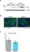
(A) Western blotting of control and Bach1-overexpressing C2C12 cells before (day 0) and after inducing differentiation (day 2). (B) Immunohistochemical staining of MHC and nuclei (DAPI) of control and Bach1-overexpressing C2C12 cells 5 days after inducing differentiation. (C) Fusion index after 5 days. (n = 3).
Fig 9
Fig 9. Senescence and HO-1 overexpression of Bach1-silenced cells are not the cause of disturbed differentiation.
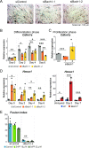
(A) Bach1-silenced and control C2C12 cells were stained for SA β-gal 6 days after inducing differentiation. (n = 3). (B, C)Expression levels of Cdkn1a (p21) measured by RT-qPCR at differentiation phase (B) and proliferation phase (C). (n = 3).(D) Expression level of Hmox1 in Bach1-silenced and control C2C12 cells before (day 0) and after inducing differentiation (Day 2, 4 and 6) (left), and of muscle of Bach1-deficient and WT before (day 0) and 3 or 7 days after muscle injury (right). (n = 3). (E) Fusion index of Bach1-silenced and control C2C12 cells 6 days after inducing differentiation. (n = 3; * p< 0.05, ** p< 0.01, *** p < 0.001).
Fig 10
Fig 10. Gene expression profiles of Bach1-silenced and control C2C12 cells.
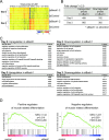
(A) Clustering analysis of microarray gene expression profiles of indicated cells before or after inducing differentiation. (B) The table shows numbers of upregulated and downregulated probes (fold change ≥ 2.0). (C) The tables show gene ontology (GO) analysis results using genes in (B). (D) GSEA enrichment plots of genes related to positive (right) and negative (left) regulators of muscle differentiation.
Fig 11
Fig 11. Bach1 decreases Smad3 protein.
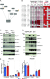
(A) Signaling pathways known to suppress MyoD and myogenin. (B) Heat map of pathway related genes from microarray results in Fig 10A. Maps show normalized signals (left), fold change (middle) and p value (right) compared Bach1-silenced to control C2C12 cells at day 0 and day 2, or compared day 2 to day 0 in Bach1-silenced or control C2C12 cells. (C) Western blotting of Bach1-silenced and control C2C12 cells before (day 0) and after inducing differentiation (day 2). (D) Western blotting of Bach1-deficient and WT mice muscle at 3 or 7 days after injury. Uninjured samples are also shown. Western blotting was performed with indicated antibodies. (E) Expression of Fbxo32 and Trim63 mRNA measured by RT-qPCR in muscle of Bach1-deficient and WT mice at 3 or 7 days after injury or before injury. (n = 3; ** p< 0.01).
Fig 12
Fig 12. Smad2 and Smad3 inhibit myoblasts differentiation of C2C12 cells.
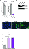
(A) Expression of Smad2 (left) and Smad3 (right) mRNA was measured by RT-qPCR in control C2C12 cells or those treated with indicated siRNA before inducing differentiation. (n = 3). (B) Western blotting of Smad2 and Smad3 in C2C12 cells treated as above after inducing differentiation (day 2). (C) Immunohistochemical staining of MHC and nuclei (DAPI) of C2C12 cells treated as above 5 days after inducing differentiation. (D) Fusion index after 5 days. (n = 3; ** p < 0.01, *** p < 0.001).
Fig 13
Fig 13. A model on the regulation of myoblast differentiation by Bach1.
Bach1 accelerates myoblast differentiation through inhibition of Smad3 and FoxO1 expression.

Similar articles

Cited by

References

    1. Hawkins RD, Hulse MA, Wilkinson C, Hodson A, Gibson M. The association football medical research programme: an audit of injuries in professional football. British Journal of Sports Medicine. 2001;35(1):43–7. Epub 2001/02/07. 10.1136/bjsm.35.1.43 - DOI - PMC - PubMed
    1. Ekstrand J, Hagglund M, Walden M. Epidemiology of muscle injuries in professional football (soccer). Am J Sports Med. 2011;39(6):1226–32. Epub 2011/02/22. 10.1177/0363546510395879 . - DOI - PubMed
    1. Brooks J, Fuller C, Kemp S, Reddin D. Epidemiology of injuries in English professional rugby union: part 2 training Injuries. British Journal of Sports Medicine. 2005;39(10):767–75. 10.1136/bjsm.2005.018408 - DOI - PMC - PubMed
    1. Ahmad CS, Dick RW, Snell E, Kenney ND, Curriero FC, Pollack K, et al. Major and Minor League Baseball Hamstring Injuries: Epidemiologic Findings From the Major League Baseball Injury Surveillance System. Am J Sports Med. 2014;42(6):1464–70. Epub 2014/04/15. 10.1177/0363546514529083 . - DOI - PubMed
    1. Opar DA, Drezner J, Shield A, Williams M, Webner D, Sennett B, et al. Acute hamstring strain injury in track-and-field athletes: A 3-year observational study at the Penn Relay Carnival. Scandinavian journal of medicine & science in sports. 2014;24(4):e254–9. Epub 2013/12/18. 10.1111/sms.12159 . - DOI - PubMed

Publication types

Substances