. 2020 Aug 10;16(8):e1008730.
doi: 10.1371/journal.ppat.1008730. eCollection 2020 Aug.

Sperm associated antigen 9 promotes oncogenic KSHV-encoded interferon regulatory factor-induced cellular transformation and angiogenesis by activating the JNK/VEGFA pathway

Affiliations

Sperm associated antigen 9 promotes oncogenic KSHV-encoded interferon regulatory factor-induced cellular transformation and angiogenesis by activating the JNK/VEGFA pathway

Wan Li et al. PLoS Pathog. .

Erratum in

Abstract

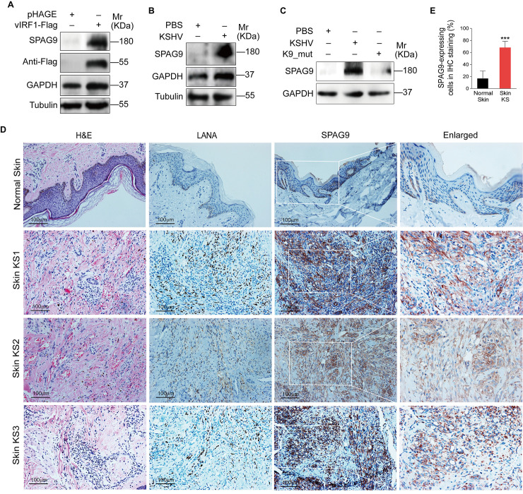
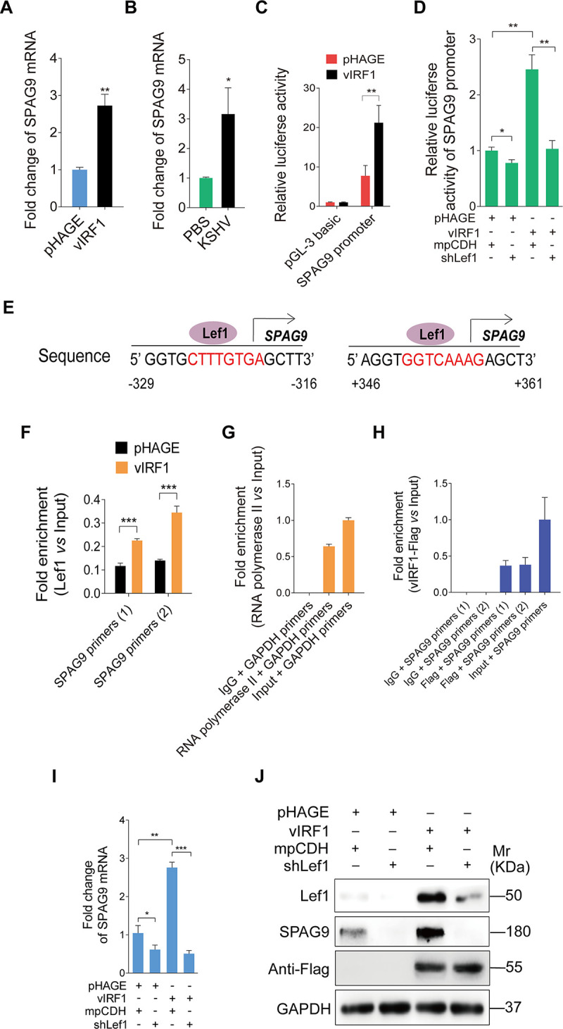
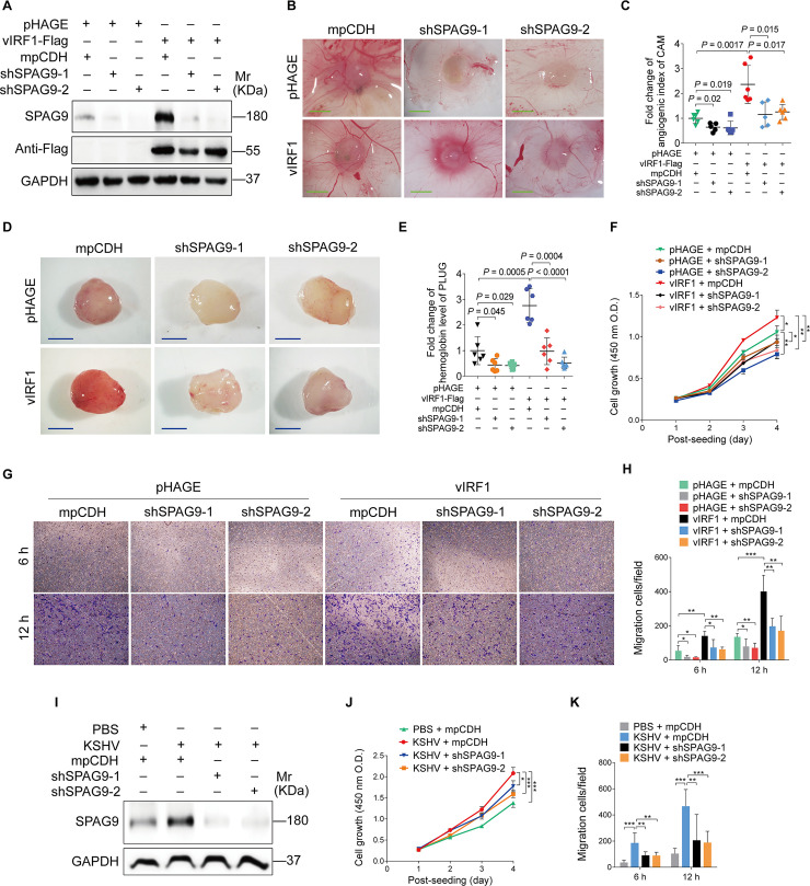
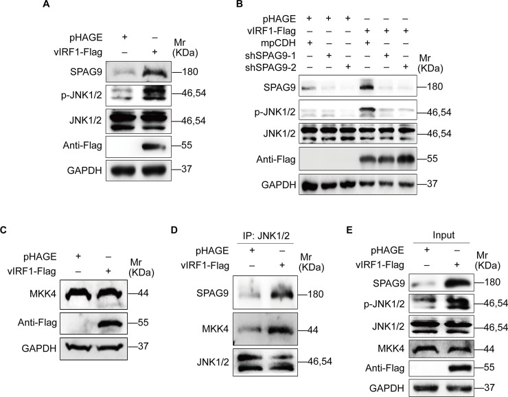
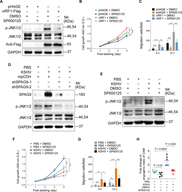
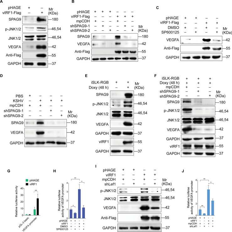
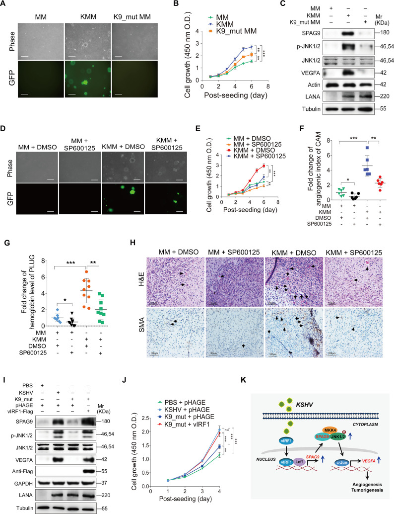
Kaposi's sarcoma (KS), caused by Kaposi's sarcoma-associated herpesvirus (KSHV), is a highly angioproliferative disseminated tumor of endothelial cells commonly found in AIDS patients. We have recently shown that KSHV-encoded viral interferon regulatory factor 1 (vIRF1) mediates KSHV-induced cell motility (PLoS Pathog. 2019 Jan 30;15(1):e1007578). However, the role of vIRF1 in KSHV-induced cellular transformation and angiogenesis remains unknown. Here, we show that vIRF1 promotes angiogenesis by upregulating sperm associated antigen 9 (SPAG9) using two in vivo angiogenesis models including the chick chorioallantoic membrane assay (CAM) and the matrigel plug angiogenesis assay in mice. Mechanistically, vIRF1 interacts with transcription factor Lef1 to promote SPAG9 transcription. vIRF1-induced SPAG9 promotes the interaction of mitogen-activated protein kinase kinase 4 (MKK4) with JNK1/2 to increase their phosphorylation, resulting in enhanced VEGFA expression, angiogenesis, cell proliferation and migration. Finally, genetic deletion of ORF-K9 from KSHV genome abolishes KSHV-induced cellular transformation and impairs angiogenesis. Our results reveal that vIRF1 transcriptionally activates SPAG9 expression to promote angiogenesis and tumorigenesis via activating JNK/VEGFA signaling. These novel findings define the mechanism of KSHV induction of the SPAG9/JNK/VEGFA pathway and establish the scientific basis for targeting this pathway for treating KSHV-associated cancers.


Conflict of interest statement

The authors have declared that no competing interests exist.

Figures

Fig 1
Fig 1. SPAG9 is upregulated in vIRF1-transduced HUVECs and KSHV-infected HUVECs.
(A). Western-blotting analysis of SPAG9 in HUVECs transduced with lentiviral-vIRF1 or its control lentiviral-pHAGE (MOI of 2). (B). Western-blotting analysis of SPAG9 in HUVECs treated with PBS (PBS) or infected by KSHV wild-type virus (KSHV) (MOI of 3). (C). Western-blotting analysis of SPAG9 expression in HUVECs treated with PBS (PBS) or infected with wild-type KSHV (KSHV_WT) (MOI of 3) or vIRF1 mutant virus (K9_mut) (MOI of 3) for 30 h. (D). Hematoxylin and eosin (H&E) staining and immunohistochemical staining (IHC) of KSHV LANA, SPAG9 in normal skin, skin KS of patient #1 (Skin KS1), patient #2 (Skin KS2), and patient #3 (Skin KS3). Magnification, ×200, ×400. (E). Results were quantified in (D). Data were shown as mean ± SD. *** P < 0.001, Student's t-test.
Fig 2
Fig 2. vIRF1 transcriptionally activates SPAG9 by interacting with Lef1.
(A). RT-qPCR analysis of mRNA level of SPAG9 in HUVECs transduced with lentiviral-vIRF1 or its control lentiviral-pHAGE. (B). RT-qPCR analysis of mRNA level of SPAG9 in treated with PBS (PBS) or infected by KSHV wild-type virus (KSHV) (MOI of 3). (C). Luciferase reporter assay of the activity of SPAG9 promoter in HUVECs transduced with lentiviral-vIRF1 or its control lentiviral-pHAGE. (D). Luciferase reporter assay of the activity of SPAG9 promoter in vIRF1-expressing HUVECs transduced with a mixture of lentivirus-mediated shRNAs targeting Lef1 (shLef1). (E). Putative binding sites of Lef1 in the promoter region of SPAG9 gene. (F). ChIP assays of SPAG9 promoter. Immunoprecipitation was performed in vIRF1- or pHAGE-transduced HUVECs with anti-Lef1 antibody. Both SPAG9 primers (1) and (2) were used to amplify the sequences of the above two putative binding sites of Lef1 in the region of SPAG9 promoter as described in (E). (G) and (H). ChIP assays of SPAG9 promoter. Immunoprecipitation was performed in vIRF1-transduced HUVECs with anti-Flag antibody. Isotype IgG, anti-RNA polymerase II antibody and amplification of GAPDH promoter were used to examined the work of system. (I). RT-qPCR analysis of mRNA expression level of SPAG9 in vIRF1-expressing HUVECs transduced with a mixture of lentivirus-mediated shRNAs targeting Lef1 (shLef1). (J). Western-blotting analysis of SPAG9 expression in vIRF1-expressing HUVECs transduced with a mixture of lentivirus-mediated shRNAs targeting Lef1 (shLef1). Data were shown as mean ± SD. * P < 0.05, ** P < 0.01, and *** P < 0.001, Student's t-test.
Fig 3
Fig 3. Knockdown of SPAG9 inhibits vIRF1-induced angiogenesis, cell proliferation and migration.
(A). Western-blotting analysis of SPAG9 expression in vIRF1-expressing HUVECs transduced with lentivirus-mediated No.1 (shSPAG9-1) and No. 2 (shSPAG9-2) shRNAs targeting SPAG9. (B). Lentiviral vIRF1- or its control pHAGE-infected endothelial cell line were transduced with lentivirus-mediated No.1 (shSPAG9-1) and No. 2 (shSPAG9-2) shRNAs targeting SPAG9, and then were subjected to chicken chorioallantoic membranes (CAMs) assay. Representative images are shown. Magnification, ×100. Scar bars, 40 μm. (C). Quantification of CAMs assay described in (B). (D). Cells treated as in (B) were mixed with the high concentration Matrigel, and then were injected into the right flanks of nude mice. Plugs were harvested 10 days after the injection and photographed using stereomicroscope. Representative images of Matrigel plug assay in mice are displayed. (E). Quantification of Matrigel plug assay in mice described in (D). (F). CCK-8 assay of HUVECs treated as in (A). (G). Transwell migration analysis of HUVECs treated as in (A). The migrated and invaded HUVECs were counted at 6 h and 12 h post seeding. (H). Quantification of Transwell migration assay described in (G). (I). Western-blotting analysis of SPAG9 expression in KSHV-infected HUVECs transduced with lentivirus-mediated No.1 (shSPAG9-1) and No. 2 (shSPAG9-2) shRNAs targeting SPAG9. (J). CCK-8 assay of HUVECs treated as in (I). (K). Transwell migration analysis of HUVECs treated as in (I). The migrated and invaded HUVECs were counted at 6 h and 12 h post seeding. Data were shown as mean ± SD. * P < 0.05, ** P < 0.01, and *** P < 0.001, Student's t-test.
Fig 4
Fig 4. vIRF1-upregulated SPAG9 promotes the binding of MKK4 to JNK1/2 to activate the JNK1/2 signaling.
(A). Western-blotting analysis of the expression levels of SPAG9, phosphorylated JNK1/2 and total JNK1/2 in vIRF1-expressing HUVECs. (B). Western-blotting analysis of phosphorylated JNK1/2 expression in vIRF1-expressing HUVECs transduced with lentivirus-mediated No.1 (shSPAG9-1) and No. 2 (shSPAG9-2) shRNAs targeting SPAG9. (C). Western-blotting analysis of MKK4 expression in HUVECs transduced with lentiviral-vIRF1 or its control lentiviral-pHAGE. (D) and (E). Immunoprecipitation analyses of the interaction of JNK1/2-SPAG9-MKK4 complex in vIRF1-transduced or its control pHAGE-transduced HUVECs.
Fig 5
Fig 5. Inhibition of the JNK signaling reduces vIRF1-induced cell proliferation, migration and angiogenesis.
(A). Western-blotting analysis of phosphorylated JNK1/2 and total JNK1/2 in vIRF1-transduced HUVECs treated with the JNK inhibitor, SP600125 (50 μM) for 48 h. (B). CCK-8 assay of HUVECs treated as in (A). (C). Transwell migration analysis of HUVECs treated as in (A). The migrated and invaded HUVECs were counted at 6 h and 12 h post seeding. (D). Western-blotting analysis of phosphorylated JNK1/2 and total JNK1/2 expression in KSHV-infected HUVECs transduced with lentivirus-mediated No.1 (shSPAG9-1) and No. 2 (shSPAG9-2) shRNAs targeting SPAG9. (E). Western-blotting analysis of phosphorylated JNK1/2 and total JNK1/2 in KSHV-infected HUVECs treated with the JNK inhibitor, SP600125 (50 μM) for 48 h. (F). CCK-8 assay of HUVECs treated as in (E). (G). Transwell migration analysis of HUVECs treated as in (E). The migrated and invaded HUVECs were counted at 6 h and 12 h post seeding. (H). Lentiviral vIRF1- or its control pHAGE-infected endothelial cell line were treated with the JNK inhibitor, SP600125 (50 μM) for 48 h, and then were subjected to chicken chorioallantoic membranes (CAMs) assay. The quantified results represent mean ± SD. * P < 0.05, ** P < 0.01, and *** P < 0.001, Student's t-test.
Fig 6
Fig 6. vIRF1 upregulates VEGFA expression by activating the SPAG9/JNK1/2 pathway.
(A). Western-blotting analysis of SPAG9, phosphorylated JNK1/2, total JNK1/2 and VEGFA expression in HUVECs transduced with lentiviral-vIRF1or its control lentiviral-pHAGE. (B). Western-blotting analysis of phosphorylated JNK1/2, total JNK1/2 and VEGFA expression in vIRF1-expressing HUVECs transduced with lentivirus-mediated No.1 (shSPAG9-1) and No. 2 (shSPAG9-2) shRNAs targeting SPAG9. (C). Western-blotting analysis of phosphorylated JNK1/2, JNK1/2 and VEGFA in vIRF1-expressing HUVECs treated with the JNK inhibitor, SP600125 (50 μM) for 48 h. (D). Western-blotting analysis of phosphorylated JNK1/2, JNK1/2 and VEGFA in KSHV-infected HUVECs treated with the JNK inhibitor, SP600125 (50 μM) for 48 h. (E). Western-blotting analysis of SPAG9, phosphorylated JNK1/2, JNK1/2 and VEGFA expression in iSLK-RGB cells treated with doxycycline (Doxy) (1 μg/ml) for 48 h. (F). Western-blotting analysis of SPAG9, phosphorylated JNK1/2, JNK1/2 and VEGFA expression in Doxy-induced iSLK-RGB cells transduced with lentivirus-mediated No.1 (shSPAG9-1) and No. 2 (shSPAG9-2) shRNAs targeting SPAG9. (G). Luciferase reporter assay of the activity of VEGFA promoter in HUVECs transduced with lentiviral-vIRF1 or its control lentiviral-pHAGE. (H). Luciferase reporter assay of the activity of VEGFA promoter in vIRF1-expressing HUVECs treated with the JNK inhibitor, SP600125 (50 μM) for 48 h. (I). Western-blotting analysis of phosphorylated JNK1/2, JNK1/2 and VEGFA expression in vIRF1-expressing HUVECs transduced with lentivirus-mediated a mixture of shRNAs targeting Lef1 (shLef1). (J). Luciferase reporter assay of the activity of VEGFA promoter in vIRF1-expressing HUVECs transduced with lentivirus-mediated a mixture of shRNAs targeting Lef1 (shLef1). The quantified results represent mean ± SD. * P < 0.05, ** P < 0.01, and *** P < 0.001, Student's t-test.
Fig 7
Fig 7. vIRF1 induces cellular transformation and angiogenesis by activating the SPAG9/JNK/VEGFA pathway.
(A). Soft agar assay of MM cells, KSHV-infected and transformed MM cells (KMM) and a mutant with a deletion of KSHV ORF-K9 infected MM cells (K9_Mut) (MOI of 3). The representative images were captured at 2 weeks post seeding. Magnification, ×100. Scar bars, 40 μm. (B). CCK-8 assay of cells treated as in (A). (C). Western-blotting analysis of SPAG9, phosphorylated JNK1/2, JNK1/2 and VEGFA expression in cells treated as in (A). (D). Soft agar assay of MM and KMM cells treated with the JNK inhibitor, SP600125 (50 μM) for 48 h. The representative images were captured at 2 weeks post seeding. Magnification, ×100. Scar bars, 40 μm. (E). CCK-8 assay of cells treated as in (D). (F). Chicken chorioallantoic membranes (CAMs) assay of cells treated as in (D). (G). Matrigel plug assay in mice of cells treated as in (D). (H). Hematoxylin and eosin (H&E) staining analysis of histologic features (up; ×400) and immunohistochemical (IHC) staining analysis of the expression of SMA (down; ×400) in plugs in mice induced by cells treated as in (D). The newly formed blood vessels and the SMA expression were pointed out by black arrows, respectively. (I). Western-blotting analysis of SPAG9, phosphorylated JNK1/2, JNK1/2 and VEGFA expression in HUVECs treated with PBS (PBS) or infected with wild-type KSHV (KSHV_WT) (MOI of 3) or vIRF1 mutant virus (K9_mut) (MOI of 3) followed by transduction with lentiviral vIRF1 at MOI 2 at 6 hpi. (J). CCK-8 assay of cells treated as in (I). (K). A schematic working model of the mechanism by which vIRF1 facilitates angiogenesis and cell transformation. vIRF1 enhanced SPAG9 transcription by interacting with Lef1 to promote the transcriptional activity of Lef1. Increased SPAG9 expression enhanced the activation of JNK1/2 pathway and VEGFA transcription contributing to KSHV-induced angiogenesis and tumorigenesis.

Similar articles

Cited by

References

    1. Chang Y, Cesarman E, Pessin MS, Lee F, Culpepper J, Knowles DM, et al. Identification of herpesvirus-like DNA sequences in AIDS-associated Kaposi's sarcoma. Science. 1994;266(5192):1865–9. Epub 1994/12/16. 10.1126/science.7997879 . - DOI - PubMed
    1. Polizzotto MN, Uldrick TS, Hu D, Yarchoan R. Clinical Manifestations of Kaposi Sarcoma Herpesvirus Lytic Activation: Multicentric Castleman Disease (KSHV-MCD) and the KSHV Inflammatory Cytokine Syndrome. Frontiers in microbiology. 2012;3:73 10.3389/fmicb.2012.00073 - DOI - PMC - PubMed
    1. Mesri EA, Cesarman E, Boshoff C. Kaposi's sarcoma and its associated herpesvirus. Nat Rev Cancer. 2010;10(10):707–19. Epub 2010/09/25. 10.1038/nrc2888 - DOI - PMC - PubMed
    1. Ye F, Lei X, Gao SJ. Mechanisms of Kaposi's Sarcoma-Associated Herpesvirus Latency and Reactivation. Advances in virology. 2011;2011 10.1155/2011/193860 - DOI - PMC - PubMed
    1. Pitha PM, Au WC, Lowther W, Juang YT, Schafer SL, Burysek L, et al. Role of the interferon regulatory factors (IRFs) in virus-mediated signaling and regulation of cell growth. Biochimie. 1998;80(8–9):651–8. 10.1016/s0300-9084(99)80018-2 . - DOI - PubMed

Publication types

MeSH terms

Substances