. 2020 Aug 11;4(15):3716-3727.
doi: 10.1182/bloodadvances.2020001699.

An engineered factor Va prevents bleeding induced by direct-acting oral anticoagulants by different mechanisms

Affiliations

An engineered factor Va prevents bleeding induced by direct-acting oral anticoagulants by different mechanisms

Annette von Drygalski et al. Blood Adv. .

Abstract

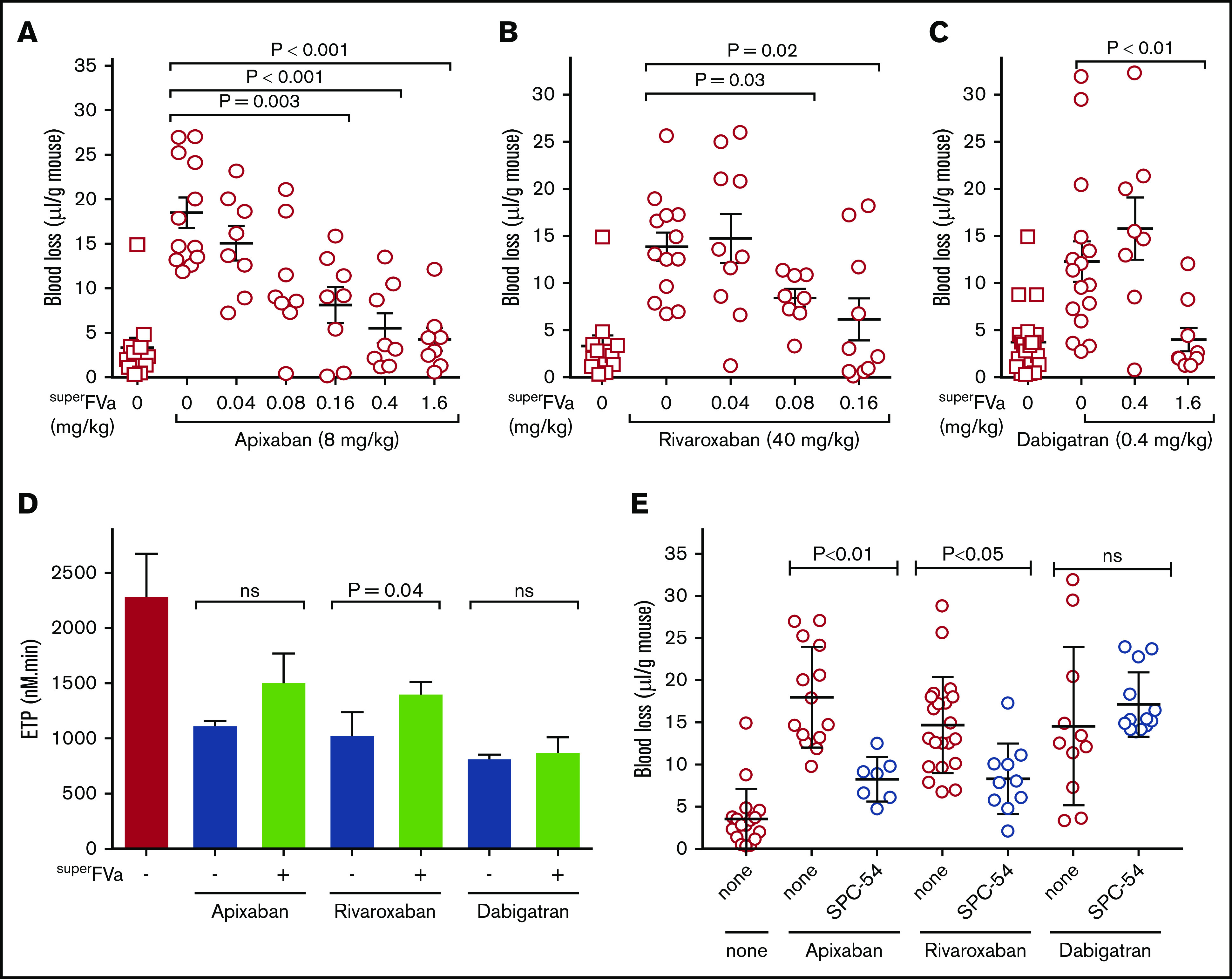
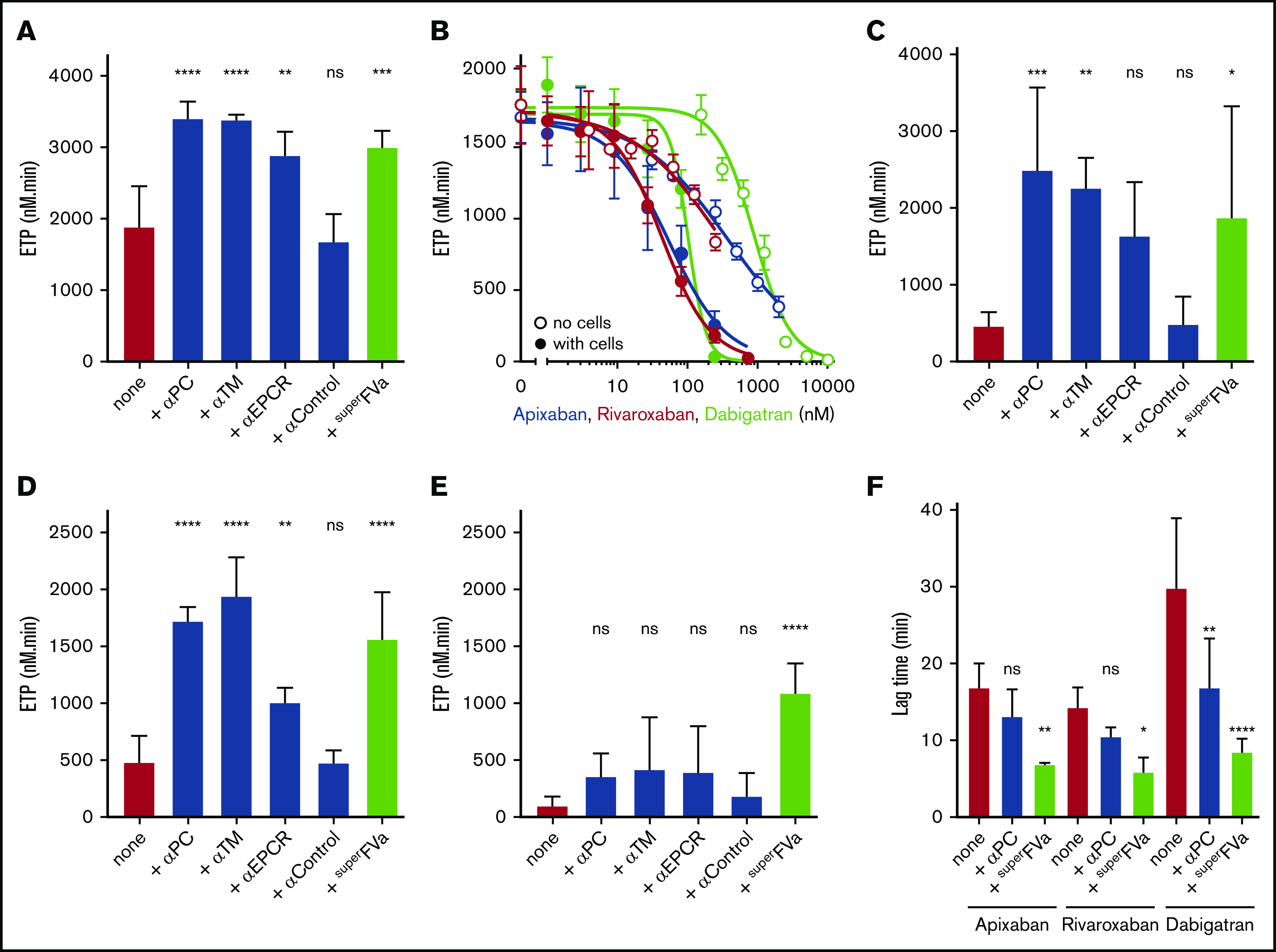
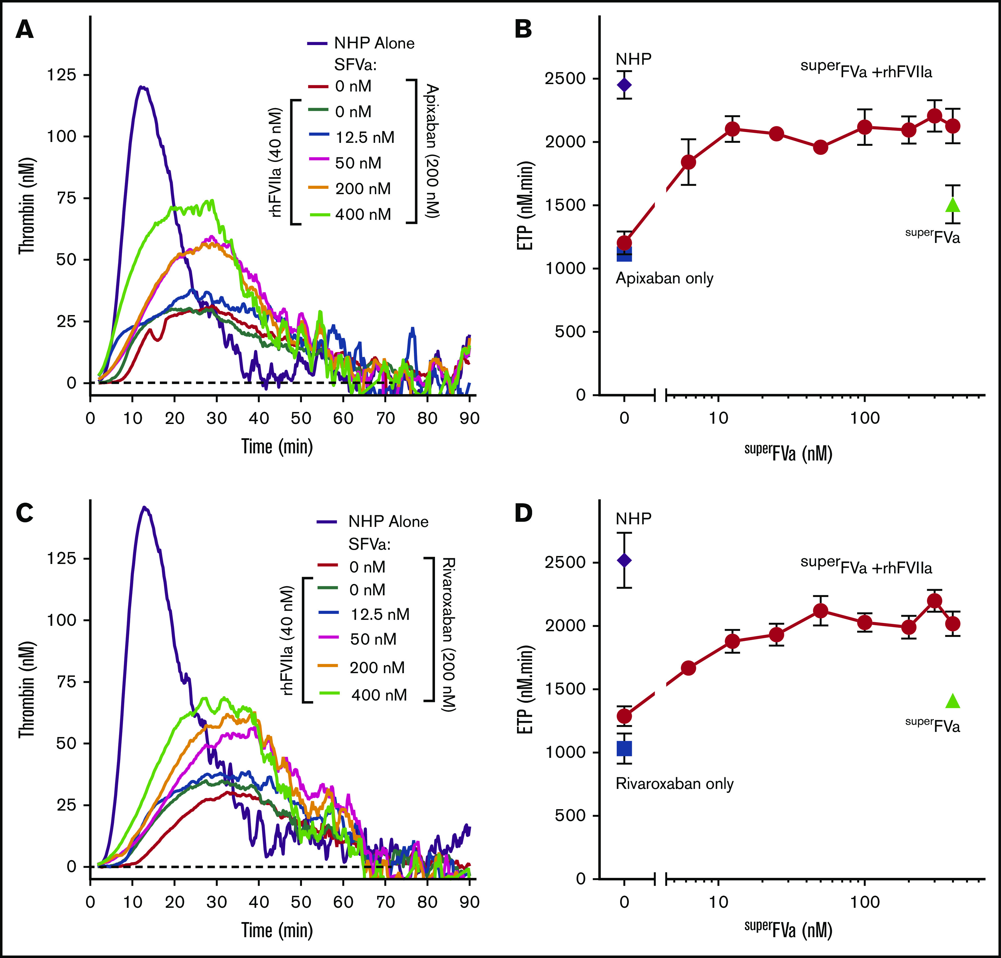
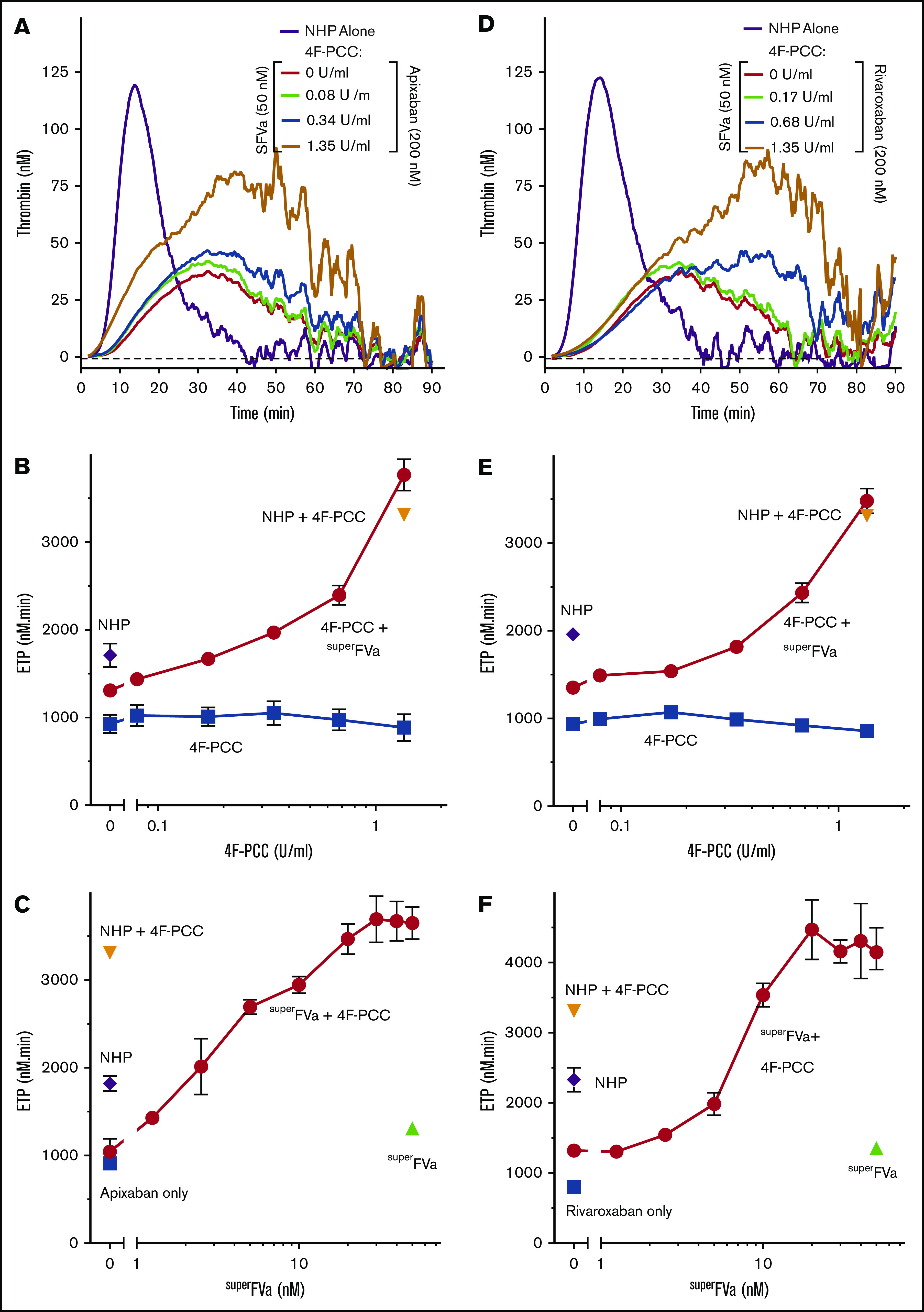
Control of bleeding with direct-acting oral anticoagulants (DOACs) remains an unmet clinical need. Activated superFactor V (superFVa) is an engineered activated protein C (APC)-resistant FVa variant with enhanced procoagulant activity resulting from an A2/A3 domain disulfide bond and was studied here for control of DOAC-induced bleeding. SuperFVa reversed bleeding induced by FXa inhibitors (rivaroxaban, apixaban), and the FIIa inhibitor dabigatran in BalbC mice. The blocking anti-protein C and APC [(A)PC] antibody SPC-54 also reduced FXa inhibitor induced bleeding similar to superFVa, whereas dabigatran-induced bleeding was not affected. This indicated that sufficient APC was generated to contribute to bleeding in the presence of FXa inhibitors, but not in the presence of dabigatran, suggesting that mechanisms contributing to bleeding differed for FXa and FIIa inhibitors. Despite different mechanisms contributing to bleeding, superFVa effectively reduced bleeding for all DOACs, indicating the versatility of superFVa's properties that contribute to its universal prohemostatic effects for DOAC associated bleeding. Supported by thrombin generation assays on endothelial cells in normal plasma spiked with DOACs and patient plasma anticoagulated with DOACs, 3 complementary mechanisms were identified by which superFVa achieved DOAC class-independent prohemostatic efficiency. These mechanisms are resistance to inactivation by APC, overcoming the FV activation threshold, and maximizing the efficiency of the prothrombinase complex when the available FXa is increased by FVIIa-based prohemostatics. In summary, it is this versatility of superFVa that delineates it from other prohemostatic agents as a promising class-independent rescue agent in bleeding situations associated with DOACs.


Conflict of interest statement

Conflict-of-interest disclosure: A.v.D. has received honoraria for participating in scientific advisory board panels, consulting, and speaking engagements for Takeda, Biomarin, Bioverativ/Sanofi, Novo Nordisk, and Uniqure, and reports research support from Pfizer and Sanofi. The University of California San Diego and The Scripps Research Institute hold intellectual property rights related to superFVa on which A.v.D., A.J.G., J.H.G., and L.O.M. are listed as inventors. A.v.D., A.J.G., and L.O.M. are founders of Hematherix LLC, a biotech company that is developing superFVa therapy for bleeding complications. A.v.D. and L.O.M. are members of the board of directors of Hematherix LLC. The remaining authors declare no competing financial interests.

Figures

None
Graphical abstract
Figure 1.
Figure 1.
Inhibition of DOAC-induced bleeding by superFVa and an anti-protein C antibody. Mice were treated with apixaban (8 mg/kg) (A), rivaroxaban (40 mg/kg) (B), or dabigatran (0.4 mg/kg) (C) by IV tail vein injection and bleeding was measured for 20 minutes after tail clip. Blood loss was expressed in microliters of blood per gram (g) mouse. (D) Human plasma with and without apixaban, rivaroxaban (both 200 nM) or dabigatran (1 μM) were treated with superFVa (100 nM) and endogenous thrombin potential was assessed. (E) Mice were injected retro-orbitally with the blocking anti-protein C and APC [(A)PC] antibody SPC-54 (5 mg/kg) 2 hours before IV treatment with apixaban (8 mg/kg), rivaroxaban (40 mg/kg), or dabigatran (0.4 mg/kg). Bleeding was measured for 20 minutes after tail clip. Blood loss was expressed in microliters of blood per gram (g) mouse. Error bars represent SEM (n = 7-14 per group). P values were determined by Kruskal-Wallis followed by a 2-tailed Mann-Whitney U test and values ≤.05 were considered statistically significant.
Figure 2.
Figure 2.
The role of superFVa and APC’s anticoagulant activity on the DOAC mediated inhibition of thrombin generation on endothelial cells. Thrombin generation was performed on EA.hy926 endothelial cells using human plasma in the absence of exogenously added tissue factor or phospholipids. (A) Thrombin generation on EA.hy926 endothelial cells in the presence of antibodies (α) against (A)PC (C1), TM, or EPCR (all 50 μg/mL) or superFVa (200 nM). (B) Thrombin generation in the presence and absence of EA.hy926 endothelial cells with different concentrations apixaban, rivaroxaban, or dabigatran. Thrombin generation in the absence of cells was initiated by 0.2 pM tissue factor and 4 μM phospholipid vesicles (PC/PS 80/20). (C) Panel A in the presence of apixaban (200 nM). (D) Panel A in the presence of rivaroxaban (200 nM). (E) Panel A in the presence of dabigatran (200 nM). (F) Lag time of thrombin generation on EA.hy926 endothelial cells was determined in the presence of all 3 direct oral anticoagulants (200 nM) with and without antibodies against (A)PC (50 μg/mL) or superFVa (200 nM). ns, not significant; TM, thrombomodulin.
Figure 3.
Figure 3.
Improvement of endothelial thrombin generation in DOAC patient plasma by superFVa. Thrombin generation was performed in plasma from patients on chronic anticoagulation with DOACs in the presence of EA.hy926 endothelial cells (○) and in the absence of cells (□). Thrombin generation in the absence of cells was initiated by 0.4 pM tissue factor and 10 μM phospholipid vesicles (PC/PS/PE 40/20/40). Shown are the endogenous thrombin potential (ETP) (A,C,E) and the lag time (B,D,F) of thrombin generation in the presence or absence of antibodies (50 μg/mL) against (A)PC (αPC; C1) or superFVa (20 nM) of patients on chronic anticoagulation with apixaban (A-B), rivaroxaban (C-D) , and dabigatran (E-F).
Figure 4.
Figure 4.
Effects of superFVa and rhFVIIa on thrombin generation in NHP spiked with FXa inhibitors. Thrombin generation was measured in NHP in the presence of the FXa inhibitors, apixaban or rivaroxaban. (A,C) Representative graphs showing the change in thrombin generation curves as the result of increasing concentrations of superFVa (0-400 nM) in the presence or absence of rhFVIIa (40 nM) in NHP spiked with apixaban (200 nM) (A) or rivaroxaban (200 nM) (C). (B,D) Enhancement of ETP by increasing concentrations of superFVa (0-400 nM) in the absence (Δ) or presence (○) of rhFVIIa (40 nM) in NHP spiked with apixaban (200 nM) (B) or rivaroxaban (200 nM) (D). Additional controls shown are NHP with (□) and without (◇) apixaban or rivaroxaban. (B,D) Peak height and lag time of are shown in supplemental Figure 4. Error bars represent standard error of the mean (n ≥ 3).
Figure 5.
Figure 5.
Effects of superFVa and 4F-PCC on thrombin generation in NHP spiked with FXa inhibitors. Thrombin generation was measured in NHP in the presence of the FXa inhibitors, apixaban or rivaroxaban. (A,D) Representative graphs showing the change in thrombin generation curves as the result of increasing concentrations of 4F-PCC (0-1.35 U/mL) in the presence or absence of superFVa (50 nM) in NHP spiked with apixaban (200 nM) (A) or rivaroxaban (200 nM) (D). (B,E) Enhancement of ETP by increasing concentrations of 4F-PCC (0-1.35 U/mL) in the absence (∎) or presence (○) of superFVa (50 nM) in NHP spiked with apixaban (200 nM) (B) or rivaroxaban (200 nM) (E). (C,F) Enhancement of ETP by increasing concentrations of superFVa (0-50 nM) in the absence (Δ) or presence (○) of 4F-PCC (1.35 U/mL) in NHP spiked with apixaban (200 nM) (C) or rivaroxaban (200 nM) (F). Peak height and lag time of (B-C) are shown in supplemental Figure 5 and of (E-F) in supplemental Figure 6. Additional controls shown are NHP with (□) and without (◇) apixaban or rivaroxaban or NHP with 1.35 U/mL 4F-PCC (∇). Error bars represent standard error of the mean (n ≥ 3).
Figure 6.
Figure 6.
Reversal of DOAC-induced bleeding with superFVa and rhFVIIa in wild-type BalbC mice. Mice were treated with apixaban (8 mg/kg) (A), rivaroxaban (40 mg/kg) (B), and dabigatran (0.4 mg/kg) (C) by IV tail vein injection and bleeding was measured for 20 minutes after tail clip. Blood loss was expressed in microliters of blood per gram (g) mouse. Increasing doses of superFVa, rhFVIIa, or rhFVIIa in combination with superFVa were injected retro-orbitally 30 minutes after DOAC administration and 5 minutes before tail clip. Error bars represent SEM (n = 8-12 per group). P values were determined by Kruskal-Wallis followed by a 2-tailed Mann-Whitney U test and values ≤.05 were considered statistically significant.

Similar articles

Cited by

References

    1. Ruff CT, Giugliano RP, Braunwald E, et al. . Comparison of the efficacy and safety of new oral anticoagulants with warfarin in patients with atrial fibrillation: a meta-analysis of randomised trials. Lancet. 2014;383(9921):955-962. - PubMed
    1. Camm AJ, Accetta G, Ambrosio G, et al. ; GARFIELD-AF Investigators . Evolving antithrombotic treatment patterns for patients with newly diagnosed atrial fibrillation. Heart. 2017;103(4):307-314. - PMC - PubMed
    1. Lee CH, Fang CC, Tsai LM, et al. . Changing treatment patterns in patients with venous thromboembolism in Taiwan. Circ J. 2020;84(2):283-293. - PubMed
    1. Connolly SJ, Ezekowitz MD, Yusuf S, et al. ; RE-LY Steering Committee and Investigators . Dabigatran versus warfarin in patients with atrial fibrillation. N Engl J Med. 2009;361(12):1139-1151. - PubMed
    1. Connolly SJ, Eikelboom J, Joyner C, et al. ; AVERROES Steering Committee and Investigators . Apixaban in patients with atrial fibrillation. N Engl J Med. 2011;364(9):806-817. - PubMed

Publication types