. 2020 Aug 11;4(15):3728-3740.
doi: 10.1182/bloodadvances.2020002326.

Sustained fetal hematopoiesis causes juvenile death from leukemia: evidence from a dual-age-specific mouse model

Affiliations

Sustained fetal hematopoiesis causes juvenile death from leukemia: evidence from a dual-age-specific mouse model

Nitza Vara et al. Blood Adv. .

Abstract

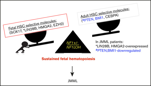
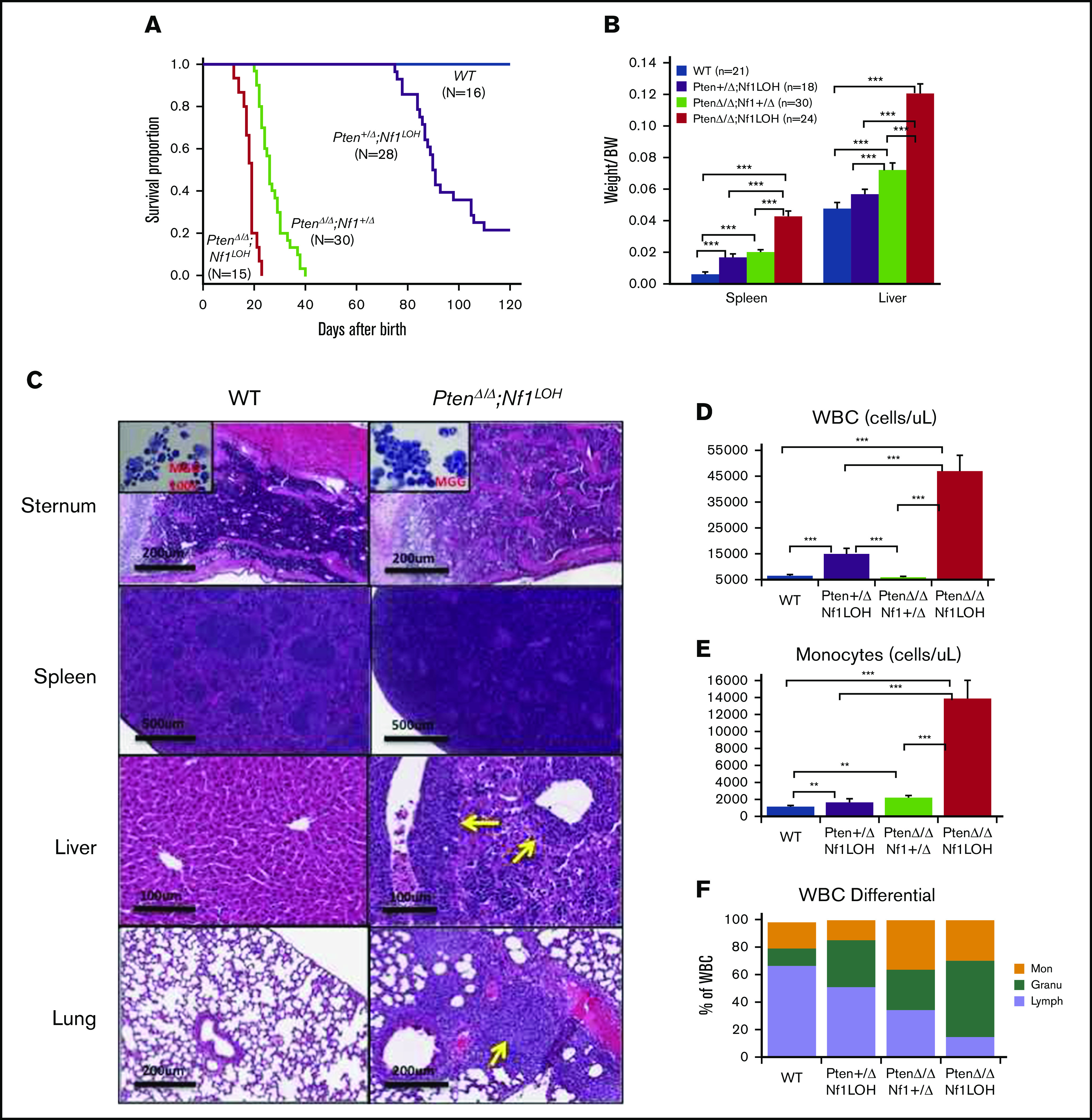
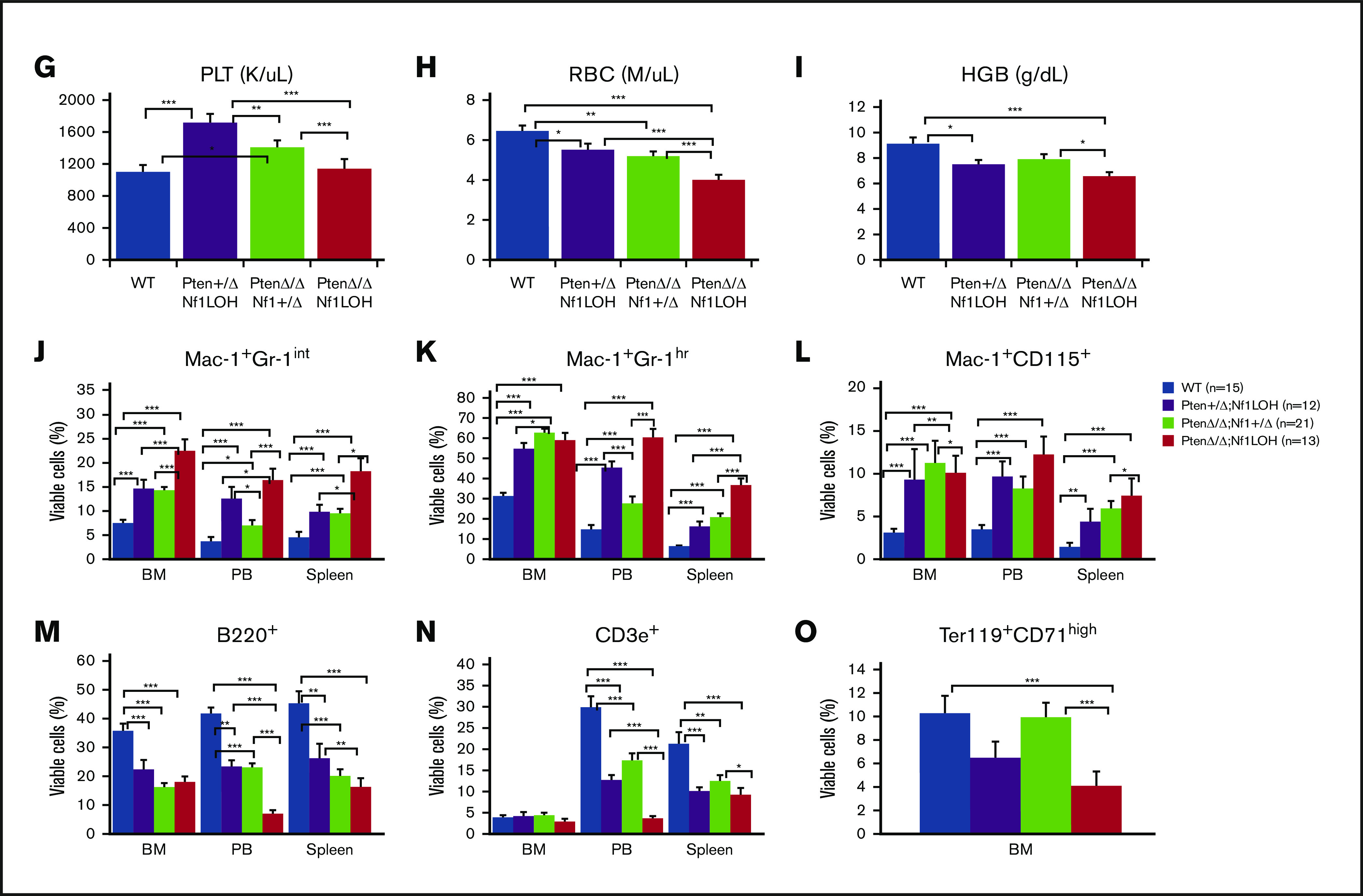
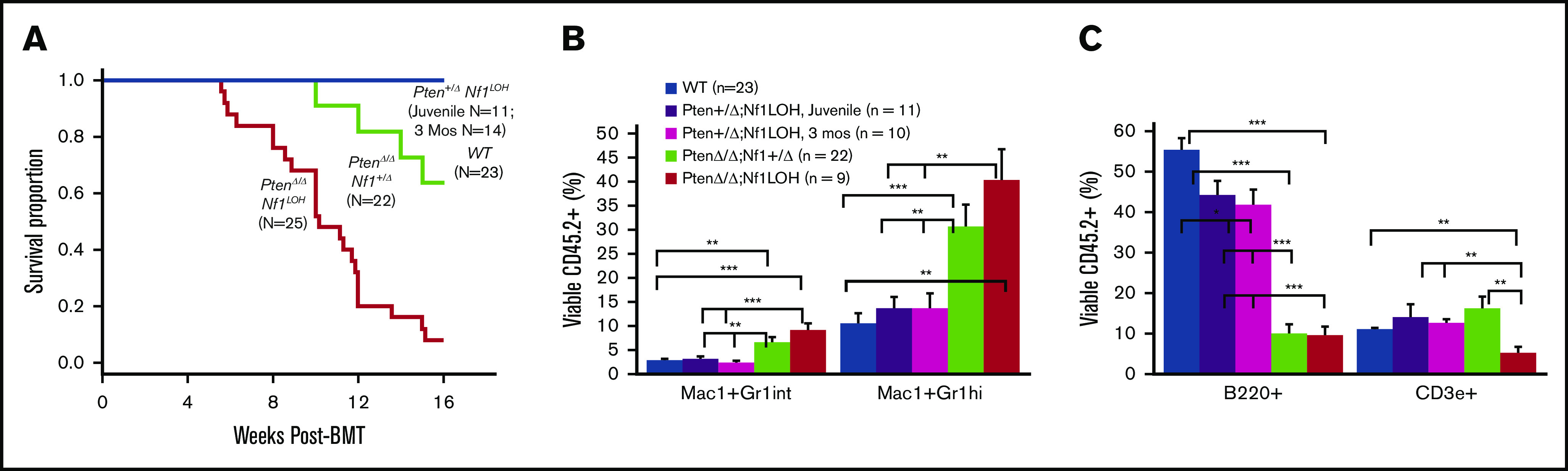
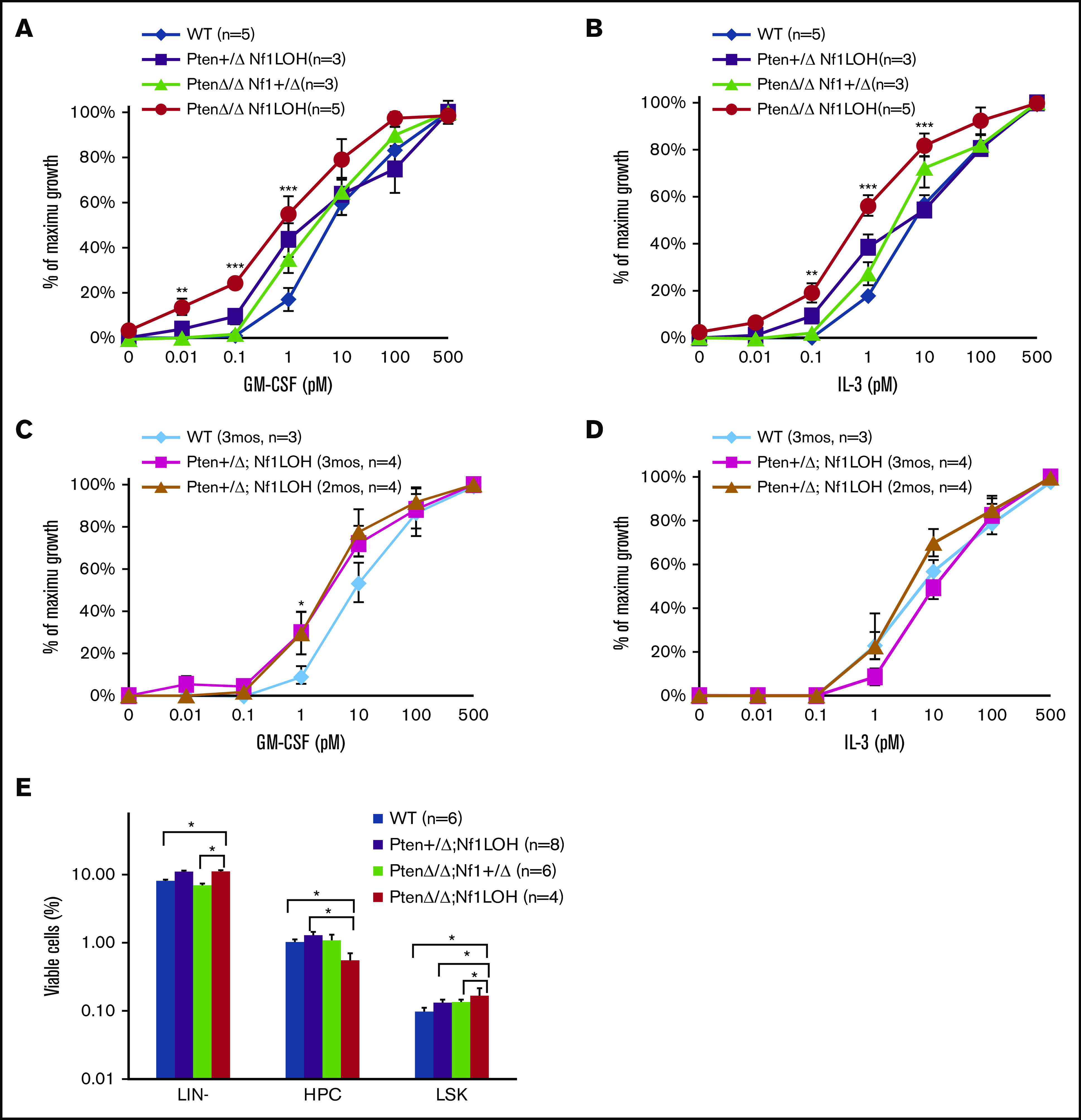
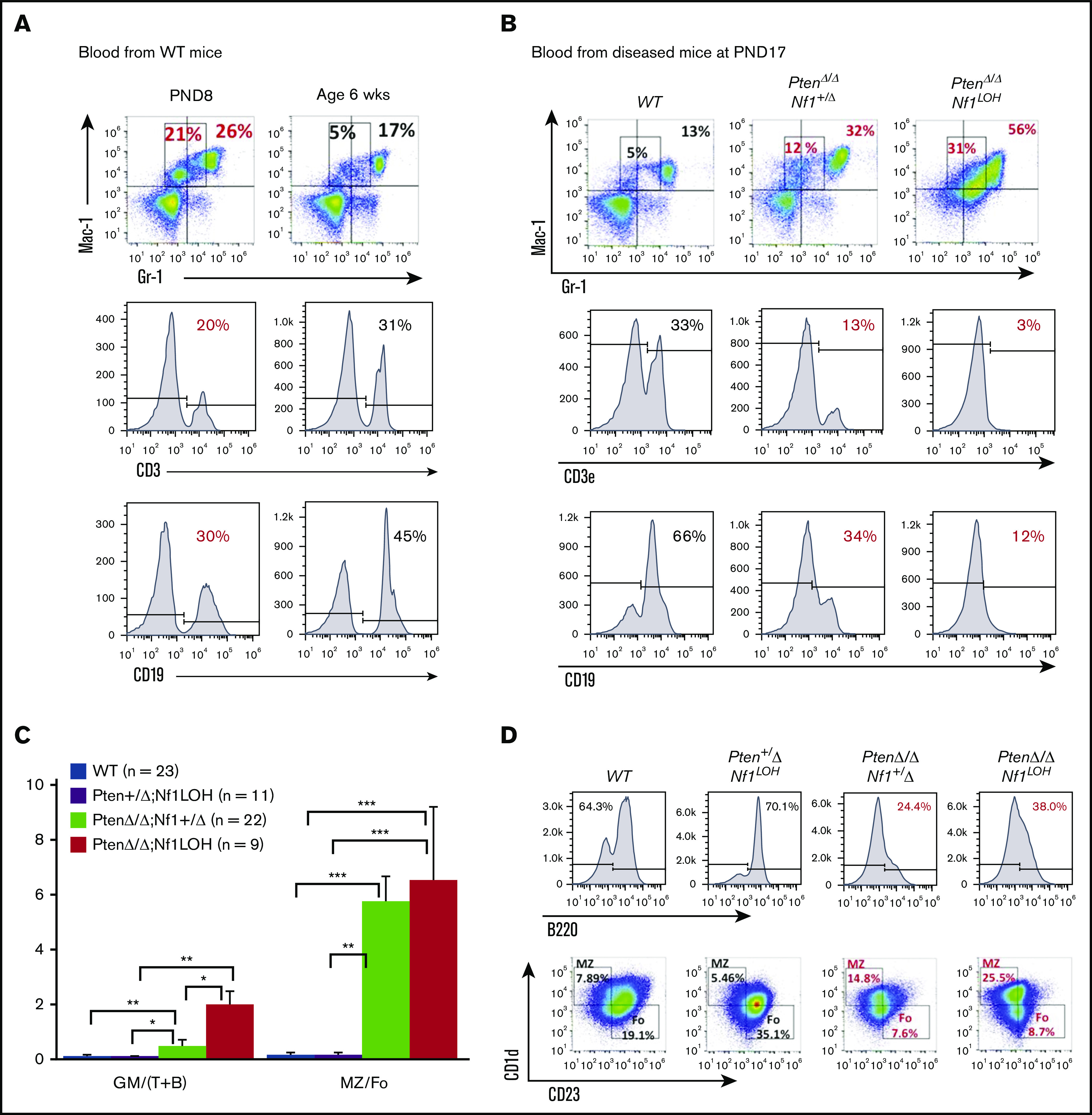
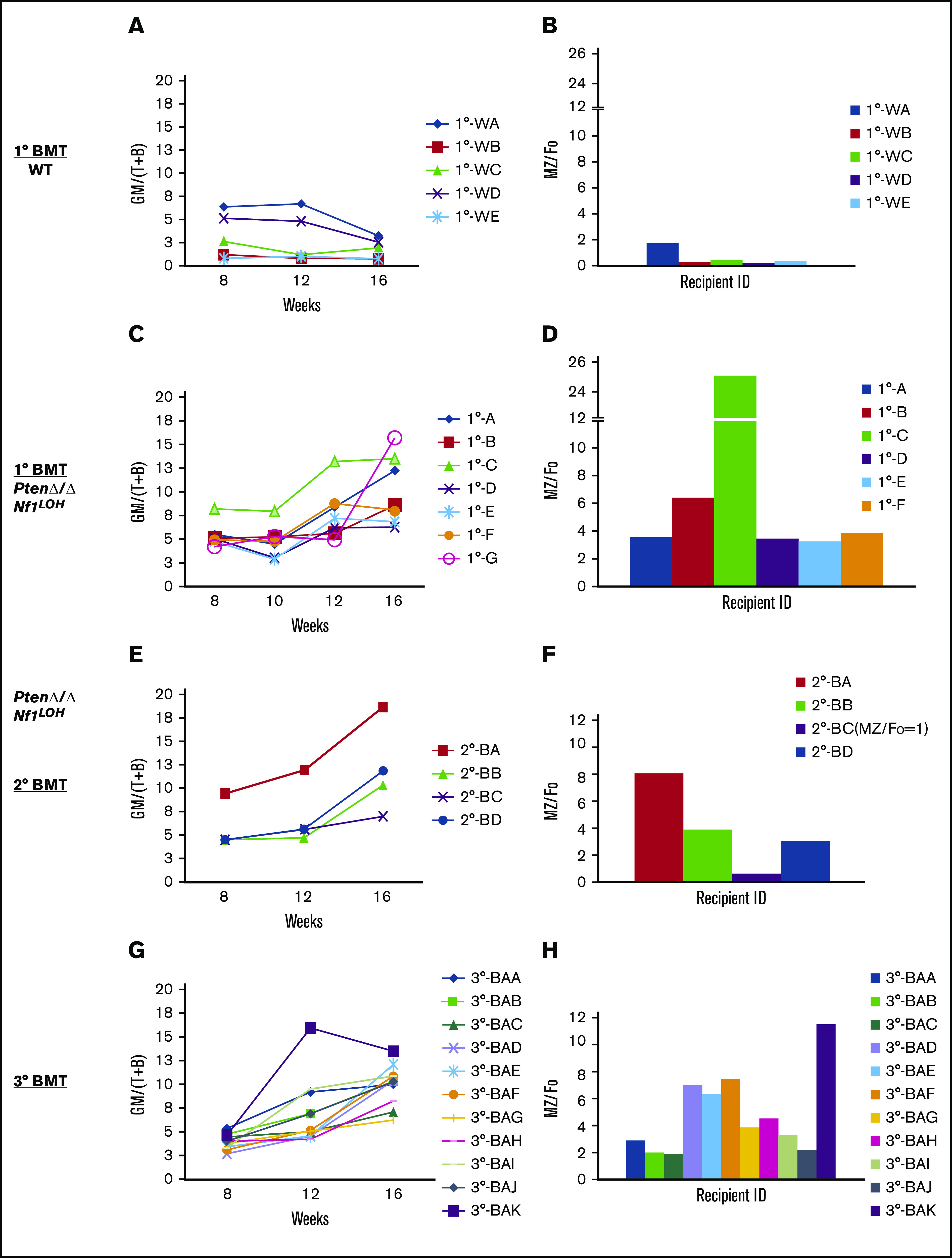
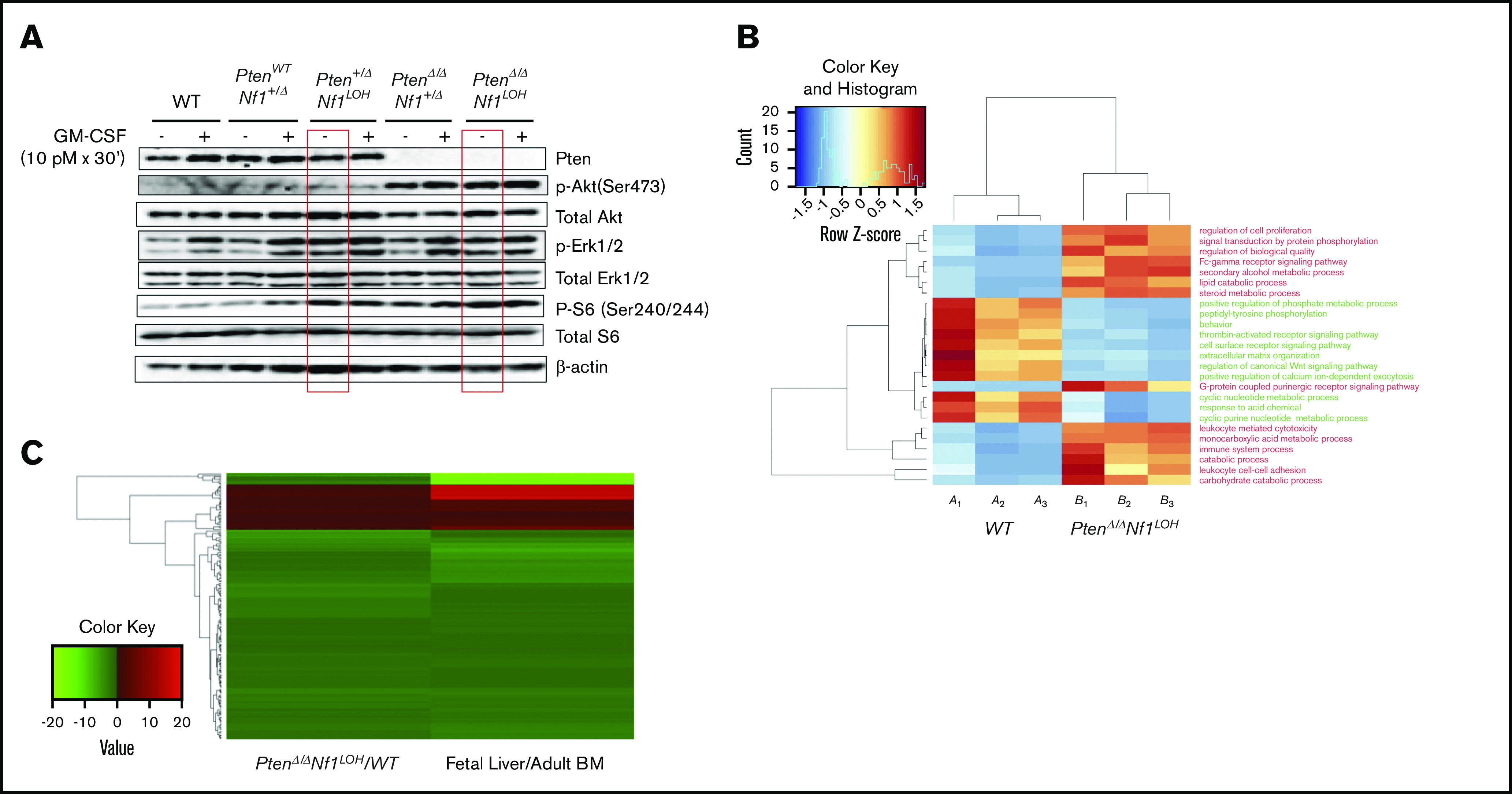
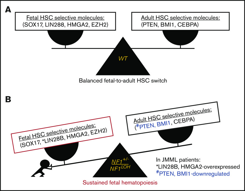
It is not clear whether disrupted age-specific hematopoiesis contributes to the complex manifestations in leukemia patients who carry identical mutations, particularly in pediatric and adult patients with similar clinical characteristics. By studying a dual-age-specific mouse model, we demonstrate that (1) loss of Pten during the fetal-to-adult hematopoiesis switch (hematopoiesis switch) causes sustained fetal hematopoiesis, resulting in death in juvenile leukemia; (2) myeloid-biased hematopoiesis in juvenile mice is associated with the sustained fetal properties of hematopoietic stem cells (HSCs); (3) the age specificity of juvenile myelomonocytic leukemia depends on the copy number of Pten and Nf1; (4) single-allelic Pten deletion during the hematopoiesis switch causes constitutive activation of MAPK in juvenile mice with Nf1 loss of heterozygosity (LOH); and (5) Nf1 LOH causes monocytosis in juvenile mice with Pten haploinsufficiency but does not cause lethality until adulthood. Our data suggest that 1 copy of Pten is sufficient to maintain an intact negative-feedback loop of the Akt pathway and HSC function in reconstitution, despite MAPK being constitutively activated in juvenile Pten+/ΔNf1LOH mice. However, 2 copies of Pten are required to maintain the integrity of the MAPK pathway in juvenile mice with Nf1 haploinsufficiency. Our data indicate that previous investigations of Pten function in wild-type mice may not reflect the impact of Pten loss in mice with Nf1 mutations or other genetic defects. We provide a proof of concept that disassociated age-specific hematopoiesis contributes to leukemogenesis and pediatric demise.


Conflict of interest statement

Conflict-of-interest disclosures: The authors declare no competing financial interests.

Figures

None
Graphical abstract
Figure 1.
Figure 1.
Correlation of phenotype and genotype in the dual-age–specific leukemia mouse model. (A) Kaplan-Meier survival curves show the relationship between survival time and genotype (P < .001). Death was counted in mice without any intervention. Male/female ratios were 2:1 in PtenΔ/ΔNf1LOH mice and Pten+/ΔNf1LOH mice and 1:1 in PtenΔ/ΔNf1+/Δ mice and WT mice. (B) Relationship between the genotype and the spleen and liver weights. Organs from littermates were collected at PND17-19 when PtenΔ/ΔNf1LOH mice with JMML were moribund. Data are presented as median ratios of spleen or liver weight/body weight (BW). (C) Representative photomicrographs of hematoxylin and eosin–stained tissue sections from PtenΔ/ΔNf1LOH mice with JMML (right panels) and WT control littermates (left panels) at PND18. Infiltrates are indicated by yellow arrows. (D-I) Blood profiles from juvenile PtenΔ/ΔNf1LOH mice and littermates at PND17-19 when diseased mice were moribund (n = 12-21). Complete blood counts were performed with a Vet Abc Hematological analyzer. Differentials were manually counted from blood smears stained with May-Grünwald-Giemsa. (J-O) Flow cytometry analysis of cell subpopulations in BM, PB, and spleen. Data are mean ± standard error. *P < .05, ** P <.01, ***P < .001. See additional supportive data in supplemental Figures 1-7. Granu, granulocytes; HGB, hemoglobin; Lymph, lymphocytes; Mon, monocytes; PLT, platelets; RBC, red blood cells.
Figure 1.
Figure 1.
Correlation of phenotype and genotype in the dual-age–specific leukemia mouse model. (A) Kaplan-Meier survival curves show the relationship between survival time and genotype (P < .001). Death was counted in mice without any intervention. Male/female ratios were 2:1 in PtenΔ/ΔNf1LOH mice and Pten+/ΔNf1LOH mice and 1:1 in PtenΔ/ΔNf1+/Δ mice and WT mice. (B) Relationship between the genotype and the spleen and liver weights. Organs from littermates were collected at PND17-19 when PtenΔ/ΔNf1LOH mice with JMML were moribund. Data are presented as median ratios of spleen or liver weight/body weight (BW). (C) Representative photomicrographs of hematoxylin and eosin–stained tissue sections from PtenΔ/ΔNf1LOH mice with JMML (right panels) and WT control littermates (left panels) at PND18. Infiltrates are indicated by yellow arrows. (D-I) Blood profiles from juvenile PtenΔ/ΔNf1LOH mice and littermates at PND17-19 when diseased mice were moribund (n = 12-21). Complete blood counts were performed with a Vet Abc Hematological analyzer. Differentials were manually counted from blood smears stained with May-Grünwald-Giemsa. (J-O) Flow cytometry analysis of cell subpopulations in BM, PB, and spleen. Data are mean ± standard error. *P < .05, ** P <.01, ***P < .001. See additional supportive data in supplemental Figures 1-7. Granu, granulocytes; HGB, hemoglobin; Lymph, lymphocytes; Mon, monocytes; PLT, platelets; RBC, red blood cells.
Figure 2.
Figure 2.
Reconstitution of BM cells from mice with JMML and littermates in competitive BMT. (A) Kaplan-Meier survival curves from recipients transplanted with BM from mice with JMML and juvenile littermates or from Pten+/Δ Nf1LOH mice with CMML at the age of 3 months when they were moribund. (B-C) Blood engraftment data from transplanted mice. Blood was collected from recipient mice at 12 to 16 weeks post-BMT. Data are mean ± standard error. *P < .05, **P < .01, ***P < .001. See additional supportive data in supplemental Figure 8.
Figure 3.
Figure 3.
Analysis of BM progenitors in diseased mice and littermates. (A-D) Colony formation assay data. Unfractionated BM nucleated cells (WBM) were collected from PtenΔ/ΔNf1LOH mice with JMML and littermates at PND17-19 or from mice with CMML at age 2 to 3 months when they were moribund. Data from CFU-GM showed that GM-CSF and IL-3 sensitivities were significantly increased in BM cells from PtenΔ/Δ Nf1LOH mice with JMML compared with juvenile littermates, whereas Pten+/ΔNf1LOH mice showed GM-CSF hypersensitivity only as adults. (E) Flow cytometry analysis of hematopoietic progenitors in BM from juvenile mice. WBMs from PtenΔ/ΔNf1LOH mice with JMML and littermates at PND17-19 were analyzed by flow cytometry; data demonstrated that lineage (LIN)-negative cells (negative for Gr1, CD3, B220, and Ter119) and LINSca1+cKit+ cells (LSK) were significantly increased in PtenΔ/ΔNf1LOH mice with JMML, whereas hematopoietic progenitor cells (HPCs; LINSca1cKit+) were decreased. Data are mean ± standard error. *P < .05, **P < .01, ***P < .001.
Figure 4.
Figure 4.
Significant fetal hematopoiesis in juvenile mice with Pten deletion and leukemia. (A) Representative flow cytometry data from WT mouse blood at PND8 and 6 weeks (n = 4 each group). (B) Representative flow cytometry data from blood of WT mice and PtenΔ/Δ mice with leukemia at age 3 weeks (≥10 mice in each group). (C) Relationship between donor-derived (CD45.2+) progenies in PB and MZ/Fo B-cell ratios in spleens of recipients. (D) Representative flow cytometry data from blood and spleens of recipient mice transplanted from donor mice with various copy numbers of Pten and Nf1. GM [blood myeloid; CD45.2+CD11b+], (T+B) [blood lymphoid lineage; sum of CD45.2+B220+ + CD45.2+CD3e+], and blood myeloid/lymphoid ratios (GM/[T+B]) were calculated as ratios of myeloid cells/lymphocytes in total WBCs with CD45.2+ in recipient mice (CD45.1+). Fo, donor-derived follicular B cells are the predominant B cells in adults; MZ, donor-derived splenic marginal zone B cells representing fetal origin HSCs. The ratios of splenic MZ (fetal origin)/Fo (adult origin) B cells represented the fetal properties of donor HSCs. Data are median ± standard error. *P < .05, **P < .01, ***P < .001. See additional supportive data in supplemental Figures 8 and 10.
Figure 5.
Figure 5.
Serial single-HSC transplantation. (A-D) 1° Transplant: single LT-HSC from juvenile WT mice and PtenΔ/ΔNf1LOH mice with JMML was transplanted with 2 × 10⁵ rescue cells into WT recipient mice with CD45.1+, respectively. Sixteen weeks posttransplant, WT donor HSCs reconstituted balanced progenies, whereas HSCs with JMML (PtenΔ/ΔNf1LOH) reconstituted various degrees of myeloid-biased progenies with elevated ratios of MZ/Fo B cells. Recipient 1°-G, which had the worst myeloid-biased blood, died before spleen tissue collection for MZ cell analysis. (E-F) 2° Transplant: single donor-derived HSC (CD45.2+) from primary recipient 1°-B was retransplanted into 2° BMT recipients. 2° Recipient mice continuously reconstituted various degrees of myeloid-biased progenies with elevated MZ/Fo B-cell ratios. Recipient 2°-BC reconstituted balanced progenies with the least splenic MZ cells. (G-H) 3° Transplant: single donor-derived HSC (CD45.2+) from recipient 2°-BA was retransplanted to 3°-BMT recipients. Recipient 3°-BAK had the worst myeloid-biased engraftment with the highest MZ/Fo B-cell ratio. Data represent ≥2 sets of paired experiments. See additional supportive data in supplemental Figures 11 and 12.
Figure 6.
Figure 6.
Dysregulated signal transduction pathways and molecular defects in mice with Pten deletion, Nf1 deficiency, and JMML. (A) Representative data from western blot analyses of the elements in GM-CSF signal transduction pathways in unfractionated BM nucleated cells from juvenile mice with various copy numbers of Pten and Nf1. Representative data were from ≥3 experiments from littermate mice at PND17-19. (B) Gene ontology (GO) analysis. Genes that were significantly over- or underexpressed in LIN BM cells from mice with JMML and WT littermates at age PND17 were used in GO analysis, together with HCA. (C) Heatmap and HCA of 182 genes that were differentially expressed in this study (PtenΔ/Δ Nf1LOH/WT) vs the data (Fetal Liver/Adult BM) reported by Manesia et al. We found a log2-fold change in either being positive or negative consistently in both studies (182 genes). See additional supportive data in supplemental Figures 13 and 14 and supplemental Tables 4 and 5 for “GO HeatMap Terms & GeneIDs” and “Combined Analysis DEG Results.”
Figure 7.
Figure 7.
Proposed leukemogenesis model of the aberrant fetal-to-adult hematopoiesis switch in JMML. Fetal and adult hematopoiesis are governed by a developmental/age-specific molecular driving force and executed by fetal-selective genes and adult-necessary genes in hematopoiesis (eg, SOX17, LIN28B, HMGA2, and EZH2 for fetal HSCs, and BMI1, PTEN, and CEBPA for adult HSCs). (A) During the neonatal period when the molecular driving force is balanced in fetal to adult hematopoiesis, healthy newborns can complete the transition from fetal to adult hematopoiesis to meet the needs for growth in normoxia. (B) When the molecular driving force is disrupted in newborns during the fetal-to-adult hematopoiesis switch by mutations or dysregulated epigenetics, fetal hematopoiesis is sustained and causes myeloid/lymphoid distribution errors and insufficient adult hematopoiesis in juveniles, resulting in pediatric demise. This may be the case in patients with JMML who are born with an NF1 mutation and hold an instable molecular driving force from fetal to adult hematopoiesis switch, such as elevated fetal-specific LIN28B and HMGA2 along with PTEN and BMI1 deficiencies. Because JMML patients cannot develop adult hematopoiesis at an age when WT juveniles normally complete their fetal-to-adult hematopoiesis switch, they die of sustained fetal hematopoiesis as juveniles.

Similar articles

Cited by

References

    1. Niemeyer CM, Arico M, Basso G, et al. ; European Working Group on Myelodysplastic Syndromes in Childhood (EWOG-MDS) . Chronic myelomonocytic leukemia in childhood: a retrospective analysis of 110 cases. Blood. 1997;89(10):3534-3543. - PubMed
    1. Alfonso A, Montalban-Bravo G, Takahashi K, et al. . Natural history of chronic myelomonocytic leukemia treated with hypomethylating agents. Am J Hematol. 2017;92(7):599-606. - PMC - PubMed
    1. Geissler K, Jäger E, Barna A, et al. . Chronic myelomonocytic leukemia patients with RAS pathway mutations show high in vitro myeloid colony formation in the absence of exogenous growth factors. Leukemia. 2016;30(11):2280-2281. - PMC - PubMed
    1. Sakaguchi H, Okuno Y, Muramatsu H, et al. . Exome sequencing identifies secondary mutations of SETBP1 and JAK3 in juvenile myelomonocytic leukemia. Nat Genet. 2013;45(8):937-941. - PubMed
    1. Hofmans M, Lammens T, Helsmoortel HH, et al. . The long non-coding RNA landscape in juvenile myelomonocytic leukemia. Haematologica. 2018;103(11):e501-e504. - PMC - PubMed

Publication types