. 2020 Aug 10;13(1):111.
doi: 10.1186/s13041-020-00649-7.

Altered dopaminergic pathways and therapeutic effects of intranasal dopamine in two distinct mouse models of autism

Affiliations

Altered dopaminergic pathways and therapeutic effects of intranasal dopamine in two distinct mouse models of autism

Owen Y Chao et al. Mol Brain. .

Abstract

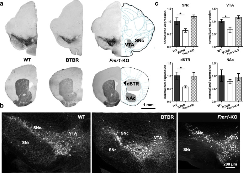
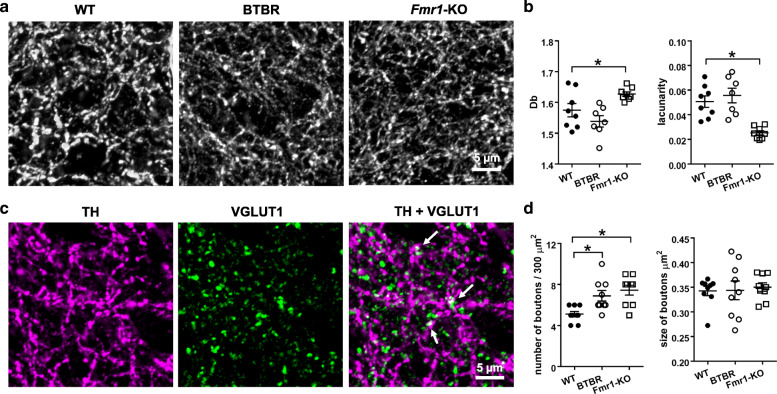
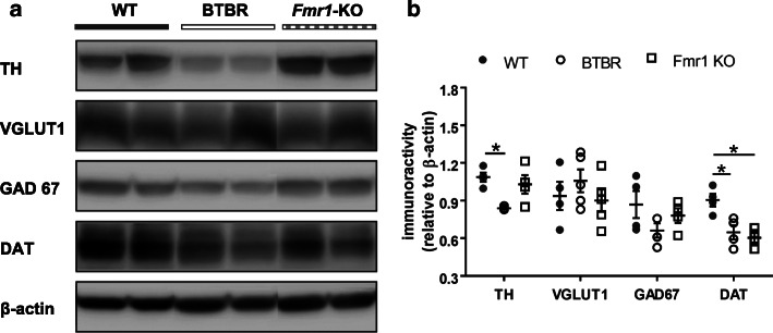
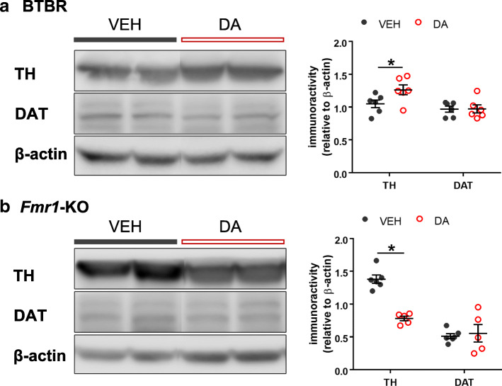
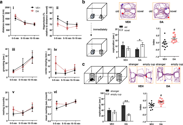
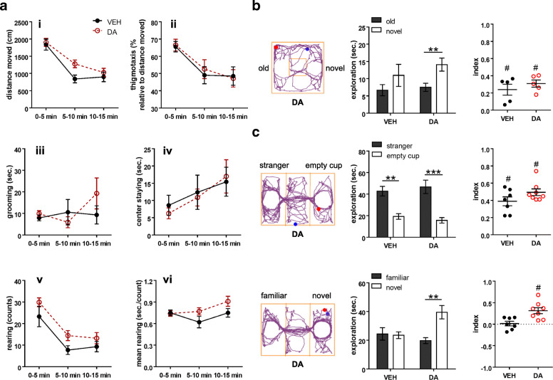
The dopamine (DA) system has a profound impact on reward-motivated behavior and is critically involved in neurodevelopmental disorders, such as autism spectrum disorder (ASD). Although DA defects are found in autistic patients, it is not well defined how the DA pathways are altered in ASD and whether DA can be utilized as a potential therapeutic agent for ASD. To this end, we employed a phenotypic and a genetic ASD model, i.e., Black and Tan BRachyury T+Itpr3tf/J (BTBR) mice and Fragile X Mental Retardation 1 knockout (Fmr1-KO) mice, respectively. Immunostaining of tyrosine hydroxylase (TH) to mark dopaminergic neurons revealed an overall reduction in the TH expression in the substantia nigra, ventral tegmental area and dorsal striatum of BTBR mice, as compared to C57BL/6 J wild-type ones. In contrast, Fmr1-KO animals did not show such an alteration but displayed abnormal morphology of TH-positive axons in the striatum with higher "complexity" and lower "texture". Both strains exhibited decreased expression of striatal dopamine transporter (DAT) and increased spatial coupling between vesicular glutamate transporter 1 (VGLUT1, a label for glutamatergic terminals) and TH signals, while GABAergic neurons quantified by glutamic acid decarboxylase 67 (GAD67) remained intact. Intranasal administration of DA rescued the deficits in non-selective attention, object-based attention and social approaching of BTBR mice, likely by enhancing the level of TH in the striatum. Application of intranasal DA to Fmr1-KO animals alleviated their impairment of social novelty, in association with reduced striatal TH protein. These results suggest that although the DA system is modified differently in the two ASD models, intranasal treatment with DA effectively rectifies their behavioral phenotypes, which may present a promising therapy for diverse types of ASD.

Keywords: Autism; BTBR; Fmr1; Fragile X syndrome; Social behavior; Striatum.


Conflict of interest statement

The authors declare no competing financial and non-financial interests in relation to this work. CM is an employee of M et P Pharma AG. However, the company did not play a role in any aspect of the study.

Figures

Fig. 1
Fig. 1
Immunohistochemical analyses of TH expression in WT, BTBR and Fmr1-KO mice. a Representative diagrams and images of anti-TH staining in substantia nigra pars compacta (SNc), ventral tegmental area (VTA), dorsal striatum (dSTR) and nucleus accumbens (NAc). b Examples of confocal images (20x) of dopaminergic neurons in WT, BTBR and Fmr1-KO mice. c Fluorescence intensity of anti-TH staining in the region of interest (ROI) was measured in an identical microscopic setting and normalized to the WT animals. The BTBR brain exhibited decreased TH-positive expression in SNc, VTA and dSTR, while the Fmr1- KO brain did not. TH: tyrosine hydroxylase; SNr: substantia nigra pars reticulata. n = 6–7 samples/group. *p < 0.05, compared to WT
Fig. 2
Fig. 2
Fractal analyses of TH-positive axons and measurements of co-labelled TH and VGLUT1 signals in the WT, BTBR and Fmr1-KO striatum. a Representative images of anti-TH staining in the dorsal striatum, taken by a confocal microscope with a 63x lens, forming a 30 × 30 μm field with 6 z-stacks of 0.35 μm steps. b Fractal dimensions Db of TH-positive axons in the Fmr1-KO striatum was higher, while the value of lacunarity was lower, than those in the WT striatum. c Representative diagrams of anti-TH (magenta), anti-VGLUT1 (green) and their merged image in the WT striatum. Merged dots (white), pointed by arrows, indicate boutons with adjacent TH and VGLUT1 labelling. d Boutons with the co-labelled signals were counted and their size was measured. The size of the boutons was similar between groups, whereas the number of the boutons was more in BTBR and Fmr1-KO mice than that in WT controls. n = 7–9 samples/group. *p < 0.05, compared to WT
Fig. 3
Fig. 3
Western blotting of TH, VGLUT1, GAD67 and DAT in the striatum. a Examples of Western blots of striatal lysates from WT, BTBR and Fmr1-KO mice. Images of protein bands were aligned for comparison. b Blot intensity was normalized to an internal standard β-actin. Decreased TH and DAT levels were found in BTBR mice, while Fmr1-KO animals only showed reduced DAT expression, as compared to the WT group. Relative quantities of other proteins were comparable among groups. VGLUT1: vesicular glutamate transporter 1; GAD67: glutamate decarboxylase 67; DAT: dopamine transporter; TH: tyrosine hydroxylase. n = 4–5 mice/group. *p < 0.05, compared to WT
Fig. 4
Fig. 4
Immunoblotting of TH and DAT in the striatum after intranasal delivery of vehicle (VEH) or dopamine (DA). a Western blots of striatal lysates from BTBR mice treated with VEH or DA. Increased TH expression was found after DA administration. b Western blots of striatal lysates from Fmr1-KO mice treated with VEH or DA. Reduced TH level was noticed after DA application. In both cases, DAT protein was unaltered by intranasal DA. TH: tyrosine hydroxylase; DAT: dopamine transporter. n = 5–6 mice/group. *p < 0.05, compared to VEH
Fig. 5
Fig. 5
Behavioral effects of intranasal dopamine on BTBR mice. a Behaviors measured in the open field test. The dopamine (DA)-treated group showed comparable distance travelled (i), self-grooming (iii), center staying (iv), and counts of rearing (v) to the vehicle (VEH)-treated group, except for a lower percentage of thigmotaxis at the time intervals 0–5 and 5–10 min (ii) and a longer duration of rearing in the first 5 min (vi). b Behaviors assessed in the object-based attention test. An example of movement tracking of a VEH- or DA-treated animal is illustrated. Blue and red dots indicate the start and end points of a test trial, respectively. Unlike the VEH-treated animals, the DA-treated ones exhibited the preference toward the novel object, and thus, showing positive values of the index. Index = [time spent on the novel object - time spent on the old object] / total exploration time. c Behaviors evaluated in the three-chamber social test. An example of movement tracking of a VEH- or DA-treated animal is presented. Blue and red dots indicate the start and end points of a sociability trial, respectively. The VEH-treated animals explored the stranger mouse and the empty cup equally, while the DA-treated ones showed the preference to the stranger mouse and had positive values of the sociable index. Index = [time for exploring the stranger mouse - time for exploring the empty cup] / total exploration time. n = 12 mice/group. *p < 0.05, **p < 0.01, compared to the respective values. #p < 0.05, compared to 0 by one sample t-test
Fig. 6
Fig. 6
Behavioral effects of intranasal dopamine on Fmr1-KO mice. a Behaviors measured in the open field test. The dopamine (DA)-treated group showed comparable distance travelled (i), thigmotaxis (ii), self-grooming (iii), center staying (iv), counts of rearing (v), and average duration of rearing (vi) to the vehicle (VEH)-treated group. b Behaviors assessed in the object-based attention test. An example of movement tracking of a DA-treated animal is shown. Blue and red dots indicate the start and end points of a test trial, respectively. The DA-treated animals exhibited the preference toward the novel object, although both groups had positive values of the index. Index = [time spent on the novel object - time spent on the old object] / total exploration time. c Behaviors evaluated in the three-chamber social test. Examples of movement tracking of a DA-treated animal in a sociability and a social novelty trial are displayed. Blue and red dots indicate the start and end points of each trial, respectively. In the sociability trial (upper panels), both groups displayed social preference toward the stranger mouse and had positive values of the sociable index. Index = [time for exploring the stranger mouse - time for exploring the empty cup] / total exploration time. In the social novelty trial (bottom panels), the DA-treated group explored the novel stranger more than the familiar one, whereas the VEH-treated group did not. The DA-treated group thus had higher values of the social novelty index. Index = [time for exploring the novel mouse - time for exploring the familiar mouse] / total exploration time. n = 7–9 mice/group (2–4 mice were excluded in the object-based attention test due to < 10 s exploration). **p < 0.01, ***p < 0.001, compared to the respective values. #p < 0.05, compared to 0 by one sample t-test

References

    1. Lai MC, Lombardo MV, Baron-Cohen S. Autism. Lancet. 2014;383(9920):896–910. - PubMed
    1. Simonoff E, Pickles A, Charman T, Chandler S, Loucas T, Baird G. Psychiatric disorders in children with autism spectrum disorders: prevalence, comorbidity, and associated factors in a population-derived sample. J Am Acad Child Adolesc Psychiatry. 2008;47(8):921–929. - PubMed
    1. Gandal MJ, Haney JR, Parikshak NN, Leppa V, Ramaswami G, Hartl C, Schork AJ, Appadurai V, Buil A, Werge TM, et al. Shared molecular neuropathology across major psychiatric disorders parallels polygenic overlap. Science. 2018;359(6376):693–697. - PMC - PubMed
    1. Bromberg-Martin ES, Matsumoto M, Hikosaka O. Dopamine in motivational control: rewarding, aversive, and alerting. Neuron. 2010;68(5):815–834. - PMC - PubMed
    1. Volkow ND, Wang GJ, Newcorn J, Telang F, Solanto MV, Fowler JS, Logan J, Ma Y, Schulz K, Pradhan K, et al. Depressed dopamine activity in caudate and preliminary evidence of limbic involvement in adults with attention-deficit/hyperactivity disorder. Arch Gen Psychiatry. 2007;64(8):932–940. - PubMed

Publication types

MeSH terms