. 2020 Aug 5;107(3):580-589.e6.
doi: 10.1016/j.neuron.2020.05.007. Epub 2020 Jun 16.

Plasticity and Spontaneous Activity Pulses in Disused Human Brain Circuits

Affiliations

Plasticity and Spontaneous Activity Pulses in Disused Human Brain Circuits

Dillan J Newbold et al. Neuron. .

Abstract

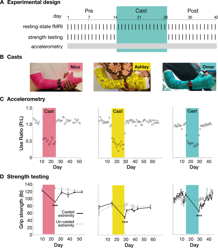
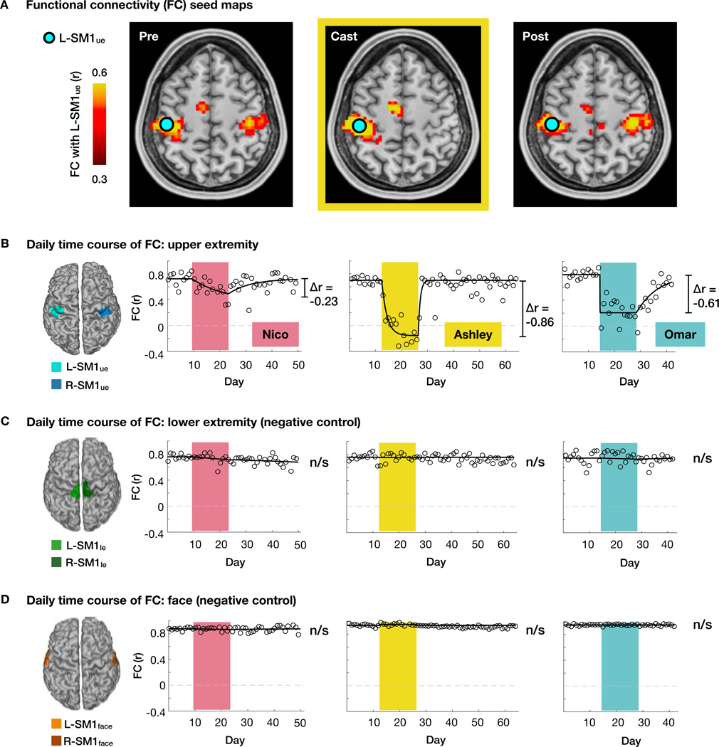
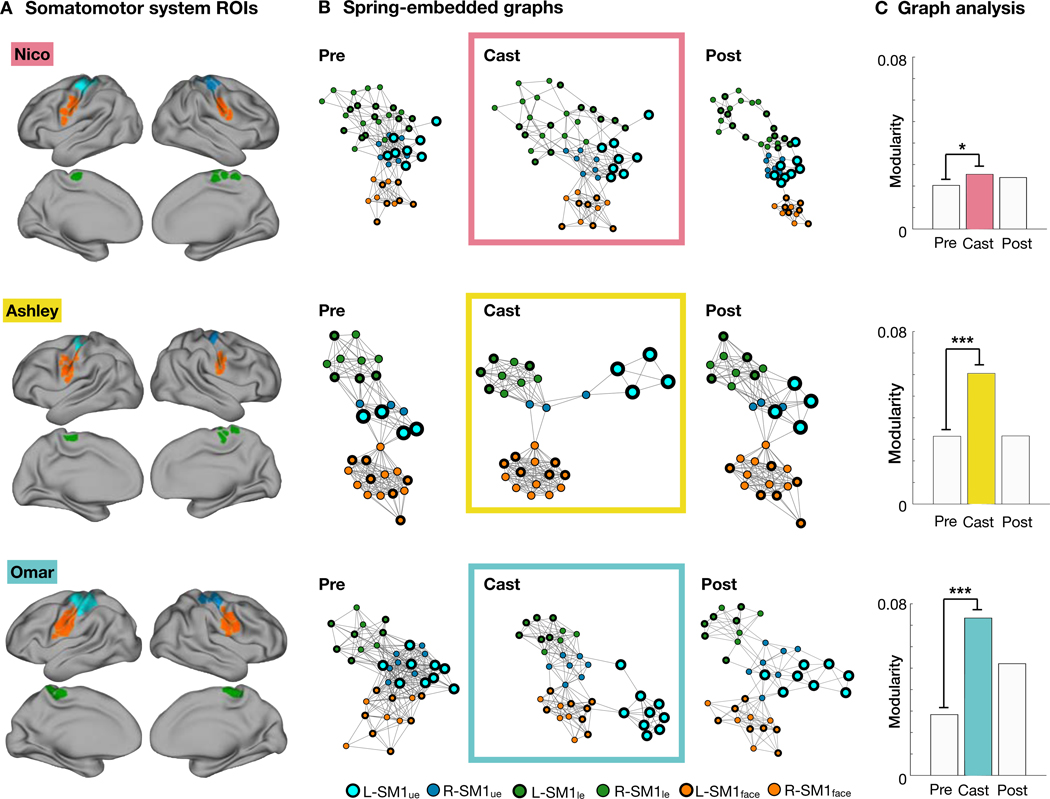
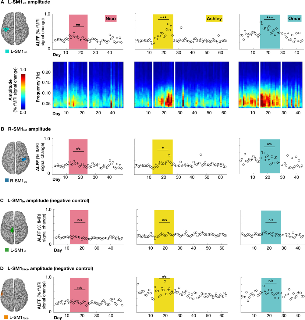
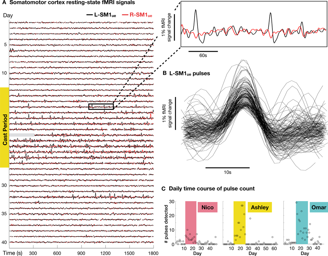
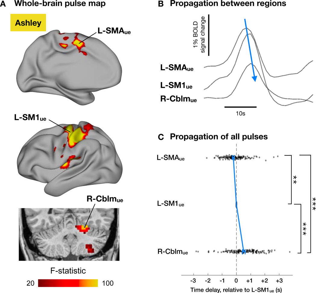
To induce brain plasticity in humans, we casted the dominant upper extremity for 2 weeks and tracked changes in functional connectivity using daily 30-min scans of resting-state functional MRI (rs-fMRI). Casting caused cortical and cerebellar regions controlling the disused extremity to functionally disconnect from the rest of the somatomotor system, while internal connectivity within the disused sub-circuit was maintained. Functional disconnection was evident within 48 h, progressed throughout the cast period, and reversed after cast removal. During the cast period, large, spontaneous pulses of activity propagated through the disused somatomotor sub-circuit. The adult brain seems to rely on regular use to maintain its functional architecture. Disuse-driven spontaneous activity pulses may help preserve functionally disconnected sub-circuits.

Keywords: ALFF; amplitude of low-frequency fluctuations; cerebellum; disuse; fMRI; functional connectivity; plasticity; primary motor cortex; resting state; spontaneous activity; supplementary motor area.


Conflict of interest statement

Declaration Of Interests The authors declare the following competing financial interest: N.U.F.D. is co-founder of NOUS Imaging.

Figures

Figure 1.
Figure 1.. Casting caused disuse and reduced strength of the dominant upper extremity.
(A) Experimental design for an example participant (Omar; all participants Figure S1). (B) Casts covered the entire dominant upper extremity. Cast colors are used in subsequent figures to identify participants: pink (Nico), yellow (Ashley), green (Omar). (C) Daily accelerometry data from both wrists, plotted as use ratios (R/L use counts). Use count = seconds each day when RMS acceleration > 0.16 m/s2. Daily use counts for each wrist shown in Figure S1. (D) Grip strength before and after casting (***P < 0.001). Error bars indicate s.e.m. across three repeated measurements.
Figure 2.
Figure 2.. Disused somatomotor cortex became functionally uncoupled from opposite hemisphere.
(A) Seed maps showing functional connectivity (FC) of each voxel with the left primary somatomotor cortex (L-SM1ue) during scans acquired before, during and after casting (Pre, Cast, Post) in an example participant (Ashley). The L-SM1ue region of interest (ROI) was defined using task functional MRI. (B) Daily time course of FC between L-SM1ue and R-SM1ue for each participant. Δr values are based on a time-varying exponential decay model (black lines, dr⁄dt = α(r − r); Nico: P = 0.002, Ashley: P < 0.001, Omar: P<0.001). (C and D) Daily time course of FC in lower extremity (C) and face (D) regions of the left and right somatomotor cortex.
Figure 3.
Figure 3.. Disused somatomotor cortex dissociated from the remainder of the somatomotor system.
(A) Regions of interest (ROIs) in the lower extremity (green), upper extremity (cyan/blue) and face (orange) subdivisions of the somatomotor system for each participant. ROIs were selected using a functional connectivity gradient-based approach (Gordon et al., 2017). (B) Spring-embedded graph representation of the somatomotor system before, during and after casting. The disused region (L-SM1ue) separated from the remainder of the somatomotor cortex during the cast period but remained internally connected. (C) Modularity quantifies the degree of dissociation between L-SM1ue and the rest of the somatomotor cortex. All participants showed increased modularity during the cast period. *P < 0.05; ***P < 0.001.
Figure 4.
Figure 4.. Disused somatomotor cortex (L-SM1ue) showed increased amplitude of low- frequency fluctuations (ALFF).
(A) Top: ALFF in resting-state functional MRI (rs-fMRI) signals recorded from L-SM1ue on each day of the experiment. All participants showed significantly increased L-SM1ue ALFF during the cast period, relative to the pre period (Nico: +22%, P < 0.01; Ashley: +81%, P < 0.001; Omar: +36%, P < 0.001). Bottom: Amplitude spectra of L-SM1ue rs-fMRI signals. (B) ALFF in R-SM1ue. Only participant Ashley showed significantly increased R-SM1ue ALFF during the cast period. (C) ALFF in lower-extremity somatomotor cortex (L-SM1le; negative control). No participants showed significant changes during the cast period. (D) ALFF in face somatomotor cortex (L-SM1face; negative control). No participants showed significant changes during the cast period.
Figure 5.
Figure 5.. Disuse pulses in somatomotor cortex.
(A) Resting-state functional MRI (rs-fMRI) signals from left and right primary somatomotor cortex (L-SM1ue and R-SM1ue) before, during and after casting in an example participant (Ashley; all participants Figure S3). During the cast period, large pulses occur in L-SM1ue (see inset at top-right). Data for first five minutes of scan 21 are unavailable (gray bar). (B) Recordings of 144 disuse pulses detected in an example participant (Ashley). (C) Number of pulses detected during each day of the experiment. All participants showed significantly more pulses per session during the cast period than during the pre period. (Nico: P = 0.002; Ashley: P < 0.001; Omar: P < 0.001)
Figure 6.
Figure 6.. Disuse pulses propagate through the disused somatomotor sub-circuit.
(A) Whole- brain analysis of variance (ANOVA) of disuse pulses in an example participant (Ashley; all participants shown in Figure S4). In addition to L-SM1ue, pulses also occur in left supplementary motor area (L-SMAue) and right cerebellum (R-Cblmue). (B) Example pulse in L-SMAue, L-SM1ue and R-Cblmue. Note the temporal offset of peaks in each region. (C) Time delays (relative to L-SM1ue) of all pulses in each region for an example participant (Ashley; all participant Figure S4). Blue lines indicate median delay ± s.e.m. in each region. Pulses occurred first in L-SMAue, then L-SM1ue, and finally in R-Cblmue. Pulse times were determined by parabolic interpolation of cross-correlations with the mean pulse time series. **P<0.01. ***P<0.001.

Comment in

References

    1. Ackman JB, Burbridge TJ, and Crair MC (2012). Retinal waves coordinate patterned activity throughout the developing visual system. Nature 490, 219–225. - PMC - PubMed
    1. Barch DM, Burgess GC, Harms MP, Petersen SE, Schlaggar BL, Corbetta M, Glasser MF, Curtiss S, Dixit S, Feldt C, et al. (2013). Function in the human connectome: task-fMRI and individual differences in behavior. Neuroimage 80, 169–189. - PMC - PubMed
    1. Behzadi Y, Restom K, Liau J, and Liu TT (2007). A component based noise correction method (CompCor) for BOLD and perfusion based fMRI. Neuroimage 37, 90–101. - PMC - PubMed
    1. Biswal B, Yetkin FZ, Haughton VM, and Hyde JS (1995). Functional connectivity in the motor cortex of resting human brain using echo-planar MRI. Magn Reson Med 34, 537–541. - PubMed
    1. Bullmore E, and Sporns O (2009). Complex brain networks: graph theoretical analysis of structural and functional systems. Nat Rev Neurosci 10, 186–198. - PubMed

Publication types