. 2020 Aug 10;10(1):13453.
doi: 10.1038/s41598-020-70428-w.

Environmentally controlled magnetic nano-tweezer for living cells and extracellular matrices

Affiliations

Environmentally controlled magnetic nano-tweezer for living cells and extracellular matrices

Christian Aermes et al. Sci Rep. .

Abstract

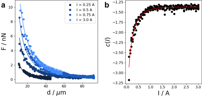
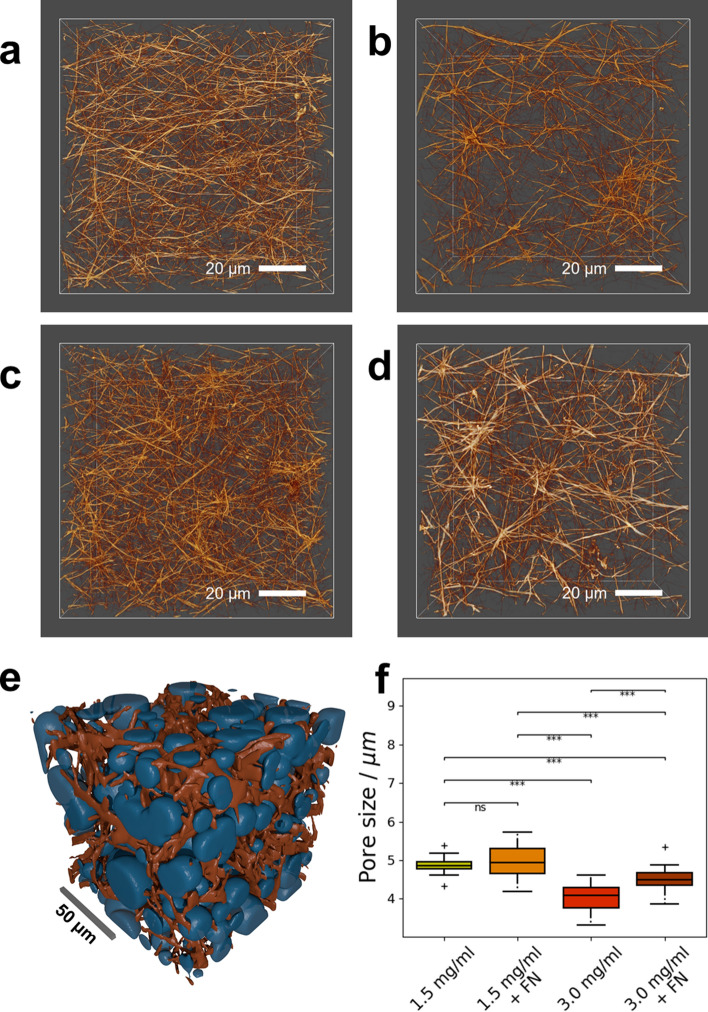
The magnetic tweezer technique has become a versatile tool for unfolding or folding of individual molecules, mainly DNA. In addition to single molecule analysis, the magnetic tweezer can be used to analyze the mechanical properties of cells and extracellular matrices. We have established a magnetic tweezer that is capable of measuring the linear and non-linear viscoelastic behavior of a wide range of soft matter in precisely controlled environmental conditions, such as temperature, CO2 and humidity. The magnetic tweezer presented in this study is suitable to detect specific differences in the mechanical properties of different cell lines, such as human breast cancer cells and mouse embryonic fibroblasts, as well as collagen matrices of distinct concentrations in the presence and absence of fibronectin crosslinks. The precise calibration and control mechanism employed in the presented magnetic tweezer setup provides the ability to apply physiological force up to 5 nN on 4.5 µm superparamagnetic beads coated with fibronectin and coupled to the cells or collagen matrices. These measurements reveal specific local linear and non-linear viscoelastic behavior of the investigated samples. The viscoelastic response of cells and collagen matrices to the force application is best described by a weak power law behavior. Our results demonstrate that the stress stiffening response and the fluidization of cells is cell type specific and varies largely between differently invasive and aggressive cancer cells. Finally, we showed that the viscoelastic behavior of collagen matrices with and without fibronectin crosslinks measured by the magnetic tweezer can be related to the microstructure of these matrices.


Conflict of interest statement

The authors declare no competing interests.

Figures

Figure 1
Figure 1
(a) Schematic image of the magnetic tweezer setup created with Inkscape. The magnetic field was generated by a coil with a ferromagnetic core made from µ-metal. A custom-built voltage driven current supply was connected to the coil to generate the magnetic field. The magnetic tweezer was connected to a micromanipulator that allowed precise placement of the needle tip close to the specimen. The whole setup was mounted on an inverted microscope and enclosed by an incubation chamber with temperature and CO2 control. Cells with super paramagnetic beads bound to them were placed in a petri dish with cell medium. The petri dish was then placed under the microscope. A CMOS camera captured live images of the bead's deflection under an acting magnetic force from the tweezer. This deflection was then tracked live over time and analyzed by a custom written program. (b) Picture of the magnetic tweezer setup. (c) To generate a high gradient field, the core was shaped conically at its tip, forming a needle. The tip had an opening angle of about 35° and a radius of curvature of about 10 µm. Scale bar is 50 µm.
Figure 2
Figure 2
Flow chart of the measurement software contains all important steps: After initialization of the hardware, the remnant magnetization of the core was automatically set to zero. The image shows a ROI selected for bead and needle position tracking. During the measurement, the coil current was continuously adapted based on the bead-to-needle distance to ensure a constant force. When a measurement was completed, the program was either set up for the next measurement or the hardware was de-initialized and finished.
Figure 3
Figure 3
Force calibration for precise control during measurements. (a) Exemplary distance dependence of the force on 4.5 µm beads is presented for four different currents. Force–distance curves were fitted with a power law. (b) The power law exponent of the fit was current-dependent and was fitted with Eq. 5.
Figure 4
Figure 4
Results of measurements on cells with a single force pulse with 1 nN amplitude and 2 s duration. (a) A representative brightfield image shows a 4.5 microm super paramagnetic bead coupled to an MCF-7 cancer cell. The red circle in all images marked the initial position of the bead. Scalebars are 10 microm. Left: Bead position just before the force was turned on. Middle: Bead position after 2 s of force application. Right: Bead position after 2 s of relaxation after the force is turned off. (b) The averaged creep response of MDA-MB-231 (n = 160, N = 5), MCF-7 (n = 108, N = 4), PINCH-1−/− (n = 97, N = 4), and PINCH-1fl/fl (n = 78, N = 3) cells over time was fitted with the Kelvin–Voigt model. Especially at lower time scales, the Kelvin–Voigt model fails to predict the creep curves correctly. This may be seen from the R2 values of the fit (MDA-MB-231: R2 = 0.9634; MCF-7: R2 = 0.9298; PINCH-1−/−: R2 = 0.9175; PINCH-1fl/fl: R2 = 0.8892). (c) The same averaged creep response was fitted with a weak power law. The creep response for all cell lines closely followed a weak power law over time. The R2 values of the power law fit are consistently higher than for the Kelvin–Voigt model for all cell lines (MDA-MB-231: R2 = 0.9978; MCF-7: R2 = 0.9978; PINCH-1−/−: R2 = 0.9968; PINCH-1fl/fl; R2 = 0.9968). The different cell lines displayed distinct differences in their creep response. (d) Stiffness values of MDA-MB-231, MCF-7, PINCH-1−/−, and PINCH-1fl/fl cells obtained from the weak power law fit. E) Cell fluidity (power law exponent β) of MDA-MB-321, MCF-7, PINCH-1−/−, and PINCH-1fl/fl cells. *p < 0.05, **p < 0.01, ***p < 0.001 and ns not significant.
Figure 5
Figure 5
(a) The application of ten consecutive pulses lead to a stiffening in PINCH-1−/− (n = 50, N = 4) and PINCH-1fl/fl cells (n = 83, N = 4), while the stiffness of MDA-MB-231 (n = 56, N = 4) and MCF-7 cells (n = 88, N = 4) was not affected by the cycle number. Each force pulse had a magnitude of 1 nN and lasted for 2 s. Between two consecutive pulses, the force was returned to 0 for 2 s. (b) Cell fluidity (power law exponent β) was unaffected by the cycle number for all four cell lines. (c) Stiffness values obtained from a weak power law fit to the creep curves in response to the application of a staircase-like force sequence. For the staircase-like sequence, the initial force was 1 nN. Each step in the sequence lasted for 2 s. After each step, the applied force was increased by 1 nN. The last step in the sequence had a magnitude of 5 nN. MDA-MB-231 (n = 68, N = 3), MCF-7 (n = 54, N = 3), PINCH-1−/− (n = 97, N = 4), and PINCH-1fl/fl (n = 78, N = 3) cells displayed a significant increase in stiffness with the applied force. (d) The power law exponent β of MDA-MB-231, MCF-7, PINCH-1−/−, and PINCH-1fl/fl cells also displayed a force dependence.
Figure 6
Figure 6
Magnetic tweezer measurements of ordinary collagen gels and gels crosslinked with fibronectin (FN). (a) Schematic representation of the measurements created with Inkscape. The gels were polymerized in a cell culture dish and submerged in PBS. Super paramagnetic beads (with a diameter of 4.5 µm) were coupled to the surface of the collagen matrix. A constant force of one nanonewton was applied for 2 s to displace the beads. (b) Averaged displacement curves of beads coupled to collagen gels with a 1.5 mg/ml (n = 275, N = 6) and 3.0 mg/ml (n = 274, N = 6) type I collagen concentration (a 2 to 1 mixture of bovine to rat collagen) and both collagen gels crosslinked with fibronectin (1.5 mg/ml + FN: n = 270, N = 6; 3.0 mg/ml + FN: n = 276, N = 6). The displacement closely followed a power law. Note: Error bars in the curves are smaller than the respective symbols. (c) Stiffness values of the collagen gels. Fibronectin increased the stiffness of collagen gels significantly. (d) Power law exponent β from the fit. Crosslinking with fibronectin did not affect the power law exponent β. (e) Comparison of the stiffness values during creep and relaxation for the different collagen gels. The significantly higher stiffness during relaxation indicates incomplete recovery behavior. f The relaxation of collagen gels displayed a more fluid-like behavior compared to the preceding creep. *p < 0.05, **p < 0.01, ***p < 0.001 and ns not significant.
Figure 7
Figure 7
Determination of pore sizes for the investigated collagen gels. (a) Representative 3D image of a 1.5 mg/ml collagen gel. (b) 3D image of a representative fibronectin-crosslinked 1.5 mg/ml collagen gel. (c) 3D image of a representative 3.0 mg/ml collagen gel. (d) 3D image of a 3.0 mg/ml collagen gel crosslinked with fibronectin. Scale bars in (a)–(d) are 20 µm. (e) Visualization of the pore-fitting algorithm. Spheres (blue) are fitted into the fluid phase of the binary segmentation of the collagen gel (orange) to determine the pore size. The scalebar is 50 µm. Segmentation and pore data were determined as described previously. Pore representations were created by drawing the respective spheres into a binary matrix using custom written Python software. Segmentation and pore representations were exported to STL surface models using Fiji, and subsequently rendered using Blender. (f) Pore sizes of the investigated collagen gels. 1.5 mg/ml collagen gels (N = 6) have a bigger pore size than 3.0 mg/ml gels (N = 6) due to the lower concentration of collagen fibers in the gel. Crosslinking with fibronectin increases the pore size of 3.0 mg/ml collagen gels (N = 6) but has no significant effect on the pore size of 1.5 mg/ml collagen gels (N = 5). ***p < 0.001 and ns not significant.

References

    1. Neuman KC, Nagy A. Single-molecule force spectroscopy: optical tweezers, magnetic tweezers and atomic force microscopy. Nat. Methods. 2008;5:491–505. - PMC - PubMed
    1. Kruithof M, Chien FT, Routh A, Logie C, Rhodes D, van Noort J. Single-molecule force spectroscopy reveals a highly compliant helical folding for the 30-nm chromatin fiber. Nat. Struct. Mol. Biol. 2009;16:534–540. - PubMed
    1. Meng H, Andresen K, van Noort J. Quantitative analysis of single-molecule force spectroscopy on folded chromatin fibers. Nucleic Acids Res. 2015;43:3578–3590. - PMC - PubMed
    1. Capitanio M, Pavone FS. Interrogating biology with force: single molecule high-resolution measurements with optical tweezers. Biophys. J. 2013;105:1293–1303. - PMC - PubMed
    1. Le S, Chen H, Cong P, Lin J, Dröge P, Yan J. Mechanosensing of DNA bending in a single specific protein-DNA complex. Sci. Rep. 2013;3:3508. - PMC - PubMed

Publication types