Review
. 2020 Oct 1;143(10):2874-2894.
doi: 10.1093/brain/awaa174.

Definitions and classification of malformations of cortical development: practical guidelines

Affiliations
Review

Definitions and classification of malformations of cortical development: practical guidelines

Mariasavina Severino et al. Brain. .

Erratum in

Abstract

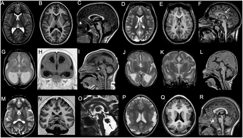
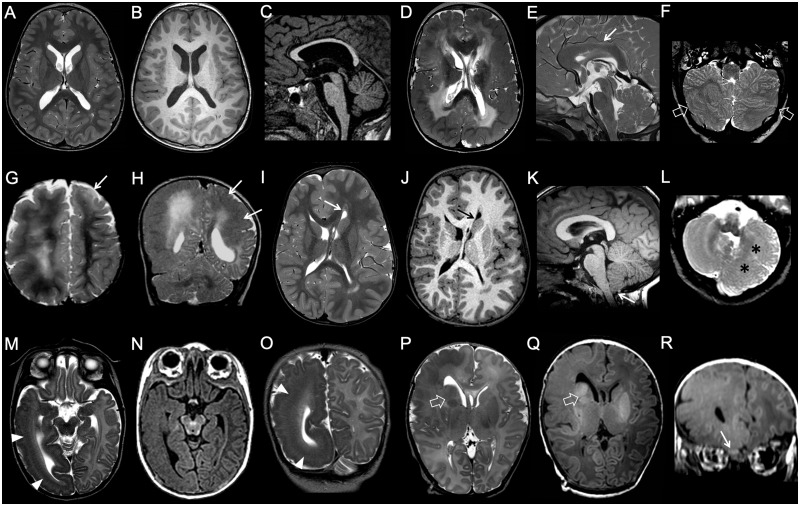
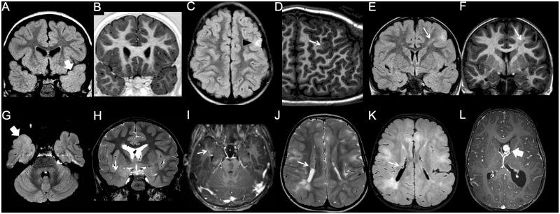
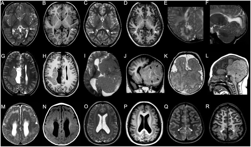
Malformations of cortical development are a group of rare disorders commonly manifesting with developmental delay, cerebral palsy or seizures. The neurological outcome is extremely variable depending on the type, extent and severity of the malformation and the involved genetic pathways of brain development. Neuroimaging plays an essential role in the diagnosis of these malformations, but several issues regarding malformations of cortical development definitions and classification remain unclear. The purpose of this consensus statement is to provide standardized malformations of cortical development terminology and classification for neuroradiological pattern interpretation. A committee of international experts in paediatric neuroradiology prepared systematic literature reviews and formulated neuroimaging recommendations in collaboration with geneticists, paediatric neurologists and pathologists during consensus meetings in the context of the European Network Neuro-MIG initiative on Brain Malformations (https://www.neuro-mig.org/). Malformations of cortical development neuroimaging features and practical recommendations are provided to aid both expert and non-expert radiologists and neurologists who may encounter patients with malformations of cortical development in their practice, with the aim of improving malformations of cortical development diagnosis and imaging interpretation worldwide.

Keywords: classification; epilepsy; malformations of cortical development; neuroimaging.


Figures

Figure 1
Figure 1
Imaging characteristics of microcephaly. (AC) Microcephaly with normal gyration. In this 9-year-old male with head circumference (50 cm) below the third centile for age, gyrification is normal. Both corpus callosum and hindbrain are also well proportioned. (DF) Microcephaly with simplified gyral pattern. The gyri are less numerous, and the sulci shallower and less deep than normal. Corpus callosum is also thin (arrow), and the vermis is mildly hypoplastic. (GI) Microcephaly with lissencephaly. In this neonate, there is profound microcephaly with an almost complete absence of gyrification and thick cortex. There is concomitant ventriculomegaly, and pontine and corpus callosum hypoplasia (case courtesy of Zoltan Patay, USA). (JL) Microcephaly with lissencephaly and agenesis of the corpus callosum. In this neonate, there is slightly less severe degree of microcephaly and simplified gyrification with thick cortex. There is associated pontine hypoplasia and corpus callosum agenesis (case courtesy of Zoltan Patay, USA). (MO) Microcephaly with lissencephaly and midbrain-hindbrain involvement. In this patient with VLDLR1 mutation, microcephaly is associated with gyral simplification and a thickened cortex. The corpus callosum is normal, and appears paradoxically larger. There is concomitant profound pontocerebellar hypoplasia, with prevalent vermis involvement (arrow). (P and R) Microcephaly with polymicrogyria. In this patient with WDR62 mutation, microcephaly is associated with bilateral perisylvian polymicrogyria and with a simplified, aberrant gyral pattern in both parietal lobes. The corpus callosum and hindbrain are normal.
Figure 2
Figure 2
Imaging characteristics of megalencephaly. (AC) Bilateral symmetrical brain overgrowth with normal-appearing cortex. Cortical thickness and convolutional patterns are normal, but global brain size is increased. The corpus callosum is thinned. (DF) Bilateral symmetrical brain overgrowth with cortical malformations. There is a diffusely dysplastic cortex with an abnormal convolutional pattern, with a pachygyric appearance involving both hemispheres in an essentially symmetric distribution. Notice abnormal shape and orientation of frontal horns, related to aberrant septal bundles, and diffuse white matter signal abnormalities. The corpus callosum is thickened (arrow), and there is a diffusely dysplastic cerebellar cortex with grossly abnormal foliation (empty arrows). (GH) Bilateral asymmetrical brain overgrowth with cortical malformations in two subjects. (G) Global enlargement of the right hemisphere with grossly abnormal, pachygyric cortex and hyperintense white matter is associated with more limited dysplasia of the left frontal pole (arrow). (H) Obvious right-sided megalencephaly is associated with left-sided pachygyria (arrows). (IL) Unilateral complete brain overgrowth (also known as hemimegalencephaly). There is marked enlargement of one hemisphere with a dysplastic cortex and typical distortion of the ipsilateral frontal horn (arrows). Ipsilateral cerebellar hypertrophy is associated (asterisks). Note the mild tonsillar ectopia (arrow). (MR) Unilateral partial brain overgrowth (also known as lobar hemimegalencephaly) in two neonates. (MO) The enlargement is limited to the right temporo-occipital lobes, with evidence of subcortical band heterotopia (arrowheads). (P and R) There is marked enlargement of the right frontal lobe with abnormal gyral pattern and white matter signal. Note the enlarged right basal ganglia (empty arrows) and olfactory bulb (arrow) (case courtesy by Tamara Meulman, The Netherlands).
Figure 3
Figure 3
Imaging characteristics of focal cortical dysplasia. (A and B) Type I FCD. The left temporal pole is slightly smaller than the contralateral, and there is a blurred grey-white matter junction, with abnormal myelination compared to the contralateral side, best seen on the FLAIR image (arrow). (C and D) Type IIa FCD. There is focal, FLAIR-hyperintense cortico-subcortical lesion in the left frontal lobe (arrowhead). Curvilinear 3D T1 reformat shows corresponding focal blurring of the grey-white matter junction (arrow). (E and F) Type IIb FCD. Focal cortico-subcortical area of hyperintensity in FLAIR, and hypointensity in T1 is associated with typical transmantle sign (arrow). (GI) Type III FCD. Slightly hypoplastic right temporal pole is associated with a blurred grey-white matter junction, better appreciated on the FLAIR image (thick arrow). Notice associated draining vein of a developmental venous anomaly (thin arrow). (JL) Tuberous sclerosis complex. Typical, diffuse cortical tubers and white matter lesions are associated with subependymal nodules (thin arrows) and a subependymal giant cell astrocytoma arising at the left foramen of Monro (thick arrow).
Figure 4
Figure 4
Imaging characteristics of grey matter heterotopia. (A and B) Single, unilateral periventricular nodular heterotopia. Single subependymal heterotopic nodule in the left paratrigonal region is isointense to grey matter on both T1- and T2-weighted images (arrow). (C and D) Multiple, bilateral periventricular nodular heterotopia. There are bilateral heterotopic nodules causing distortion of the bilateral trigonal margins (arrows). (E and F) Laminar heterotopia. There is a smooth, curvilinear heterotopic layer (arrows) associated with white matter abnormalities and overlying gyral simplification. (G and H) Subcortical, transmantle curvilinear heterotopia. In this patient with concurrent corpus callosum agenesis and a interhemispheric cyst, there is a large, convoluted grey matter heterotopia that curves across the whole thickness of right cerebral hemisphere. (I and J) Brain in brain malformation. Giant, gyriform heterotopia replaces most of left hemisphere. Overlying cortex is also abnormal (arrowheads) and there is concurrent corpus callosum agenesis. (K and L) Aventriculy. Grossly malformed, uncleaved telencephalon with extensive cortical malformation is associated with absence of the lateral and third ventricles (case courtesy of William B Dobyns, USA). (M and N) Diffuse band heterotopia. In this female with DCX mutation, grey matter heterotopia band extends across the white matter of both hemispheres in a symmetric fashion. Both outer and inner heterotopia margins are smooth. Overlying gyrification is simplified. (O and P) Posterior band heterotopia. Smooth-marginated heterotopia bands are present only in the parietal regions, in between normally myelinated white matter. Overlying gyrification is slightly simplified. (Q and R) Ribbon-like heterotopia. There is an undulated subcortical grey matter ribbon involving both hemispheres symmetrically. Overlying cortex is polymicrogyric, and there is corpus callosum agenesis. EML1 mutations have been recently described to cause this pattern (case courtesy of Jana Rydland, Norway).
Figure 5
Figure 5
Imaging characterisitcs of lissencephaly. (AC) Agyria. In this patient with a LIS1 mutation there is complete absence of sulcation with a figure-of-eight appearance on axial images. Presence of a sparse cell layer zone (arrowheads) between thick arrested neuronal layer and thin superficial molecular layer. Note hypoplastic corpus callosum. (DF) Pachygyria, frontal form. Gyration is somewhat simplified in the frontal lobes, with mild cortical thickening (arrows). (GI) Pachygyria, posterior prevalence. In this patient with a LIS1 mutation, there is almost complete agyria in the parietal lobes (with a sparse cell layer) and less severe pachygyria in the frontal lobes, establishing a typical postero-anterior gradient. Note grey matter heterotopia in the subcortical white matter (arrows). (JL) Anterior pachygyria posterior double cortex. In this male patient with DCX mutation, there is frontal pachygyria associated with parietal SBH. Notice rudimentary convolutions overlying the heterotopic band. (MO) In this patient with TUBA1A mutation there is bilateral perisylvian pachygyria associated with a less pronounced frontal pachygyria. Dysmorphic basal ganglia (asterisks) and frontal horns (arrows) that seem to wrap around the caudate heads are typical of tubulinopathies. There is associated vermian hypoplasia with a dysmorphic brainstem.
Figure 6
Figure 6
Imaging characteristics of cobblestone malformation. (AC) Walker-Warburg phenotype. There is a severely thinned cerebral mantle with a complete lack of sulcation. The cortex is thin as most neurons have migrated in the subpial region. There is marked ventriculomegaly. Notice marked pontocerebellar hypoplasia with the characteristic brainstem kink (arrow) (case courtesy of Anna Nastro, Italy). (DF) Muscle-eye-brain disease. Cobblestone pattern with a polymicrogyria-like appearance is associated with diffusely abnormal myelination and dysmorphic ventriculomegaly. Notice concurrent characteristic pontine cleft and cerebellar hypoplasia (arrowhead) with multiple microcysts (arrows) (case courtesy of Zoran Rumboldt, Croatia). (GL) Posterior cobblestone in two subjects. (GI) Cobblestone pattern involves both parietal lobes with a polymicrogyric appearance (arrows), with a flat surface and an irregular, lumpy-bumpy grey-white matter interface. Notice corresponding regional dysmyelination (asterisks). (JL) In this patient with LAMA2 mutation, there is a limited cobblestone cortex in the mesial temporo-occipital lobes (arrows) associated with cerebellar microcysts (arrowhead). Notice associated pontine hypoplasia (thin arrow).
Figure 7
Figure 7
Imaging characteristics of polymicrogyria and schizencephaly. (A and B) Unilateral, diffuse polymicrogyria. There is unilateral perisylvian polymicrogyria, characterized by tightly packed microconvolutions and associated with anomalous orientation of the sylvian fissure (arrows). (C and D) Unilateral, focal polymicrogyria. There is a focal area with packed convolutions and abnormal gyration in the left frontal lobe (arrow). (E and F) Bilateral, regional polymicrogyria in two subjects. There is symmetric abnormal convolutional pattern involving the parietal (E, arrows) or frontal lobes (F, arrows), while the rest of the brain is unaffected. (GI) Bilateral, diffuse polymicrogyria (fronto-parietal and perisylvian). There are multiple small convolutions diffusely involving the frontal and parietal lobes bilaterally, with abnormal orientation of the sylvian fissures (arrows). (J and K) Unilateral schizencephaly, open lips. There is a wide left transhemispheric cleft bordered by polymicrogyric cortex (open arrows). The septum pellucidum is absent, and there is contralateral periventricular heterotopia (arrow). (L and M) Unilateral schizencephaly, closed lips. There is a narrow right transhemispheric cleft bordered by polymicrogyric cortex. A dimple along the margin of the lateral ventricle signals the emergence of the cleft (arrow). The septum pellucidum is absent, and there is a concurrent contralateral polymicrogyric infolding. (N) Bilateral schizencephaly. There is right closed lips schizencephaly and left open lips schizencephaly associated with septal agenesis.
Figure 8
Figure 8
Imaging characteristics of dysgyria. (A and B) Primary diffuse dysgyria, bilateral, asymmetric form. The gyrification is bilaterally and asymmetrically abnormal, with an anomalous orientation of the sulci. The cortex, however, has normal thickness. (C and D) Primary focal dysgyria, bilateral, symmetric form, in two subjects. (C) In this patient with achondroplasia due to FGFR3 mutation there is an abnormal orientation of the sulci along the inferomedial temporal lobes (arrows). (D) In this patient with Angelman syndrome there is abnormal orientation of the sulci in the occipital lobes (arrowheads). (EH) Secondary dysgyria in three subjects. (E and F) In this patient with a Chiari II malformation there is crowding of the convolutions in the medial occipital and parietal lobes (arrows). The condition is known as ‘stenogyria’ and is not associated to cortical structural abnormalities. (G and H) In these two patients with ACTA2 mutation, orientation of mesial sulci is abnormal secondary to arterial wall stiffness and straightening. Notice, in particular, absence of a clearly defined cingulate gyrus in both (empty arrows) (images courtesy of Felice D’Arco, UK).

References

    1. Adachi Y, Poduri A, Kawaguch A, Yoon G, Salih MA, Yamashita F, et al.Congenital microcephaly with a simplified gyral pattern: associated findings and their significance. AJNR Am J Neuroradiol 2011; 32: 1123–9. - PMC - PubMed
    1. Alcantara D, ÓDriscoll M.. Congenital microcephaly. Am J Med Genet C Genet 2014; 166C: 124–39. - PubMed
    1. Aronica E, Mühlebner A.. Neuropathology of epilepsy. Handb Clin Neurol 2017; 145: 193–216. - PubMed
    1. Ashwal S, Michelson D, Plawner L, Dobyns WB.. Quality Standards Subcommittee of the American Academy of Neurology and the Practice Committee of the Child Neurology Society. Practice parameter: evaluation of the child with microcephaly (an evidence-based review): report of the Quality Standards Subcommittee of the American Academy of Neurology and the Practice Committee of the Child Neurology Society. Neurology 2009; 73: 887–97. - PMC - PubMed
    1. Astick M, Vanderhaeghen P.. From human pluripotent stem cells to cortical circuits. Curr Top Dev Biol 2018; 129: 67–98. - PubMed

Publication types

MeSH terms