Case Reports
. 2020 Aug 1;143(8):2406-2420.
doi: 10.1093/brain/awaa206.

Loss of supervillin causes myopathy with myofibrillar disorganization and autophagic vacuoles

Affiliations
Case Reports

Loss of supervillin causes myopathy with myofibrillar disorganization and autophagic vacuoles

Carola Hedberg-Oldfors et al. Brain. .

Erratum in

Abstract

The muscle specific isoform of the supervillin protein (SV2), encoded by the SVIL gene, is a large sarcolemmal myosin II- and F-actin-binding protein. Supervillin (SV2) binds and co-localizes with costameric dystrophin and binds nebulin, potentially attaching the sarcolemma to myofibrillar Z-lines. Despite its important role in muscle cell physiology suggested by various in vitro studies, there are so far no reports of any human disease caused by SVIL mutations. We here report four patients from two unrelated, consanguineous families with a childhood/adolescence onset of a myopathy associated with homozygous loss-of-function mutations in SVIL. Wide neck, anteverted shoulders and prominent trapezius muscles together with variable contractures were characteristic features. All patients showed increased levels of serum creatine kinase but no or minor muscle weakness. Mild cardiac manifestations were observed. Muscle biopsies showed complete loss of large supervillin isoforms in muscle fibres by western blot and immunohistochemical analyses. Light and electron microscopic investigations revealed a structural myopathy with numerous lobulated muscle fibres and considerable myofibrillar alterations with a coarse and irregular intermyofibrillar network. Autophagic vacuoles, as well as frequent and extensive deposits of lipoproteins, including immature lipofuscin, were observed. Several sarcolemma-associated proteins, including dystrophin and sarcoglycans, were partially mis-localized. The results demonstrate the importance of the supervillin (SV2) protein for the structural integrity of muscle fibres in humans and show that recessive loss-of-function mutations in SVIL cause a distinctive and novel myopathy.

Keywords: SVIL; cardiac disease; costameric protein; myopathy; supervillin.


Figures

Figure 1
Figure 1
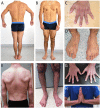
Clinical picture of Family 1. (A–D) Patient III:5. (E–H) Patient III:1. (A and B) Hypertrophic trapezius muscles and wide neck. Limb muscles are hypertrophic proximally as compared to distally in both arms and legs and the lumbar back muscles are prominent. (C and D) Syndactyly is present between all fingers and the feet show sandal gap sign. (E) Wide neck, anteverted shoulders, prominent trapezius and latissimus dorsi muscles and a tendency to thoracic kyphosis. (F) The lower legs are thin with reduced size of the medial parts of the gastrocnemius muscles and a tendency to sandal gap widening between the first and second toe. (G and H) Slight syndactyly is present between fingers and slight contractures of the long finger flexors.
Figure 2
Figure 2
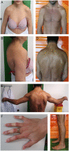
Clinical picture of Family 2. (A, B and F) Patient III:2. (CE and G) Patient III:3. Wide neck due to hypertrophic M. trapezius, malposition of shoulder girdle with anteverted shoulders, hyperkyphosis (AD), limited range of motion and contractures of elbows (E), contractures of long finger flexors (F) and contractures of knees and Achilles tendons (G).
Figure 3
Figure 3
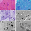
Biopsy from the vastus lateralis muscle of Patient III:1 in Family 1, as visualized by light (A–C) and electron microscopy (D–F). (A) Marked structural changes in groups of muscle fibres with internalized nuclei, eosinophilic protein aggregates (arrows) and multiple small vacuoles (arrowheads) (haematoxylin and eosin). (B) Some protein aggregates are purple in Gomori trichrome staining and others are dark green (arrows). (C) The intermyofibrillar network is deranged and partly unusually coarse (arrows). Many fibres appear lobulated which was the overall major alteration in the muscle biopsy (nicotinamide adenine dinucleotide tetrazolium reductase, NADH-TR). (D) In groups of muscle fibres, there are numerous autophagic vacuoles with membranous degradation products (arrows). (E) Focal disruption of the normal sarcomere structures (arrow) and autophagic vacuoles appearing together. (F) Large amorphous and partly fibrillar protein aggregates (arrows).
Figure 4
Figure 4
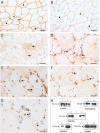
Immunohistochemical staining of muscle biopsy from Patient III:1 and western blot analysis of Patients III:1 and III:5 in Family 1. (A) α-Sarcoglycan appears focally accumulated in the subsarcolemmal region (arrow heads) and also internally (arrows) in several fibres. (B) Caveolin-3 shows variable and reduced staining at the sarcolemma (arrow heads) compared to a control (inset) and is also located in the interior of some fibres (arrows). (C) Dystrophin is distributed diffusely in the interior of several fibres (arrows) in the same region as illustrated in A. (D) Desmin is accumulated in the subsarcolemmal region in many fibres (arrows). (E) LAMP2 is present in association with several vacuoles indicating lysosomal origin (arrows). (F) LC3 is upregulated and present in the interior of some fibres (arrows). (G) Supervillin is completely absent compared to controls where it can be detected mainly in the sarcolemmal region (arrows). (H) Western blot analysis showing that supervillin is present in two isoforms, 250 kDa and 205 kDa, respectively, in controls but is completely absent in Patients III:1 and III:5. No truncated proteins were detected in the patients.
Figure 5
Figure 5
Biopsy from the deltoid muscle of Patient III:2 in Family 2, as visualized by light (A–C) and electron microscopy (D–G). (A) A considerable number of partially atrophic lobulated muscle fibres with pointed, occasionally cap-like subsarcolemmal deposits (arrows) (H&E). (B) Prominent oxidative enzyme activity in these fibres (arrows) (NADH). (C) Atrophic lobulated fibres are almost exclusively type 1 fibres (arrows); no neurogenic pattern is observed (mATPase at pH 9.4). (D) Subsarcolemmal accumulation of degraded myofibrils, glycogen, pleomorphic material and autophagy-associated (arrows) organelles. (E) Heterogeneous lipoprotein deposits including maturing lipofuscin (arrows). (F) Sporadically, nemaline rods are seen (arrow). (G) Three rods in transverse section (arrows).
Figure 6
Figure 6
Immunohistochemical staining and western blot analysis of muscle biopsy from Patient III:3 in Family 2. (A) α-Sarcoglycan immunoreactivity of the pointy sarcolemma-associated structures in the lobulated muscle fibres (arrowheads) and internally in some fibres (arrows). (B) Irregular, reduced caveolin-3 staining mostly of smaller, partially atrophic fibres (arrowheads). (C) Abnormal non-sarcolemmal dysferlin immunoreactivity in several fibres (arrows). Subsarcolemmal accumulation of desmin (arrows) (D) and LC3 (arrows) (E) immunoreactivity in affected partially atrophic fibres. (F) LC3-positive granular and vacuolar structures (arrows) were also present in muscle fibres of normal size and in hypertrophic fibres. (G) Subsarcolemmal p62-immunoreactive subsarcolemmal material in affected lobulated fibres and occasional strongly stained p62-positive autophagic vacuoles (arrows). (H) Selected immunoblot results. 1 = index case; X and K = control cases. Scale bar = 40 µm in AC, 25 µm in DG.
Figure 7
Figure 7
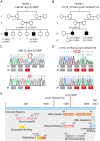
Pedigrees and molecular genetics. (A) Pedigree of Family 1. (B) Pedigree of Family 2. (C) Sequencing chromatograms demonstrating the homozygous mutation identified in Family 1 on genomic DNA, c.4812C>A; p.Tyr1604*. (D) Sequencing chromatograms demonstrating the homozygous mutation identified in Family 2 on genomic DNA, c.3578_3579del; p.Val1193Glufs*46. (E) Overview of the protein structure of supervillin isoform 2 (SV2, p250) and localization of the SVIL mutations described in this study. AR = androgen receptor; FA = focal adhesion; M = mutated sequence; N = normal sequence.

References

    1. Bhuwania R, Cornfine S, Fang Z, Kruger M, Luna EJ, Linder S.. Supervillin couples myosin-dependent contractility to podosomes and enables their turnover. J Cell Sci 2012; 125: 2300–14. - PMC - PubMed
    1. Bonnemann CG. The collagen VI-related myopathies Ullrich congenital muscular dystrophy and Bethlem myopathy. Handb Clin Neurol 2011; 101: 81–96. - PMC - PubMed
    1. Chen Y, Takizawa N, Crowley JL, Oh SW, Gatto CL, Kambara T, et al.F-actin and myosin II binding domains in supervillin. J Biol Chem 2003; 278: 46094–106. - PubMed
    1. Chen X, Yang H, Zhang S, Wang Z, Ye F, Liang C, et al.A novel splice variant of supervillin, SV5, promotes carcinoma cell proliferation and cell migration. Biochem Biophys Res Commun 2017; 482: 43–9. - PubMed
    1. Crowley JL, Smith TC, Fang Z, Takizawa N, Luna EJ.. Supervillin reorganizes the actin cytoskeleton and increases invadopodial efficiency. MBoC 2009; 20: 948–62. - PMC - PubMed

Publication types