. 2021 May;11(4):488-499.
doi: 10.1177/2192568220912706. Epub 2020 Mar 20.

Anterior Cervical Osteophyte Resection for Treatment of Dysphagia

Affiliations

Anterior Cervical Osteophyte Resection for Treatment of Dysphagia

Joshua M Kolz et al. Global Spine J. 2021 May.

Abstract

Study design: This was a retrospective cohort study.

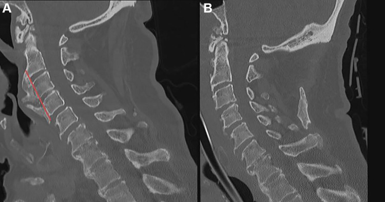
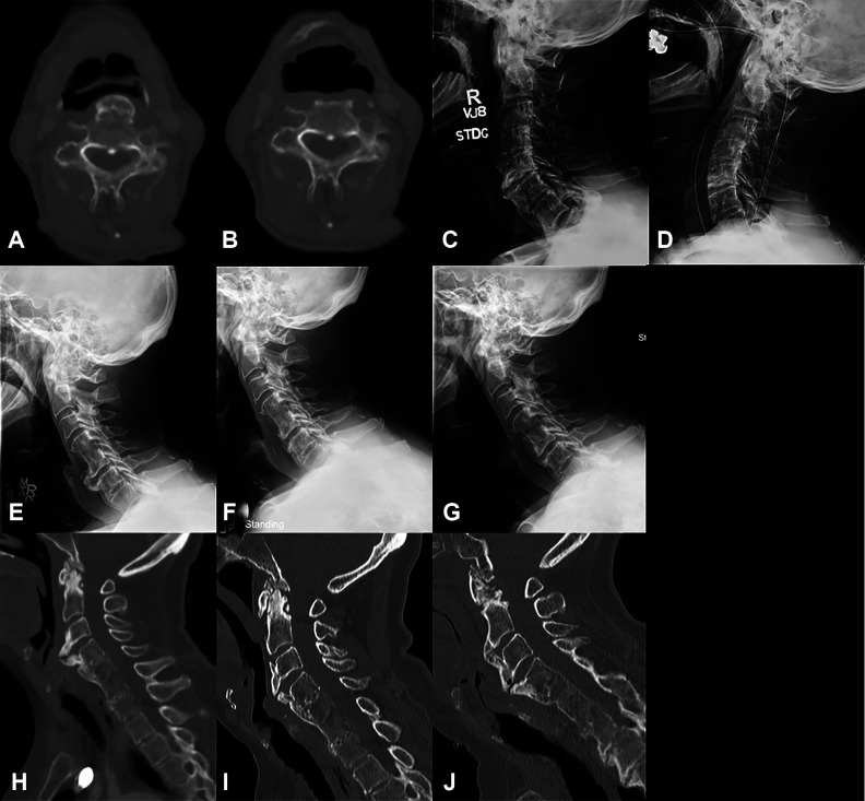
Objectives: When anterior cervical osteophytes become large enough, they may cause dysphagia. There is a paucity of work examining outcomes and complications of anterior cervical osteophyte resection for dysphagia.

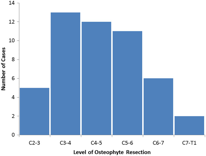
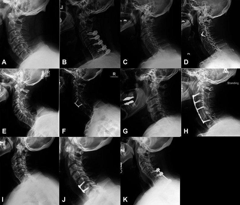
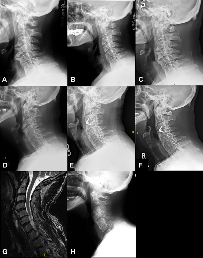
Methods: Retrospective review identified 19 patients who underwent anterior cervical osteophyte resection for a diagnosis of dysphagia. The mean age was 71 years and follow-up, 4.7 years. The most common level operated on was C3-C4 (13, 69%).

Results: Following anterior cervical osteophyte resection, 79% of patients had improvement in dysphagia. Five patients underwent cervical fusion; there were no episodes of delayed or iatrogenic instability requiring fusion. Fusion patients were younger (64 vs 71 years, P = .05) and had longer operative times (315 vs 121 minutes, P = .01). Age of 75 years or less trended toward improvement in dysphagia (P = .09; OR = 18.8; 95% CI 0.7-478.0), whereas severe dysphagia trended toward increased complications (P = .07; OR = 11.3; 95% CI = 0.8-158.5). Body mass index, use of an exposure surgeon, diffuse idiopathic skeletal hyperostosis diagnosis, surgery at 3 or more levels, prior neck surgery, and fusion were not predictive of improvement or complication.

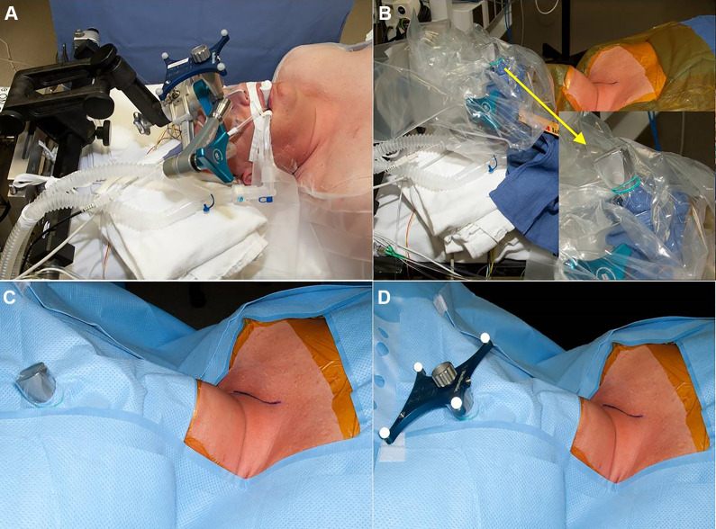
Conclusions: Anterior cervical osteophyte resection improves swallowing function in the majority of patients with symptomatic osteophytes. Spinal fusion can be added to address stenosis and other underlying cervical disease and help prevent osteophyte recurrence, whereas intraoperative navigation can be used to ensure complete osteophyte resection without breaching the cortex or entering the disc space. Because of the relatively high complication rate, patients should undergo thorough multidisciplinary workup with swallow evaluation to confirm that anterior cervical osteophytes are the primary cause of dysphagia prior to surgery.

Keywords: DISH; cervical spine; cheilectomy; dysphagia; osteophyte; syndesmophyte.


Conflict of interest statement

Declaration of Conflicting Interests: The author(s) declared no potential conflicts of interest with respect to the research, authorship, and/or publication of this article.

Figures

Figure 1.
Figure 1.
Levels of anterior osteophyte resection in 19 patients undergoing surgery for a diagnosis of dysphagia.
Figure 2.
Figure 2.
Example of draping for stereotactic navigation setup for cervical spine surgery. A Mayfield cranial stabilization system (Integra LifeSciences, Plainsboro, NJ) is used to hold the skull and cervical spine in place during the operation. The reference frame attachment for the stereotactic navigation setup is connected directly to the Mayfield (A). A sterile drape is then placed over the attachment and rubber banded in place (B). The patient is then draped in the usual sterile fashion, and a hole is cut in the drape to allow the attachment to come into the surgical field (C). The sterile reference frame is then placed on the attachment. This setup allows for sterility and excellent working space between the navigational tools and the reference frame.
Figure 3.
Figure 3.
Intraoperative stereotactic navigation can be used to determine the junction between the pathological osteophyte and the native anterior cortex and disc space, thereby ensuring complete but not overresection. Either of these 2 extremes is easily obtained without some measure of image guidance to ensure that the osteophyte is completely resected, whereas the native spinal column elements are preserved for stability. Image (A) demonstrates patients with anterior cervical syndesmophytes with significant compression on the esophagus at C3-4. The red line is the planned resection of osteophytes to remove esophageal compression without entering the disc space and destabilizing the cervical spine. Image (B) demonstrates resection of syndesmophytes after surgical resection using stereotactic navigation intraoperatively.
Figure 4.
Figure 4.
Imaging of patients who did not see significant improvement in their dysphagia after anterior cervical osteophyte resection. Axial computed tomography (CT) scan through the C4 vertebral body preoperatively (A) and 2 months postoperatively of patient 1 showing minimal residual anterior osteophyte and decreased esophageal compression. Lateral X-rays of patient 13, preoperatively (C) and 1 month postoperatively (D) showing full resection of osteophytes. Radiographic imaging of patient 5 who had a 16-mm osteophyte at C5-6 preoperatively (E), 6 mm postoperatively (F), and osteophyte regrowth to 11.5 mm 2 years postoperatively (G). CT imaging of patient 15 who had a 16.5 mm osteophyte at C3-4 preoperatively (H), 8 mm 14 months postoperatively (I), and regrowth to 13 mm at 6.5 years after osteophyte resection (J).
Figure 5.
Figure 5.
Five patients underwent concurrent cervical fusion surgery along with osteophyte resection. Lateral preoperative (A) and 2-year postoperative (B) X-rays for patient 3 who underwent C3-7 osteophyte resection and posterior decompression and fusion for concurrent myelopathy. Preoperative (C) and 5-year postoperative (D) lateral X-rays of patient 10 who underwent C3-4 anterior cervical discectomy and fusion (ACDF) for critical cervical stenosis at the time of osteophyte removal. Preoperative (E) and 2-year postoperative (F) X-rays of patient 12, who underwent prophylactic C4-5 ACDF to help prevent recurrent osteophyte regrowth. Lateral preoperative (G) and 5-years postoperative (H) X-rays of patient 17 who underwent C5 corpectomy with C2-6 anterior cervical fusion for cervical stenosis from OPLL along with osteophyte removal of dysphagia and sleep apnea. Preoperative (I) and postoperative (J) X-rays of patient 4 who underwent C5-6 ACDF for C6 radiculopathy along with C2-T1 cheilectomy and cricopharyngeal myotomy for dysphagia. The patient developed a postoperative infection with osteomyelitis, requiring a 2-stage operation with anterior debridement, with partial C5 and C6 corpectomy, revision C5-6 ACDF, and posterior C5-T1 spinal fusion, which showed good alignment 4 years postoperatively (K).
Figure 6.
Figure 6.
Preoperative (A) radiograph of patient 2, who underwent C3-5 anterior osteophytectomy (21 mm) with postoperative radiograph (B) demonstrating complete excision of the C3-4 and C4-5 osteophyte and subsequent asymptomatic regrowth of osteophytes to 11 mm (C) at the 5-year follow-up. Preoperative (D) radiographs of patient 10 who underwent C3-4 anterior cervical discectomy and fusion and cheilectomy (E) who had regrowth of osteophytes (6 mm) proximally at the C2-3 level at the 5-year follow-up. Preoperative T2 magnetic resonance image (G) of patient 8 who underwent C4-5 osteophyte resection (13 mm) and had asymptomatic regrowth of osteophytes at C4-5 of 7 mm at the 3.5-year follow-up (H).

Similar articles

Cited by

References

    1. Parker MD. Dysphagia due to cervical osteophytes: a controversial entity revisited. Dysphagia. 1989;3:157–160. - PubMed
    1. Klaassen Z, Tubbs RS, Apaydin N, Hage R, Jordan R, Loukas M. Vertebral spinal osteophytes. Anat Sci Int. 2011;86:1–9. doi:10.1007/s12565-010-0080-8 - PubMed
    1. Vodicar M, Kosak R, Vengust R. Long-term results of surgical treatment for symptomatic anterior cervical osteophytes: a case series with review of the literature. Clin Spine Surg. 2016;29:E482–E487. doi:10.1097/BSD.0b013e31829046af - PubMed
    1. Resnick D, Niwayama G. Radiographic and pathologic features of spinal involvement in diffuse idiopathic skeletal hyperostosis (DISH). Radiology. 1976;119:559–568. doi:10.1148/119.3.559 - PubMed
    1. Strasser G, Schima W, Schober E, Pokieser P, Kaider A, Denk DM. Cervical osteophytes impinging on the pharynx: importance of size and concurrent disorders for development of aspiration. AJR Am J Roentgenol. 2000;174:449–453. doi:10.2214/ajr.174.2.1740449 - PubMed

LinkOut - more resources