. 2020 Oct 30;126(6):1099-1107.
doi: 10.1093/aob/mcaa135.

Functional-morphological analyses of the delicate snap-traps of the aquatic carnivorous waterwheel plant (Aldrovanda vesiculosa) with 2D and 3D imaging techniques

Affiliations

Functional-morphological analyses of the delicate snap-traps of the aquatic carnivorous waterwheel plant (Aldrovanda vesiculosa) with 2D and 3D imaging techniques

Anna S Westermeier et al. Ann Bot. .

Abstract

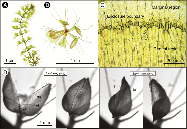
Background and aims: The endangered aquatic carnivorous waterwheel plant (Aldrovanda vesiculosa) catches prey with 3-5-mm-long underwater snap-traps. Trapping lasts 10-20 ms, which is 10-fold faster than in its famous sister, the terrestrial Venus flytrap (Dionaea muscipula). After successful capture, the trap narrows further and forms a 'stomach' for the digestion of prey, the so-called 'sickle-shaped cavity'. To date, knowledge is very scarce regarding the deformation process during narrowing and consequent functional morphology of the trap.

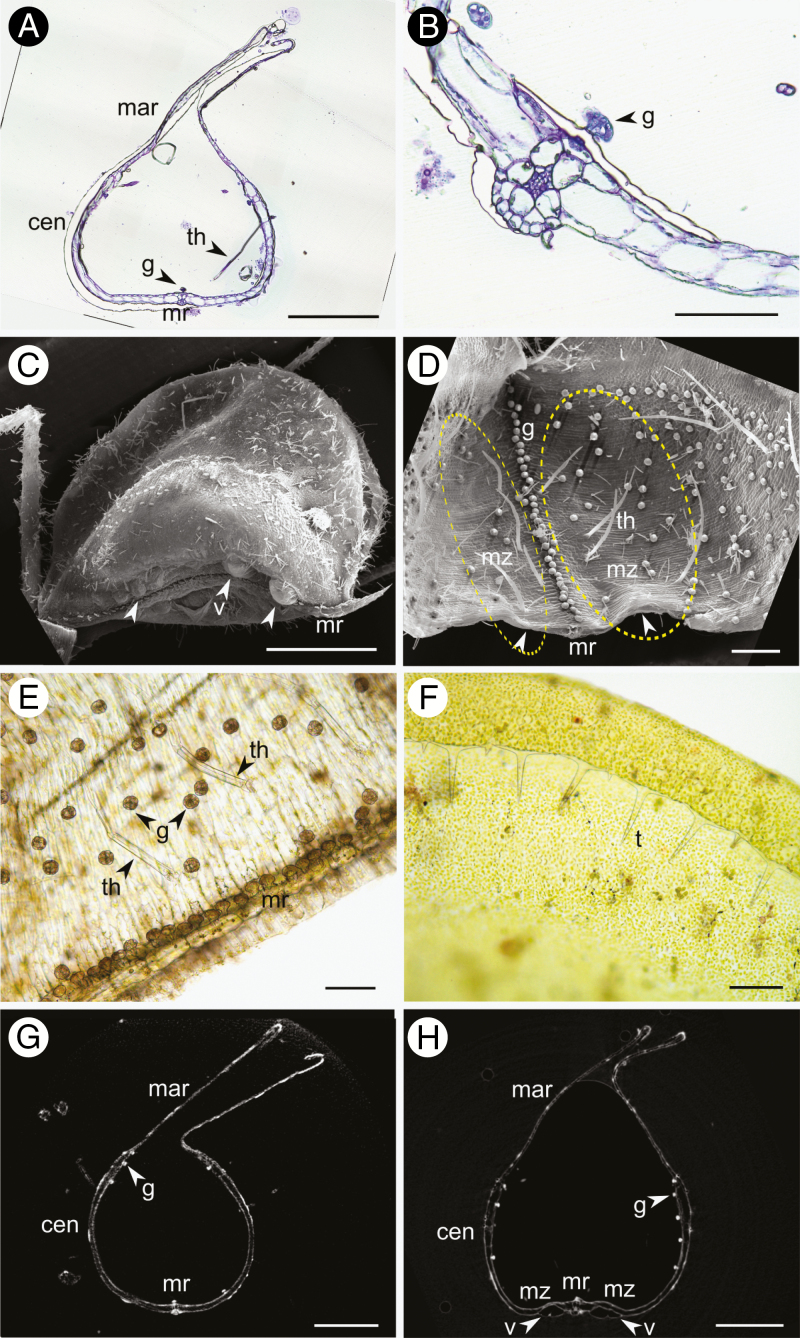
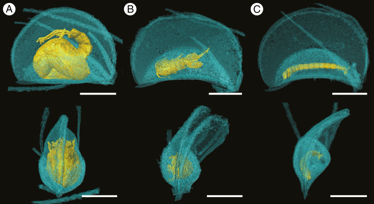
Methods: We performed comparative analyses of virtual 3D histology using computed tomography (CT) and conventional 2D histology. For 3D histology we established a contrasting agent-based preparation protocol tailored for delicate underwater plant tissues.

Key results: Our analyses reveal new structural insights into the adaptive architecture of the complex A. vesiculosa snap-trap. In particular, we discuss in detail the arrangement of sensitive trigger hairs inside the trap and present actual 3D representations of traps with prey. In addition, we provide trap volume calculations at different narrowing stages. Furthermore, the motile zone close to the trap midrib, which is thought to promote not only the fast trap closure by hydraulics but also the subsequent trap narrowing and trap reopening, is described and discussed for the first time in its entirety.

Conclusions: Our research contributes to the understanding of a complex, fast and reversible underwater plant movement and supplements preparation protocols for CT analyses of other non-lignified and sensitive plant structures.

Keywords: Carnivorous plant; functional morphology; micro-CT; plant movement; snap-trap.


Figures

Fig. 1.
Fig. 1.
Aldrovanda vesiculosa morphology and trap movements. (A) Stereo microscopy image of an A. vesiculosa shoot with whorls of snap-traps. (B) Stereo microscopy image of the first whorl, as seen from the apex, which possesses juvenile, not yet opened narrowed snap-traps. (C) Light microscopy image of the inner, untreated trap surface, showing the transition from the central region with numerous glands to the marginal region. (D) Snap-trap movements. The first image shows the open trap in the ready-to-catch state. After triggering, the trap closes within 28 ms at 13 °C (second image). After further stimulation, the trap changes to the narrowed state (third image, 12 min after closure) and finally reaches the very narrowed state (fourth image, 30 min after closure). The sickle-shaped cavity has formed, where prey is digested. During narrowing, only the free-side lobe inverts its curvature. Abbreviations: cr = central region, br = bristle-side lobe, eb = enclosure boundary, fr = free-side lobe, g = glandular hair, mar = marginal region.
Fig. 2.
Fig. 2.
Aldrovanda vesiculosa trap morphology. (A) Light microscopy image of a transverse microtome section of a narrowed trap embedded in Technovit. The central and marginal regions, glands and trigger hairs on the inner trap surface, and the midrib are visible. (B) Light microscopy image of a transverse microtome section of the midrib. (C) SEM image of a trap with prominent deformation of the motor zone upon methanol treatment. Numerous vesicles on the outer epidermis of the motor zone are visible (arrowheads). (D) View into the trap interior with SEM. The distribution of glands and trigger hairs in the trap interior can be observed. In the area of the motor zone, the trap is dented to a greater extent on the trap outside (lower epidermis) than on the inside (indicated by arrows, compare to non-dented trap of A). The needle-like structures are filamentous algae. (E) Light micrograph of an untreated fresh hand-section of the inner trap surface. The midrib, glands and trigger hairs can be seen. (F) Light micrograph of a fresh hand-section of the trap lobe margin, where numerous ‘teeth’ are located. (G) Transverse CT reconstruction of a narrowed trap fixated and scanned in FM with 1 % PTA staining. The midrib and central and marginal regions can be seen. The trap does not show deformation resulting from preparation. (H) Transverse CT reconstruction of a narrowed trap fixed in methanol, CP-dried and scanned in epoxy-glue with 1 % PTA staining. The different trap regions, glands and the midrib are visible. The lower epidermis of the motor zone is undulated and vesicles are present. Scale bars: A, G, H = 500 µm; B, E, F = 100 µm; C, D = 200 µm. Abbreviations: cen = central region, g = gland, mar = marginal region, mr = midrib, mz = motor zone, t = tooth, th = trigger hair, v = vesicle.
Fig. 3.
Fig. 3.
Lateral and apical views of rendered CT scans of liquid-scanned Aldrovanda vesiculosa traps containing prey, stained with 1 % PTA. (A) Trap with half-digested mosquito larva (Supplementary Data Video S1). (B) Trap with female copepod (Video S2). (C) Trap with a Chironomidae larva (Video S3). Scale bars = 1 mm.
Fig. 4.
Fig. 4.
Volume rendering of CP-dried Aldrovanda vesiculosa trap, stained with 1 % PTA and scanned in epoxy-glue (Supplementary Data Video S4). (A) Transverse cut showing the deformation of the motor zone with vesiculation (indicated by arrowheads), trigger hairs and glands can be seen. The gap between the lobes is indicative of the treatment with methanol and contrasting agent, which leads to a deformation of the trap lobes. (B) View of the outer surface of a trap with the midrib and the vesicles (see arrowheads; ablation of the outer epidermal cell wall upon methanol dehydration) on the motor zone being clearly visible. (C) View into trap interior in which the glands on the midrib trigger hairs next to the midrib and the deformation of the motor zone can be seen. The motor zone is highlighted by yellow dashed lines. Abbreviations: mr = midrib with glands on the trap inside, th = trigger hair. Scale bars = 500 µm.

References

    1. Adamec L. 1999. The biology and cultivation of red Australian Aldrovanda vesiculosa. Carnivorous Plant Newsletter 28: 128–132.
    1. Adamec L. 2007. Oxygen concentrations inside the traps of the carnivorous plants Utricularia and Genlisea (Lentibulariaceae). Annals of Botany 100: 849–856. - PMC - PubMed
    1. Adlassnig W, Koller-Peroutka M, Bauer S, Koshkin E, Lendl T, Lichtscheidl IK. 2012. Endocytotic uptake of nutrients in carnivorous plants. The Plant Journal 71: 303–313. - PubMed
    1. Akeret B. 1993. Ein neuer Fundort von Aldrovanda vesiculosa L. in der Nordschweiz und einige Bemerkungen zu Stratiotes aloides L. Botanica Helvetica 103: 193–199.
    1. Ashida J. 1934. Studies on the leaf movement of Aldrovanda vesiculosa L. Memoirs of the College of Sciience, University of Kyoto, Series B 9: 141–244.

Publication types