. 2020 Nov;57(11):4704-4719.
doi: 10.1007/s12035-020-02034-w. Epub 2020 Aug 11.

Hyperphosphorylation Renders Tau Prone to Aggregate and to Cause Cell Death

Affiliations

Hyperphosphorylation Renders Tau Prone to Aggregate and to Cause Cell Death

Mengyu Liu et al. Mol Neurobiol. 2020 Nov.

Abstract

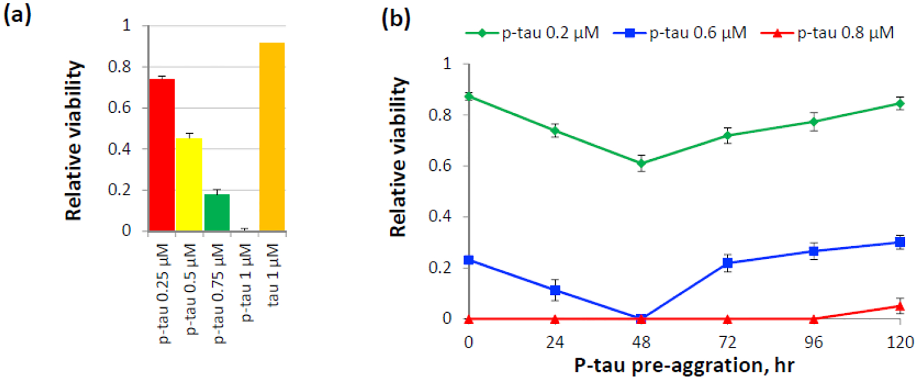
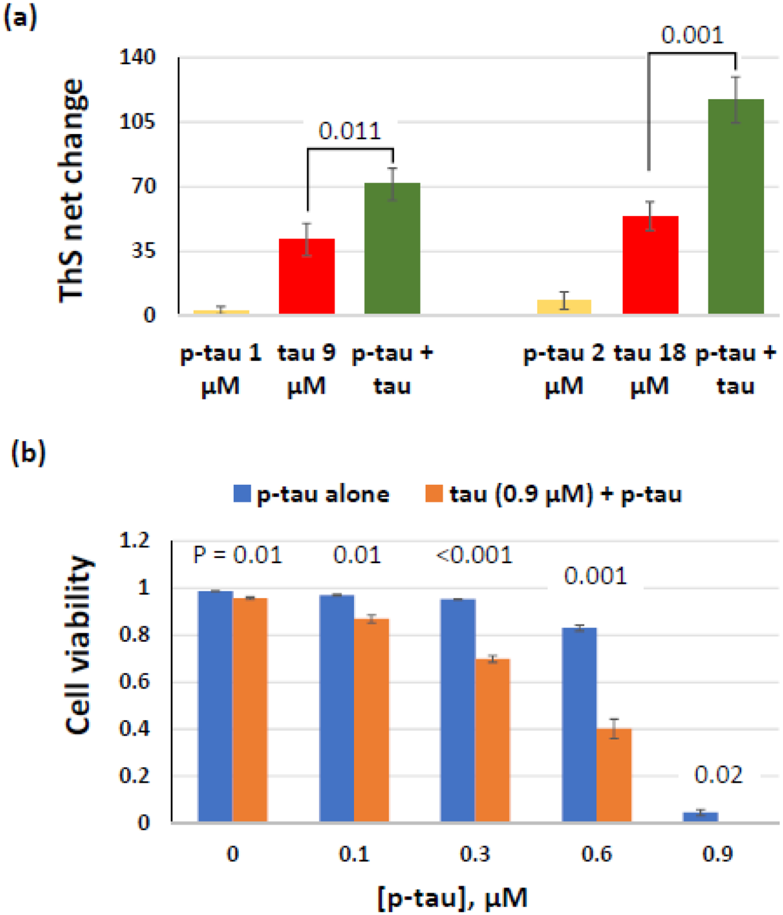
Alzheimer's disease (AD) is a neurodegenerative disorder without a cure or prevention to date. Hyperphosphorylated tau forms the neurofibrillary tangles (NFTs) that correlate well with the progression of cognitive impairments. Animal studies demonstrated the pathogenic role of hyperphosphorylated tau. Understanding how abnormal phosphorylation renders a normal tau prone to form toxic fibrils is key to delineating molecular pathology and to developing efficacious drugs for AD. Production of a tau bearing the disease-relevant hyperphosphorylation and molecular characters is a pivotal step. Here, we report the preparation and characterization of a recombinant hyperphosphorylated tau (p-tau) with strong relevance to disease. P-tau generated by the PIMAX approach resulted in phosphorylation at multiple epitopes linked to the progression of AD neuropathology. In stark contrast to unmodified tau that required an aggregation inducer, and which had minimal effects on cell functions, p-tau formed inducer-free fibrils that triggered a spike of mitochondrial superoxide, induced apoptosis, and caused cell death at sub-micromolar concentrations. P-tau-induced apoptosis was suppressed by inhibitors for reactive oxygen species. Hyperphosphorylation apparently caused rapid formation of a disease-related conformation. In both aggregation and cytotoxicity, p-tau exhibited seeding activities that converted the unmodified tau into a cytotoxic species with an increased propensity for fibrillization. These characters of p-tau are consistent with the emerging view that hyperphosphorylation causes tau to become an aggregation-prone and cytotoxic species that underlies diffusible pathology in AD and other tauopathies. Our results further suggest that p-tau affords a feasible tool for Alzheimer's disease mechanistic and drug discovery studies.

Keywords: Alzheimer’s disease; Hyperphosphorylated tau; Neurofibrillary tangle; Tauopathy.


Conflict of interest statement

Conflict of interest: The authors declare that they have no conflicts of interest with the contents of this article.

Figures

Figure 1.
Figure 1.
Hyperphosphorylated tau (p-tau) produced by the PIMAX system possesses a disease-relevant phosphorylation pattern. (a) Purified 1N4R tau, p-tau (phosphorylated by GSK-3β) and p-tau bearing Cys-to-Ser mutations were resolved by an 8% SDS-PAGE gel and visualized by Coomassie blue staining. (b) Distribution of high-confidence p-tau phosphorylation sites (i.e., 4- and 3-hitters by MS mapping). The number designation conforms to the 2N4R isoform. N1 and N2 are the two N’ domains subjected to alternative splicing. R1 – R4 are the four microtubules binding repeats. The 1N4R isoform used throughout this study lacks N2 but maintains all four microtubules binding repeats. Bold fonts represent 4-hitters that were detected by four MS analyses. The remainders are 3-hitters. Red underlines indicate phosphoepitopes for neurofibrillary tangle staging according to references [6] and [45].
Figure 2.
Figure 2.
Inducer-free and redox-independent fibrillization of p-tau. (a) Aggregation of p-tau, tau, and K18 with or without heparin was quantified by thioflavin S (ThS) fluorescence changes through the entire course of assays and (b) by calculating the net changes of ThS signals after 17 hours of reaction. Numbers above each bar are the P values for the comparison with p-tau without the use of heparin (leftmost column). These were one-tailed Student’s t tests; n = 3. (c) P-tau aggregation does not require the reducing agent DTT and is independent of Cys291 and 322. Shown are the net changes of ThS fluorescence from aggregation of wildtype and the C291S C322S mutant p-tau in the presence or absence of 1 mM DTT. P values were obtained from two-tailed Student’s t tests; n = 3.
Figure 3.
Figure 3.
Immunological and biophysical characterization of p-tau aggregation. (a) Immunoblotting of p-tau with the PHF-specific MC-1 antibody [54] and a general tau antibody MAB3494 (R&D Systems). Duration of aggregation (0 – 120 hours) are shown on the left of the corresponding slots. Parallel blots were probed with the two indicated antibodies. (b) Transmission electron microscopy of p-tau aggregated for the indicated period. (c) PICUP (photo-induced cross-linking of unmodified protein) assays detecting high-molecular weight p-tau species. Shown are overnight aggregation products. (d) FTIR (Fourier transformation infrared) spectroscopy analysis of p-tau after aggregation. Spectral shifts after aggregation are indicated by two arrows.
Figure 4.
Figure 4.
P-tau aggregates are toxic to cells. (a) Viability of cells after 20-hr treatment of 0.25 – 1 μM of p-tau or 1 μM of unmodified tau pre-aggregated for 20 hrs. Cells were double-stained by fluorescein diacetate (FDA) and propidium iodide (PI) for the quantification of relative viability. Error bars are standard deviation. n = 3. This graph is a representative of more than three biological repeats. (b) P-tau cytotoxicity is influenced by the extent of aggregation. P-tau subjected to standard aggregation reactions was collected at the indicated time points and used to treat the neuroblastoma SH-SY5Y cells for 16 hours before FDA/PI double staining and microscopic quantification. 0.8 μM of p-tau (red curve) caused quantitative cell death except the 120-hr pre-aggregation sample.
Figure 5.
Figure 5.
P-tau potentiates tau aggregation and cytotoxicity. (a) Inducer-free aggregation reactions with tau, p-tau, or a mixture of these two proteins were monitored for 16 hours. Comparison of ThS net changes is shown. (b) P-tau converts tau into a cytotoxic species. HEK 293T cells were treated with 0.9 μM of tau supplemented with 0 – 0.9 μM of p-tau for 24 hours before FDA and PI staining. Percentage of live cells is shown here. Note Errors are standard deviation; n = 3. P values (one-tailed Student’s t tests) of each pair of p-tau vs. (p-tau + tau) are shown above the corresponding columns.
Figure 6.
Figure 6.
P-tau activates apoptosis and triggers mitochondrial superoxide accumulation. (a) Examination of apoptosis after p-tau treatment. SH-SY5Y cells treated by 0.6-μM p-tau for 12 hours were examined by FITC-annexin V and PI double staining. Here shows the existence of both early apoptotic (annexin V positive and PI negative) and late apoptotic or already dead (annexin V and PI both positive) cells. DIC: differential interference contrast. (b) and (c) P-tau raises mitochondrial superoxide levels. SH-SY5Y cells treated by 0.6 μM of p-tau for 5 hours were stained by MitoSOX for for microscopy (b) and FACS analysis (c). 20 mM Tris (pH 7.4) buffer was used as mock treatment. The mean fluorescence intensity (MFI) is shown in the bar graph. Error bars show range of two independent assays. (d) Examination of the effect of ROS scavengers on apoptosis after p-tau treatment. SH-SY5Ycells grown in the presence of two concentrations of NAC, TEMPOL or Trolox for 24 hours were treated by 0.4-μM p-tau for 5 hours. The cells were then double-stained by MitoSOX and FITC-annexin V. The percentage of cells stained by annexin V and those exhibiting strong MitoSOX signals are shown. Error bars are standard deviation. n = 3.

References

    1. Glenner GG & Wong CW (1984) Alzheimer’s disease: initial report of the purification and characterization of a novel cerebrovascular amyloid protein, Biochem Biophys Res Commun. 120, 885–90. - PubMed
    1. Grundke-Iqbal I, Iqbal K, Tung YC, Quinlan M, Wisniewski HM & Binder LI (1986) Abnormal phosphorylation of the microtubule-associated protein tau (tau) in Alzheimer cytoskeletal pathology, Proc Natl Acad Sci U S A. 83, 4913–7. - PMC - PubMed
    1. Hardy J & Selkoe DJ (2002) The amyloid hypothesis of Alzheimer’s disease: progress and problems on the road to therapeutics, Science. 297, 353–6. - PubMed
    1. Mehta D, Jackson R, Paul G, Shi J & Sabbagh M (2017) Why do trials for Alzheimer’s disease drugs keep failing? A discontinued drug perspective for 2010–2015, Expert opinion on investigational drugs. 26, 735–739. - PMC - PubMed
    1. Braak H, Alafuzoff I, Arzberger T, Kretzschmar H & Del Tredici K (2006) Staging of Alzheimer disease-associated neurofibrillary pathology using paraffin sections and immunocytochemistry, Acta Neuropathologica. 112, 389–404. - PMC - PubMed

LinkOut - more resources