. 2020 Aug 3;61(10):19.
doi: 10.1167/iovs.61.10.19.

Longitudinal Analysis of Structural and Functional Changes in Presence of Reticular Pseudodrusen Associated With Age-Related Macular Degeneration

Affiliations

Longitudinal Analysis of Structural and Functional Changes in Presence of Reticular Pseudodrusen Associated With Age-Related Macular Degeneration

Marlene Sassmannshausen et al. Invest Ophthalmol Vis Sci. .

Abstract

Purpose: To examine longitudinal changes of retinal thickness and retinal sensitivity in patients with intermediate age-related macular degeneration (iAMD) and predominantly reticular pseudodrusen (RPD).

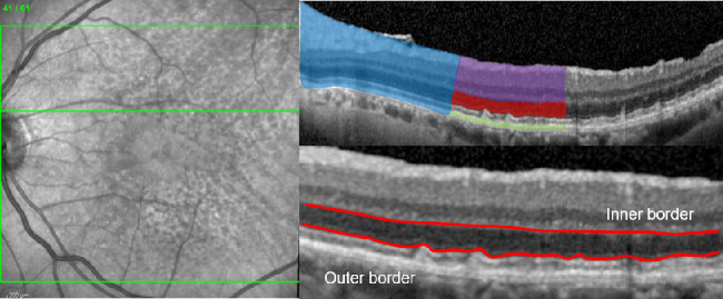
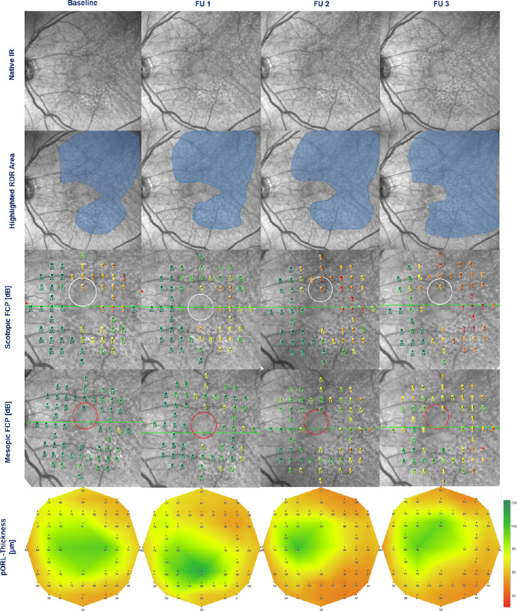
Methods: At baseline 30 eyes of 25 iAMD patients underwent optical coherence tomography imaging, mesopic and scotopic fundus-controlled perimetry (FCP) with follow-up examinations at month 12 (20 eyes), 24 (12 eyes), and 36 (11 eyes). Thicknesses of different retinal layers and results of FCP testing (n = 56 stimuli) were spatially and longitudinally analyzed using linear mixed-effects models.

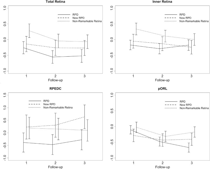
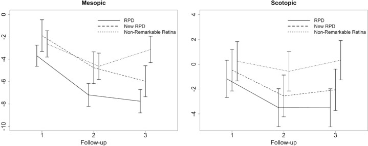
Results: At baseline, the thickness of the partial outer retinal layer (pORL, 70.21 vs. 77.47 µm) and both mesopic (16.60 vs. 18.72 dB) and scotopic (12.14 vs. 18.67 dB) retinal sensitivity were decreased in areas with RPD compared with unremarkable areas (P < 0.001). Over three years, mean change of pORL was -0.66 normative standard deviation (SD; i.e., z-score, P < 0.001) for regions with existing RPD, -0.40 SD (P < 0.001) for regions with new occurring RPD, and -0.17 SD (P = 0.041) in unremarkable regions. Decrease of scotopic and mesopic sensitivity over three years was more pronounced in areas with existing (-3.51 and -7.76 dB) and new occurring RPD (-2.06 and -5.97 dB). Structure-function analysis revealed that 1 SD decrease of pORL thickness was associated with a sensitivity reduction of 3.47 dB in scotopic and 0.79 dB in mesopic testing.

Conclusions: This study demonstrates progressive outer retinal degeneration and impairment of photoreceptor function in eyes with iAMD and RPD over three years. Preservation of outer retinal thickness and reduction of RPD formation may constitute meaningful surrogate endpoints in interventional trials on eyes with AMD and RPD aiming to slow outer retinal degeneration.


Conflict of interest statement

Disclosure: M. Sassmannshausen, Heidelberg Engineering (F); Carl Zeiss Meditec (F); Optos (F); CenterVue (F); M. Pfau, Heidelberg Engineering (F); Carl Zeiss Meditec (C, F); Optos (F); CenterVue (F); S. Thiele, Heidelberg Engineering (F, R); Carl Zeiss Meditec (F); Optos (F); CenterVue (F); Bayer (R); Novartis (R); R. Fimmers, None; J. Steinberg, None; M. Fleckenstein, Heidelberg Engineering (F); Novartis (C, F); Roche/Genentech (C); US20140303013A1 (P); F. Holz, Acucela (C, F, R); Allergan (F, R); Appelis (C, R); Bayer (C, F, R); Boehringer-Ingelheim (C); Bioeq/Formycon (F, C); CenterVue (F); Ellex (R); Roche/Genentech (C, F, R); Geuder (C); Grayburg Vision (C, R); Heidelberg Engineering (C, F, R); Kanghong (C, F); LinBioscience (C, R); NightStarX (F); Novartis (C, F, R); Optos (F); Pixium Vision (C, F, R); Oxurion (C, R); Stealth BioTherapeutics (C, R); Carl Zeiss Meditec (F, R); S. Schmitz-Valckenberg, Acucela (F); Alcon/Novartis (C, F, R); Allergan (C, F, R); Bayer (F, R); Bioeq/Formycon (F, C); Carl Zeiss MedicTec (F, R); CenterVue (F); Galimedix (C); Genentech/Roche (F, R); Heidelberg Engineering (F); Katairo (F); Optos (F)

Figures

Figure 1.
Figure 1.
Spectral-domain optical coherence tomography raster scan (left) with the segmented retinal layers (top right) of the total retina (blue), the inner retina (violet), the pORL (red), and the RPEDC (green), as well as the boundaries (bottom right—red colored) for the assessment of the pORL thickness.
Figure 2.
Figure 2.
Graphical illustration of the longitudinal thickness changes in SD of the control group of the total retina, the inner retina, the RPEDC, and the pORL thickness. For each of the four layers, the changes are shown for the three different groups: (a) regions of existing RPD at baseline, (b) regions of newly developing RPD, and (c) in non-remarkable regions of the retina. Data of the retinal thickness change were plotted in normative SD. The x-axis shows the number of the follow-up visit.
Figure 3.
Figure 3.
Representative example of a 72-year-old AMD patient with RPD in his left eye. Structural and functional data are illustrated for each visit (from left to right: baseline, follow-up 1 [FU 1], follow-up 2 [FU 2], and follow-up 3 [FU 3]). Extension area of RPD is shown by near-infrared cSLO imaging (first row: native images, second row: area involved highlighted by blue color). Corresponding functional testing is shown for mesopic (third row) and scotopic (fourth row) FCP. Thickness of the pORL (fifth row) is illustrated by a “heat map.” Note the increase in area extension of RPD over time, spatially and temporally corresponding to progressive thinning of the pORL and increasing loss of mesopic and scotopic retinal sensitivity.
Figure 4.
Figure 4.
Graphical illustration of the longitudinal sensitivity changes [estimate ± standard error] in dB for mesopic and scotopic FCP testing at each follow-up visit (FU 1–3). Results are presented for retinal regions with RPD, newly developing RPD at follow-up visit, and regions with nonremarkable pathological alterations.

References

    1. Lim LS, Mitchell P, Seddon JM, Holz FG, Wong TY. Age-related macular degeneration. The Lancet. 2012; 379: 1728–1738. - PubMed
    1. Ferris FL, Wilkinson CP, Bird A, et al. .. Clinical classification of age-related macular degeneration. Ophthalmology. 2013; 120: 844–851. - PMC - PubMed
    1. Schmitz-Valckenberg S, Steinberg JS, Fleckenstein M, Visvalingam S, Brinkmann CK, Holz FG. Combined confocal scanning laser ophthalmoscopy and spectral-domain optical coherence tomography imaging of reticular drusen associated with age-related macular degeneration. Ophthalmology. 2010; 117: 1169–1176. - PubMed
    1. Zweifel SA, Spaide RF, Curcio CA, Malek G, Imamura Y. Reticular pseudodrusen are subretinal drusenoid deposits. Ophthalmology. 2010; 117: 303–312.e1. - PubMed
    1. Curcio CA, Messinger JD, Sloan KR, McGwin G, Medeiros NE, Spaide RF. Subretinal drusenoid deposits in non-neovascular age-related macular degeneration: morphology, prevalence, topography, and biogenesis model. Retina. 2013; 33: 265–276. - PMC - PubMed

Publication types

MeSH terms