Review
. 2020 Dec;18(12):3142-3153.
doi: 10.1111/jth.15052. Epub 2020 Sep 9.

Anticoagulant and signaling functions of antithrombin

Affiliations
Review

Anticoagulant and signaling functions of antithrombin

Alireza R Rezaie et al. J Thromb Haemost. 2020 Dec.

Abstract

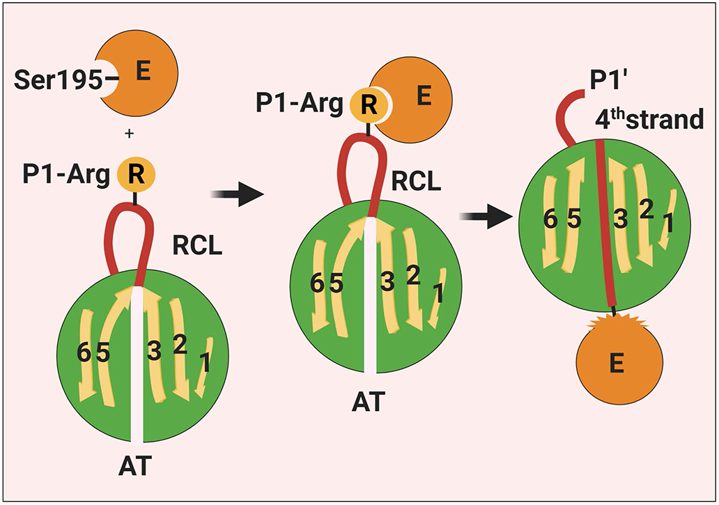
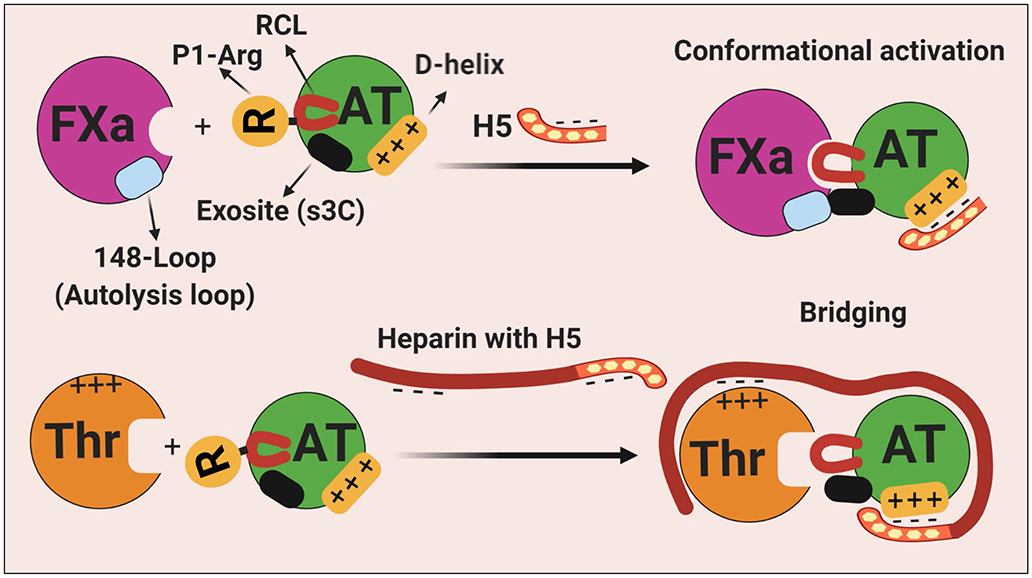
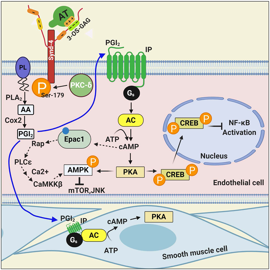
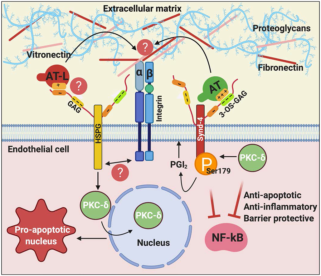
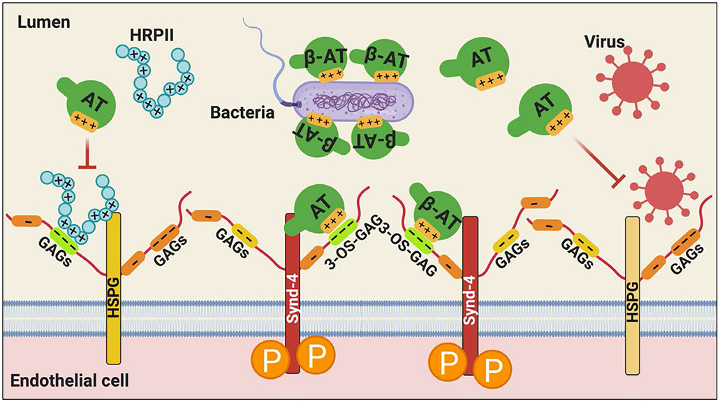
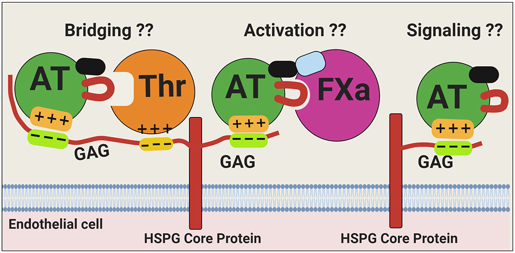
Antithrombin (AT) is a major plasma glycoprotein of the serpin superfamily that regulates the proteolytic activity of the procoagulant proteases of both intrinsic and extrinsic pathways. Two important structural features that participate in the regulatory function of AT include a mobile reactive center loop that binds to active site of coagulation proteases, trapping them in the form of inactive covalent complexes, and a basic D-helix that binds to therapeutic heparins and heparan sulfate proteoglycans (HSPGs) on vascular endothelial cells. The binding of D-helix of AT by therapeutic heparins promotes the reactivity of the serpin with coagulation proteases by several orders of magnitude by both a conformational activation of the serpin and a template (bridging) mechanism. In addition to its essential anticoagulant function, AT elicits a potent anti-inflammatory signaling response when it binds to distinct vascular endothelial cell HSPGs, thereby inducing prostacyclin synthesis. Syndecans-4 has been found as a specific membrane-bound HSPG receptor on endothelial cells that relays the signaling effect of AT to the relevant second messenger molecules in the signal transduction pathways inside the cell. However, following cleavage by coagulation proteases and/or by spontaneous conversion to a latent form, AT loses both its anti-inflammatory activity and high-affinity interaction with heparin and HSPGs. Interestingly, these low-affinity heparin conformers of AT elicit potent proapoptotic and antiangiogenic activities by also binding to specific HSPGs by unknown mechanisms. This review article will summarize current knowledge about mechanisms through which different conformers of AT exert their serine protease inhibitory and intracellular signaling functions in these biological pathways.

Keywords: anti-inflammatory; anticoagulant; antithrombin; heparan sulfate; heparin.


Conflict of interest statement

Disclosure of Conflict of Interests

The authors declare no conflict of interests.

Figures

Figure 1.
Figure 1.
Schematic representation of the interaction of a coagulation protease with AT. The binding of the P1-Arg residue on RCL of AT to the active-site pocket of a coagulation protease induces acylation/cleavage of the P1-P1’ bond which then triggers a large scale conformational change in the serpin that leads to the insertion of the RCL into β-sheet A as a central 4th strand, thereby the RCL dragging the covalently-bound protease to the opposite pole of the serpin. The conformational change also results in disruption of the catalytic machinery of the protease. Thus, the protease gets trapped as an acylated complex with no catalytic function. See the text for more details. RCL, reactive center loop; E, enzyme; AT, antithrombin. Figure was prepared by software provided by Biorender.com.
Figure 2.
Figure 2.
Schematic representation of heparin-mediated activation and promotion of protease inhibition by AT. (Top) In the native conformation of AT, the RCL is less exposed and a cryptic exosite (on s3C) is not available for productive interaction with the protease. The binding of the pentasaccharide fragment of heparin on D-helix results in the expulsion of the RCL that is coupled to the alteration of the cryptic exosite outside of the RCL. The conformationally altered exosite on AT interacts with a complementary exosite (the basic 148-loop, also called autolysis loop) on FXa (also on FIXa). (Bottom) Long-chain high molecular weight heparin bind both thrombin (on basic exosite-2) and D-helix of AT to promote the inhibition of the protease by a template (bridging mechanism). Thrombin is incapable of recognizing the activated conformer of AT because it lacks the complementary exosite site (the autolysis loop is negatively charged in thrombin) to interact with the heparin-exposed cryptic site on AT. See the text for more details. H5, pentasaccharide; s3C, strand 3 of β-sheet C; RCL, reactive center loop; AT, antithrombin; Thr, thrombin; FXa, factor Xa. Figure was prepared by software provided by Biorender.com.
Figure 3.
Figure 3.
Hypothetical model of the anti-inflammatory signaling mechanism of AT. The binding of AT via its D-helix on 3-OS containing GAGs, covalently attached to Synd-4, recruits PKC-δ to the membrane, thereby leading to phosphorylation of the cytoplasmic domain of the Synd-4 at Ser-179. This process is linked to induction of PGI2 by a PLA2 hydrolyzing arachidonylated phospholipids to produce arachidonic acid followed by its metabolism to PGI2 by Cox-2. PGI2 binds to its Gs-protein coupled receptor, thereby activating adenylyl cyclase and mediating the synthesis of cAMP and activation of protein kinase A (PKA) in both vascular and smooth muscle cells. AT-mediated cAMP signaling leads to phosphorylation of cAMP responsive element binding-protein and its transport to the nucleus, thereby modulating gene expression, including NF-κB inhibition. In addition to PKA, cAMP can also signal through Epac-1, thereby activating CaMKKβ and AMPK in cardiomyocytes. It is not known which one the two pathways is involved in AT-mediated AMPK signaling. See the text for more details. PL, plasmolegen; PLA2, phospholipase A2; AA, arachidonic acid; Cox-2, cyclooxygenase-2; PGI2, prostacyclin; IP, PGI2 receptor; AC, adenylyl cyclase; PKA, protein kinase A; CREB, cAMP responsive element binding-protein; Epac-1, exchange protein directly activated by cAMP; AMPK, adenosine monophosphate kinase; CaMKKβ, Ca2+/calmodulin-dependent protein kinase kinase β; mTOR, mammalian target of rapamycin; JNK, c-Jun N-terminal protein kinase. Figure was prepared by software provided by Biorender.com.
Figure 4.
Figure 4.
Hypothetical model of cytoprotective and pro-apoptotic signaling functions of native and latent forms of AT. The binding of native AT on 3-OS containing GAGs on Synd-4 recruits PKC-δ to the membrane, thereby phosphorylating the cytoplasmic domain of the receptor and inducing PGI2 synthesis. PGI2 signaling elicits anti-apoptotic, anti-inflammatory and barrier protective signaling responses. The binding of the low-affinity conformer of AT, latent AT, on vascular GAGs induces perinuclear/nuclear localization of PKC-δ, thereby eliciting pro-apoptotic/antiangiogenic signaling responses. We hypothesize that a crosstalk between HSPGs and different types of integrins modulates different mechanisms of AT function in these pathways by interacting with extracellular matrix proteins (i.e., vitronectin and fibronectin). See the text for more details. AT-L, latent AT; GAG, glycosaminoglycan; 3-OS, 3-O-sulfate. Figure was prepared by software provided by Biorender.com.
Figure 5.
Figure 5.
Hypothetical model of the protective effect of AT against infectious microorganisms. AT (particularly β-AT) can neutralize Gram-negative bacteria by binding via its D-helix to negatively charged molecules of the bacterial cell wall. AT can also inhibit the HSPG-dependent binding of certain family of viruses to cell surface GAGs, thereby preventing their entry into the host cell. AT can inhibit HRPII-dependent upregulation of pro-inflammatory and procoagulant responses, mediated by the Plasmodium falciparum-derived secretory protein, HRPII, in vascular endothelial cells not only by its anti-inflammatory signaling function but also by a competitive mechanism. See the text for more details. HSPG, heparan sulfate proteoglycan; Synd-4, syndecan-4; GAG, glycosaminoglycan; 3-OS, 3-O-sulfate; HRPII, histidine rich protein II. Figure was prepared by software provided by Biorender.com.
Figure 6.
Figure 6.
Hypothetical model of the interaction of AT with different vascular GAGs. Vascular HSPGs containing GAGs with different chain-lengths can bind D-helix of AT to elicit intracellular signaling responses and/or promote the serpin inhibition of coagulation proteases by a conformational activation (FXa) or by a template (thrombin) mechanism. These mechanistic concepts have been firmly established in cellular and in in vitro assays using therapeutic heparins. However, the relevance/significance of AT interaction with vascular GAGs to physiological functions (signaling or protease inhibitory function) of AT remains unknown (??) and requires further investigation. Figure was prepared by software provided by Biorender.com.

References

    1. Olson ST, Richard B, Izaguirre G, Schedin-Weiss S, Gettins PG. Molecular mechanism of antithrombin regulation of blood clotting proteinases. A paradigm for understanding proteinase regulation by serpin family protein proteinase inhibitors. Biochimie. 2010; 92:1587–1596. - PMC - PubMed
    1. Gettins PG. Serpins structure, mechanism, and function. Chem Rev. 2002; 102: 4751–4803. - PubMed
    1. Damus PS, Hicks M, Rosenberg RD. Anticoagulant action of heparin. Nature. 1973; 246: 355–357. - PubMed
    1. Carrell RW, Skinner R, Jin L, Abrahams JP. Structural Mobility of Antithrombin and its Modulation by Heparin. Thromb Haemost. 1997; 78: 516–519. - PubMed
    1. Leon M, Aiach M, Coezy E, Gunennec JY, Feissinger JN. Antithrombin III synthesis in rat liver parenchymal cells. Thromb Res. 1983; 30: 369–375. - PubMed

Publication types