. 2020 Aug 6;12(8):1758.
doi: 10.3390/polym12081758.

Poly(ε-caprolactone) Titanium Dioxide and Cefuroxime Antimicrobial Scaffolds for Cultivation of Human Limbal Stem Cells

Affiliations

Poly(ε-caprolactone) Titanium Dioxide and Cefuroxime Antimicrobial Scaffolds for Cultivation of Human Limbal Stem Cells

Mirna Tominac Trcin et al. Polymers (Basel). .

Abstract

Limbal Stem Cell Deficiency (LSCD) is a very serious and painful disease that often results in impaired vision. Cultivation of limbal stem cells for clinical application is usually performed on carriers such as amniotic membrane or surgical fibrin gel. Transplantation of these grafts is associated with the risk of local postoperative infection that can destroy the graft and devoid therapeutic benefit. For this reason, electrospun scaffolds are good alternatives, as proven to mimic the natural cells surroundings, while their fabrication technique is versatile with regard to polymer functionalization and scaffolds architecture. This study considers the development of poly(ε-caprolactone) (PCL) immune-compatible and biodegradable electrospun scaffolds, comprising cefuroxime (CF) or titanium dioxide (TiO2) active components, that provide both bactericidal activity against eye infections and support of limbal stem cells growth in vitro. The PCL/CF scaffolds were prepared by blend electrospinning, while functionalization with the TiO2 particles was performed by ultrasonic post-processing treatment. The fabricated scaffolds were evaluated in regard to their physical structure, wetting ability, static and dynamic mechanical behaviour, antimicrobial efficiency and drug release, through scanning electron microscopy, water contact angle measurement, tensile testing and dynamic mechanical analysis, antimicrobial tests and UV-Vis spectroscopy, respectively. Human limbal stem cells, isolated from surgical remains of human cadaveric cornea, were cultured on the PCL/CF and PCL/TiO2 scaffolds and further identified through immunocytochemistry in terms of cell type thus were stained against p63 marker for limbal stem cells, a nuclear transcription factor and cytokeratin 3 (CK3), a corneal epithelial differentiation marker. The electrospun PCL/CF and PCL/TiO2 successfully supported the adhesion, proliferation and differentiation of the cultivated limbal cells and provided the antimicrobial effect against Pseudomonas aeruginosa, Staphylococcus aureus and Candida albicans.

Keywords: antimicrobial activity; cefuroxime; electrospinning; limbal stem cell deficiency; polycaprolactone; scaffolds; tissue engineering; titanium dioxide.


Conflict of interest statement

The authors declare no conflict of interest.

Figures

Figure 1
Figure 1
3D printed collector geometry—NX sketch.
Figure 2
Figure 2
Tensile testing of the electrospun scaffolds.
Figure 3
Figure 3
Scanning electron microscopy (SEM) photomicrographs of the electrospun scaffolds: (a) poly(ε-caprolactone) (PCL) with randomly beaded fibers, (b) PCL/5 wt % CF and (c) PCL/25 wt % CF with uniform fibers, (d) PCL/n-TiO2 and (e) PCL/m-TiO2 with surface particle agglomerations.
Figure 4
Figure 4
Fiber diameter distribution of the electrospun scaffolds.
Figure 5
Figure 5
Electrospun scaffolds total porosity.
Figure 6
Figure 6
Water contact angle measurement on the surface of the electrospun scaffolds: (a) PCL, (b) PCL/n-TiO2, (c) PCL/m-TiO2, (d) PCL/5 wt % CF (1 s), (e) PCL/5 wt % CF (5 s), (f) PCL/25 wt % CF.
Figure 7
Figure 7
Tensile stress-strain curves of the electrospun scaffolds.
Figure 8
Figure 8
Young’s moduli of the electrospun scaffolds.
Figure 9
Figure 9
Storage modulus as a function of temperature of the electrospun scaffolds.
Figure 10
Figure 10
Damping as a function of temperature of the electrospun scaffolds.
Figure 11
Figure 11
Antimicrobial activity of the electrospun scaffolds against: (a) Pseudomonas aeruginosa 3024, (b) Staphylococcus aureus 3048 and (c) Candida albicans 11.
Figure 12
Figure 12
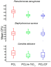
Antimicrobial efficacy of the electrospun PCL, PCL/m-TiO2 and PCL/5 wt % CF scaffolds against Pseudomonas aeruginosa, Staphylococcus aureus and Candida albicans.
Figure 13
Figure 13
Release of the cefuroxime in percentages from the electrospun PCL/CF scaffolds.
Figure 14
Figure 14
SEM photomicrographs of the adhered Limbal stem cells (LSCs) on the electrospun scaffolds: (a) PCL, (b) PCL/5 wt % CF and (c) PCL/m-TiO2.
Figure 15
Figure 15
Percentage of viable cells measured by the 3-(4,5-dimethylthiazol-2-yl)-2,5-diphenyltetrazolium bromide (MTT) assay.
Figure 16
Figure 16
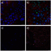
Immunofluorescence analysis of limbal stem cells cultured on PCL scaffold. Cells positive on cornea marker CK3 have cytoplasm coloured red (a,b) and cell positive on stem cell marker p63 show nuclei stained with turquoise. Cytoskeleton is stained red with phalloidin- tetramethylrhodamine B isothiocyanate (TRITC) (c,d). All nuclei are counterstained with blue stain 4′,6-diamidino-2-phenylindole (DAPI).
Figure 17
Figure 17
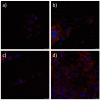
Immunofluorescence analysis of limbal stem cells cultured on PCL/5 wt % CF (a,b) and PCL/25 wt % CF (c,d) scaffold. Cells stained against cornea marker CK3 have cytoplasm coloured with red (a,c). Cell stained against stem cell marker p63 show nuclei stained with turquoise. Cytoskeleton is stained red with phalloidin-tetramethylrhodamine B isothiocyanate (TRITC) (b,d). All nuclei are counterstained with blue stain 4′,6-diamidino-2-phenylindole (DAPI).
Figure 18
Figure 18
Immunofluorescence analysis of limbal stem cells cultured on PCL/n-TiO2 (a,b) and PCL/m-TiO2 (c,d) scaffold. Cells stained against cornea marker CK3 have cytoplasm coloured with red (a,c). Cell stained against stem cell marker p63 show nuclei stained with turquoise. Cytoskeleton is stained red with phalloidin-tetramethylrhodamine B isothiocyanate (TRITC) (b,d). All nuclei are counterstained with blue stain 4′,6-diamidino-2-phenylindole (DAPI).

References

    1. Giedraitis A., Arnoczky S.P., Bedi A. Allografts in Soft Tissue Reconstructive Procedures. Sports Health A Multidiscip. Approach. 2014;6:256–264. doi: 10.1177/1941738113503442. - DOI - PMC - PubMed
    1. Langer R., Vacanti J.P. Tissue Engineering. Science. 1993;260:920–926. doi: 10.1126/science.8493529. - DOI - PubMed
    1. Vacanti C.A. History of Tissue Engineering and a Glimpse into Its Future. Tissue Eng. 2006;12:1137–1142. doi: 10.1089/ten.2006.12.1137. - DOI - PubMed
    1. Zafar M.S., Khurshid Z., Almas K. Oral tissue engineering progress and challenges. Tissue Eng. Regen. Med. 2015;12:387–397. doi: 10.1007/s13770-015-0030-6. - DOI
    1. Castells-Sala C., Alemany-Ribes M., Fernández-Muiños T., Recha-Sancho L., López-Chicón P., Aloy-Reverté C., Caballero-Camino J., Márquez-Gil A., Semino C.E. Current applications of tissue engineering in biomedicine. J. Biochips. Tiss. Chip. 2013;1 doi: 10.4172/2153-0777. - DOI

LinkOut - more resources