. 2020 Aug 11;11(1):4016.
doi: 10.1038/s41467-020-17376-1.

The genetic architecture of human brainstem structures and their involvement in common brain disorders

Torbjørn Elvsåshagen #  1   2   3 Shahram Bahrami #  4 Dennis van der Meer  5   6 Ingrid Agartz  7   8   9 Dag Alnæs  5 Deanna M Barch  10 Ramona Baur-Streubel  11 Alessandro Bertolino  12   13 Mona K Beyer  14   15 Giuseppe Blasi  12   13 Stefan Borgwardt  16   17   18 Birgitte Boye  19   20 Jan Buitelaar  21   22 Erlend Bøen  19 Elisabeth Gulowsen Celius  23   14 Simon Cervenka  8 Annette Conzelmann  24 David Coynel  25   26 Pasquale Di Carlo  13 Srdjan Djurovic  27   28 Sarah Eisenacher  29   30 Thomas Espeseth  31   32 Helena Fatouros-Bergman  8 Lena Flyckt  8 Barbara Franke  33 Oleksandr Frei  5 Barbara Gelao  12 Hanne Flinstad Harbo  23   14 Catharina A Hartman  34 Asta Håberg  35   36 Dirk Heslenfeld  37   38 Pieter J Hoekstra  39 Einar A Høgestøl  23   14 Rune Jonassen  31   40 Erik G Jönsson  5   14   8 Karolinska Schizophrenia Project (KaSP) consortiumPeter Kirsch  41   42 Iwona Kłoszewska  43 Trine Vik Lagerberg  5 Nils Inge Landrø  7   31 Stephanie Le Hellard  28 Klaus-Peter Lesch  44   45   46 Luigi A Maglanoc  5   31 Ulrik F Malt  14 Patrizia Mecocci  47 Ingrid Melle  5   14 Andreas Meyer-Lindenberg  29 Torgeir Moberget  5   31 Jan Egil Nordvik  48 Lars Nyberg  49 Kevin S O' Connell  5 Jaap Oosterlaan  37   50 Marco Papalino  13 Andreas Papassotiropoulos  25   26   51   52 Paul Pauli  11 Giulio Pergola  13 Karin Persson  53   54 Dominique de Quervain  25   26   51 Andreas Reif  55 Jaroslav Rokicki  5   31 Daan van Rooij  21 Alexey A Shadrin  5   14 André Schmidt  16 Emanuel Schwarz  29 Geir Selbæk  14   53   54 Hilkka Soininen  56   57 Piotr Sowa  15 Vidar M Steen  28   58 Magda Tsolaki  59 Bruno Vellas  60 Lei Wang  61 Eric Westman  17   62 Georg C Ziegler  44 Mathias Zink  29   63 Ole A Andreassen  5   14 Lars T Westlye  5   31 Tobias Kaufmann  64   65
Collaborators, Affiliations

The genetic architecture of human brainstem structures and their involvement in common brain disorders

Torbjørn Elvsåshagen et al. Nat Commun. .

Abstract

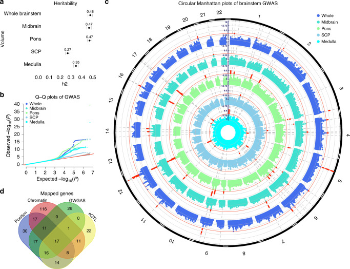
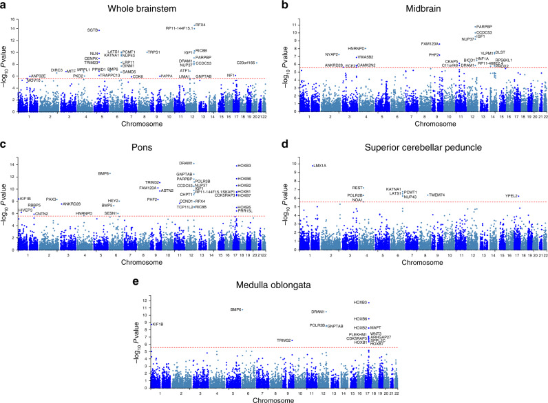
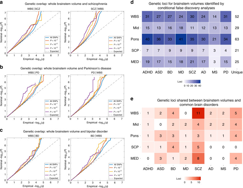
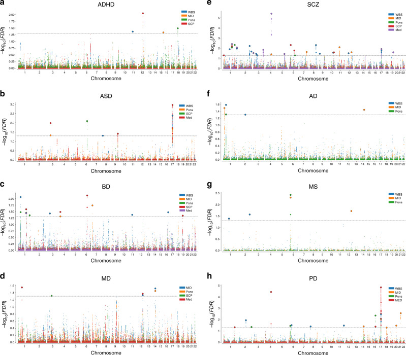
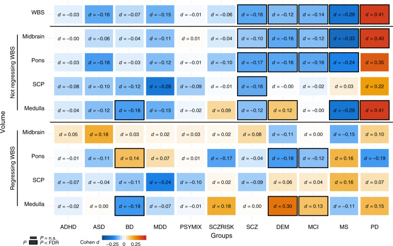
Brainstem regions support vital bodily functions, yet their genetic architectures and involvement in common brain disorders remain understudied. Here, using imaging-genetics data from a discovery sample of 27,034 individuals, we identify 45 brainstem-associated genetic loci, including the first linked to midbrain, pons, and medulla oblongata volumes, and map them to 305 genes. In a replication sample of 7432 participants most of the loci show the same effect direction and are significant at a nominal threshold. We detect genetic overlap between brainstem volumes and eight psychiatric and neurological disorders. In additional clinical data from 5062 individuals with common brain disorders and 11,257 healthy controls, we observe differential volume alterations in schizophrenia, bipolar disorder, multiple sclerosis, mild cognitive impairment, dementia, and Parkinson's disease, supporting the relevance of brainstem regions and their genetic architectures in common brain disorders.


Conflict of interest statement

Some authors received speaker’s honoraria from Lundbeck (T.E., G.B., and O.A.A.), Janssen Cilag (T.E.), Merck (E.H.), Sanofi Genzyme (E.H.), and Synovion (O.A.A). A.B. received speaker’s honoraria from Lundbeck, Otsuka, and Janssen Cilag and consultation fees from Biogen and Roche. J.B. has been a consultant to, member of advisory board of, and/or speaker for Shire, Roche, Medice, and Servier. E.G.C. has received personal fees from Almirall, Biogen, Merck, Roche and Teva, and grants and personal fees from Novartis and Sanofi. S.C. has received grant support from AstraZeneca as a coinvestigator and has served as a speaker for Otsuka. H.F.H. has received travel support, honoraria for advice or lecturing from Biogen Idec, Sanofi Genzyme, Merck, Novartis, Roche, and Teva and an unrestricted research grant from Novartis. N.I.L. has received consultation fees and travel support from Lundbeck. H.S. has received fees for advisory boards from ACImmune, Merck, and Novo Nordisk. P.S. has received honoraria for lecturing and travel support from Merck. M.T. has been member of advisory boards for Merck, IASIS Healthcare, ELPEN and FarmaSyn. M.Z. has received speaker fees for lectures, travel support and membership in advisory boards from Janssen Cilag, Lundbeck, Otsuka, Ferrer Pharma, Trommsdorff, Servier, and Roche. None of these external parties had any role in the analysis, writing or decision to publish this work. All other authors declare no competing interests.

Figures

Fig. 1
Fig. 1. Analysis of GWAS discovery sample identifies 61 loci associated with brainstem volumes.
a GCTA-based heritability estimates for the brainstem volumes in the discovery sample of n = 27,034 participants from the UK Biobank. All brainstem volumes showed substantial heritability, with higher estimates for the whole brainstem (h2 = 0.48), midbrain (h2 = 0.47), and pons (h2 = 0.47) and lower for the medulla oblongata (h2 = 0.35) and SCP (h2 = 0.27). Error bars, s.e. b Q−Q plots for the brainstem volumes of the discovery sample. c Circular Manhattan plots of GWAS for brainstem volumes of the discovery sample. The outermost plot in blue reflects the GWAS of whole brainstem volume, whereas, from the periphery to center, the turquoise, green, gray/blue, and cyan plots indicate the GWAS of the midbrain, pons, SCP, and medulla oblongata volumes, respectively. Red circular lines indicate genome-wide significance and the red radial lines are significant loci (two-sided P < 5e−8). d Venn diagram showing number of genes mapped by the four different strategies in the discovery sample, i.e., positional gene (blue), expression quantitative trait loci (eQTL; yellow), and chromatin interaction mapping (red), and identification by the GWGAS (green). Seventeen genes were identified by all four approaches. Whole whole brainstem. SCP superior cerebellar peduncle. Medulla medulla oblongata. GW(G)AS genome-wide (gene-based) association analyses. GCTA genome-wide complex trait analysis.
Fig. 2
Fig. 2. Manhattan plots from the genome-wide gene-based association analyses for volumes of the whole brainstem.
a midbrain b pons c superior cerebellar peduncle d, and medulla oblongata e in the discovery sample. Thirty-six genes were associated with whole brainstem volume and 22, 37, 10, and 17 genes were associated with volumes of the midbrain, pons, superior cerebellar peduncle, and the medulla oblongata, respectively. Twenty-two of the genes were only associated with whole brainstem volume, whereas 13, 14, 6, 5 genes were only significant for volumes of the midbrain, pons, superior cerebellar peduncle, and the medulla oblongata. The red horizontal lines indicate genome-wide significance threshold of two-sided P < 2.7e−6, i.e., 0.05/18,447 genes.
Fig. 3
Fig. 3. Genetic overlap between brainstem volumes and common brain disorders.
a Conditional Q–Q plots for whole brainstem volume conditioned on SCZ (left) and vice versa (right), demonstrating genetic overlap. b Conditional Q–Q plots for whole brainstem volume conditioned on PD (left) and vice versa (right), showing genetic overlap between these phenotypes. c Conditional Q–Q plots for whole brainstem volume conditioned on BD (left) and vice versa (right), demonstrating genetic overlap. d Enhanced discovery of genetic loci for each of the brainstem volumes when conditional false discovery rate analyses were run for each of the brainstem volumes conditioned on the eight brain disorders. These analyses revealed a total of 208 genetic loci for whole brainstem volume, and 111, 270, 55, and 125 loci for volumes of the midbrain, pons, SCP, and medulla oblongata, respectively. These genetic regions were located in 52 unique genetic loci for whole brainstem volume, and 29, 63, 21, and 25 unique loci for volumes of the midbrain, pons, SCP, and medulla oblongata. e conjunctional false discovery rate analysis detected shared genetic loci across brainstem volumes and the eight clinical conditions. The largest numbers of shared loci were found for SCZ (31), BD (14), and PD (17), whereas 8, 4, 6, 9, and 5 genetic loci were jointly shared for ASD, ADHD, MD, AD, and MS, respectively, and the brainstem volumes, when applying a conjunctional FDR threshold of 0.05. WBS whole brainstem. MID midbrain. SCP superior cerebellar peduncle. MED medulla oblongata. ADHD attention-deficit/hyperactivity disorder. ASD autism spectrum disorders. BD bipolar disorder. MD major depression. SCZ schizophrenia. AD Alzheimer’s disease. MS multiple sclerosis. PD Parkinson’s disease.
Fig. 4
Fig. 4. Manhattan plots for genetic loci shared between brainstem volumes and eight common brain disorders.
a 4 shared loci in ADHD, b 9 shared loci in ASD, c 14 shared loci in BD, d 6 shared loci in MD, e 31 shared loci in SCZ, f 5 shared loci in AD, g 5 shared loci in MS, and h 17 shared loci in PD. WBS whole brainstem. MID midbrain. SCP superior cerebellar peduncle. MED medulla oblongata. ADHD attention-deficit/hyperactivity disorder. ASD autism spectrum disorders. BD bipolar disorder. MD major depression. SCZ schizophrenia. AD Alzheimer’s disease. MS multiple sclerosis. PD Parkinson’s disease. The black horizontal lines indicate conjunctional false discovery rate threshold of 0.05.
Fig. 5
Fig. 5. Volumes of brainstem structures in individuals with common brain disorders compared with healthy controls.
Linear models were run covarying for sex, age, age², intracranial volume, and scanner site. The analyses for volumes of midbrain, pons, SCP, and medulla oblongata were run both with and without covarying for whole brainstem volume. The figure depicts the resulting case-control differences in Cohen’s d, whereas group differences in mm3 are presented in Supplementary Fig. 14. There were differential volumetric alterations in individuals with BD, SCZ, DEM, MCI, MS, and PD as indicated by black frames, after controlling for multiple testing using false discovery rate (Benjamini–Hochberg, accounting for all 99 tests). ADHD attention-deficit/hyperactivity disorder. ASD autism spectrum disorders. BD bipolar disorder. MDD major depressive disorder. PSYMIX non-SCZ psychosis spectrum diagnoses. SCZRISK prodromal SCZ or at risk mental state. SCZ schizophrenia. DEM dementia. MCI mild cognitive impairment. MS multiple sclerosis. PD Parkinson’s disease. WBS whole brainstem. SCP superior cerebellar peduncle. Medulla medulla oblongata.

References

    1. Guyenet PG. The sympathetic control of blood pressure. Nat. Rev. Neurosci. 2006;7:335–346. - PubMed
    1. Del Negro CA, Funk GD. Breathing matters. Nat. Rev. Neurosci. 2018;19:351–367. - PMC - PubMed
    1. Damasio A, Carvalho GB. The nature of feelings: evolutionary and neurobiological origins. Nat. Rev. Neurosci. 2013;14:143–152. - PubMed
    1. Fisman M. The brain stem in psychosis. Br. J. Psychiatry. 1975;126:414–422. - PubMed
    1. Williams DR, Lees AJ. Progressive supranuclear palsy: clinicopathological concepts and diagnostic challenges. Lancet Neurol. 2009;8:270–279. - PubMed

Publication types