. 2020 Aug 11;7(1):262.
doi: 10.1038/s41597-020-00608-w.

An annotated fluorescence image dataset for training nuclear segmentation methods

Affiliations

An annotated fluorescence image dataset for training nuclear segmentation methods

Florian Kromp et al. Sci Data. .

Abstract

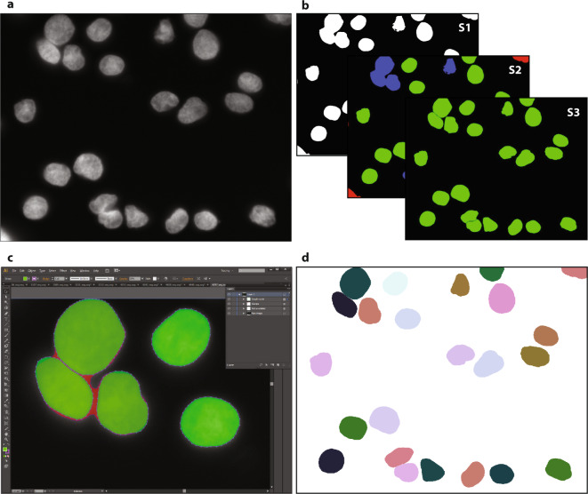
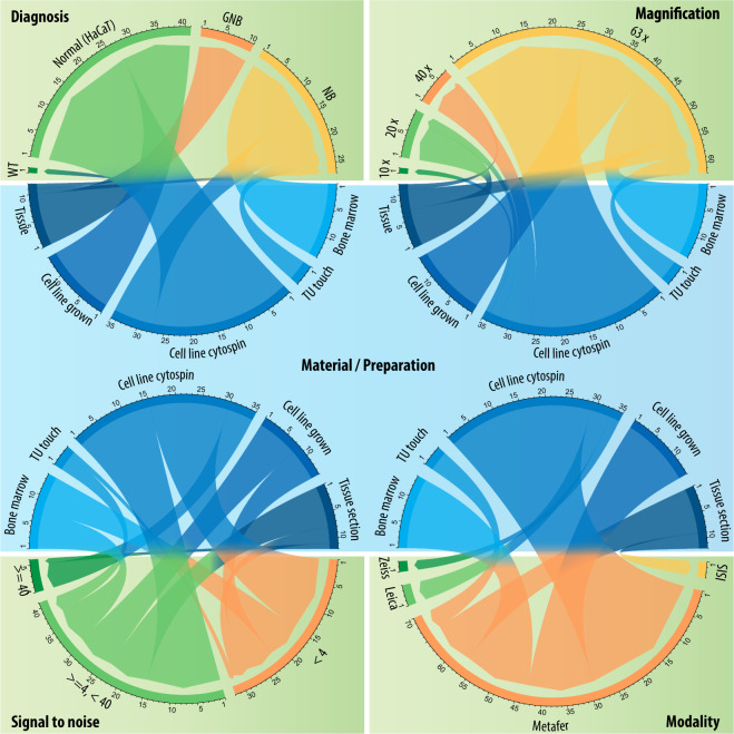
Fully-automated nuclear image segmentation is the prerequisite to ensure statistically significant, quantitative analyses of tissue preparations,applied in digital pathology or quantitative microscopy. The design of segmentation methods that work independently of the tissue type or preparation is complex, due to variations in nuclear morphology, staining intensity, cell density and nuclei aggregations. Machine learning-based segmentation methods can overcome these challenges, however high quality expert-annotated images are required for training. Currently, the limited number of annotated fluorescence image datasets publicly available do not cover a broad range of tissues and preparations. We present a comprehensive, annotated dataset including tightly aggregated nuclei of multiple tissues for the training of machine learning-based nuclear segmentation algorithms. The proposed dataset covers sample preparation methods frequently used in quantitative immunofluorescence microscopy. We demonstrate the heterogeneity of the dataset with respect to multiple parameters such as magnification, modality, signal-to-noise ratio and diagnosis. Based on a suggested split into training and test sets and additional single-nuclei expert annotations, machine learning-based image segmentation methods can be trained and evaluated.


Conflict of interest statement

The authors declare no competing interests.

Figures

Fig. 1
Fig. 1
Workflow for ground truth image annotation. (a) Raw image visualizing HaCaT cytospinned nuclei. (b) A machine learning framework was used to annotate the raw image, learning from user interaction within three consecutive steps: S1. foreground extraction, S2. connected component classification (red = non-usable objects, blue = nuclei aggregations, green = single nuclei) and S3. splitting of aggregated objects into single nuclei, resulting in an annotation mask. (c) Zoom-in of the SVG-file showing the nuclear image overlaid with polygons representing each annotated nucleus. Polygons were modified by expert biologists to fit effective nuclear borders. Challenging decisions on how to annotate nuclei, mainly occurring due to aggregated or overlapped nuclei, were presented to an expert pathologist and corrected to obtain the final ground truth. (d) The curated SVG-file was transformed into a labeled nuclear mask.
Fig. 2
Fig. 2
Heterogeneity of the proposed dataset with respect to the type of preparation. GNB: ganglioneuroblastoma, NB: neuroblastoma, TU touch: tumor touch imprint, Tissue: tissue section.

References

    1. Irshad H, Veillard A, Roux L, Racoceanu D. Methods for Nuclei Detection, Segmentation, and Classification in Digital Histopathology: A Review - Current Status and Future Potential. IEEE Rev. Biomed. Eng. 2014;7:97–114. doi: 10.1109/RBME.2013.2295804. - DOI - PubMed
    1. Meijering E, Carpenter AE, Peng H, Hamprecht FA, OlivoMarin JC. Imagining the future of bioimage analysis. Nat. Biotechnol. 2016;34:1250–1255. doi: 10.1038/nbt.3722. - DOI - PubMed
    1. Ching, T. et al. Opportunities and obstacles for deep learning in biology and medicine. J. R. Soc. Interface15 (2018). - PMC - PubMed
    1. Blom S, et al. Systems pathology by multiplexed immunohistochemistry and whole-slide digital image analysis. Sci. Rep. 2017;7:1–13. doi: 10.1038/s41598-017-15798-4. - DOI - PMC - PubMed
    1. Waters JC. Accuracy and precision in quantitative fluorescence microscopy. J. Cell Biol. 2009;185:1135–1148. doi: 10.1083/jcb.200903097. - DOI - PMC - PubMed

Publication types