. 2020 Jan 23;6(1):249.
doi: 10.18063/ijb.v6i1.249. eCollection 2020.

Graphene Oxide Induces Ester Bonds Hydrolysis of Poly-l-lactic Acid Scaffold to Accelerate Degradation

Affiliations

Graphene Oxide Induces Ester Bonds Hydrolysis of Poly-l-lactic Acid Scaffold to Accelerate Degradation

Cijun Shuai et al. Int J Bioprint. .

Erratum in

  • ERRATUM.
    [No authors listed] [No authors listed] Int J Bioprint. 2020 Sep 17;6(4):309. doi: 10.18063/ijb.v6i4.309. eCollection 2020. Int J Bioprint. 2020. PMID: 33102924 Free PMC article.

Abstract

Poly-l-lactic acid (PLLA) possesses good biocompatibility and bioabsorbability as scaffold material, while slow degradation rate limits its application in bone tissue engineering. In this study, graphene oxide (GO) was introduced into the PLLA scaffold prepared by selective laser sintering to accelerate degradation. The reason was that GO with a large number of oxygen-containing functional groups attracted water molecules and transported them into scaffold through the interface microchannels formed between lamellar GO and PLLA matrix. More importantly, hydrogen bonding interaction between the functional groups of GO and the ester bonds of PLLA induced the ester bonds to deflect toward the interfaces, making water molecules attack the ester bonds and thereby breaking the molecular chain of PLLA to accelerate degradation. As a result, some micropores appeared on the surface of the PLLA scaffold, and mass loss was increased from 0.81% to 4.22% after immersing for 4 weeks when 0.9% GO was introduced. Besides, the tensile strength and compressive strength of the scaffolds increased by 24.3% and 137.4%, respectively, due to the reinforced effect of GO. In addition, the scaffold also demonstrated good bioactivity and cytocompatibility.

Keywords: Degradation property; Ester bonds hydrolysis; GO; Poly-l-lactic acid scaffold.


Figures

Figure 1
Figure 1
(A-C) SEM of PLLA, GO and PLLA/GO composite powders, (D-F) axis view, top view, and front view of the PLLA scaffold and (G-I) axis view, top view and front view of the PLLA/GO scaffold fabricated by SLS. PLLA and PLLA/GO scaffolds had similar shape sizes and interconnected porous structures.
Figure 2
Figure 2
(A) Raman spectra of PLLA and PLLA/GO, and (B) partial magnification in spectra a. (C) Water contact angle of the PLLA scaffolds with 0%, 0.3%, 0.6%, 0.9%, and 1.2 wt % GO. (D) Water absorption rate of the PLLA scaffolds with 0%, 0.3%, 0.6%, 0.9%, and 1.2 wt % GO after immersing in aqueous solution for different time. GO was successfully introduced into the PLLA scaffold and interacted with PLLA. Hydrophilicity and water absorption capacity increased with increasing GO.
Figure 3
Figure 3
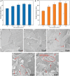
(A-E) The mass loss ratio and degradation morphology of PLLA scaffolds with 0%, 0.3%, 0.6%, 0.9%, and 1.2 wt % GO after immersing in PBS solution for 1, 2, 3, and 4 weeks, respectively. (F-J) Three-dimensional surface morphologies of PLLA/GO samples with 0%, 0.3%, 0.6%, 0.9%, and 1.2% GO after degradation for 4 weeks. (J) Surface roughness (Ra, Rq, and Rz) date of different ratios of PLLA/GO samples. Degradation of PLLA accelerated and surface roughness increased with increasing GO content.
Figure 4
Figure 4
Schematic of GO promoting the degradation of PLLA. GO accelerated the degradation of PLLA could be divided into three parts. a1: Hydrophilic GO attracted water molecules. a2: The interface channels between GO and PLLA facilitated the intrusion of water molecules and discharged of degraded low molecular products. a3: The hydrogen bonding between PLLA and GO induced the ester bond to deflect toward the interfaces, making the water molecules easily to attack the ester bond.
Figure 5
Figure 5
(A) Compressive strength and (B) compressive modulus of PLLA and PLLA/GO scaffolds with 0.3%, 0.6%, 0.9%, and 1.2 wt% GO. (C-G) Surface dispersion state of GO in PLLA substrate with 0%, 0.3%, 0.6%, 0.9%, and 1.2 wt% GO. (H) Linescan 1 showed the distribution of elements C and O. The compressive strength and modulus increased with increasing GO content, while there was a slight decrease when 1.2% GO was introduced. GO was uniformly dispersed in the PLLA matrix, but agglomerates formed when the GO content was further increased.
Figure 6
Figure 6
(A) Tensile strength and (B) tensile modulus of PLLA and PLLA/GO scaffolds with 0.3%, 0.6%, 0.9%, and 1.2 wt% GO. (C-G) Fracture morphology of GO in PLLA substrate with 0%, 0.3%, 0.6%, 0.9%, and 1.2 wt% GO. Tensile strength and modulus increased first and then decreased with increasing GO content. The fracture surface of PLLA was smooth, the introduction of GO rendered the fracture surface rough, and more GO was embedded in the PLLA matrix with the increase of GO.
Figure 7
Figure 7
(A-F) Formation of a calcium-phosphorus layer of PLLA and PLLA/GO scaffolds with 0.3%, 0.6%, 0.9%, and 1.2 wt% GO after immersing in the SBF solution for 4 weeks. Samples containing GO possessed the ability of forming a calcium-phosphorus layer, while PLLA had no calcium-phosphorus layer formation.
Figure 8
Figure 8
(A,C,E) The fluorescence staining of MG63 cells fostered on the PLLA scaffolds for 1, 3, and 5 days, (B,D,F) the fluorescence staining of MG63 cells fostered on the PLLA/0.9 GO scaffolds for 1, 3, and 5 days, respectively. (G) CCK-8 experiment of MG63 cells fostered on the PLLA and PLLA/0.9 GO scaffolds for 1, 3, and 5 days, respectively. Cells on PLLA/0.9 GO samples had better cell morphology and more cell number compared to PLLA.

References

    1. An J, Teoh JE, Suntornnond R, et al. Design and 3D Printing of Scaffolds and Tissues. Engineering. 2015;1:261–268.
    1. Liu F, Mishbak H, Bartolo PJ. Hybrid Polycaprolactone/Hydrogel Scaffold Fabrication and in-process Plasma Treatment Using PABS. Int J Bioprint. 2019;5:174. - PMC - PubMed
    1. Cardoso GB, Perea GN, D'Avila MA, et al. Initial Study of Electrospinning PCL/PLLA Blends. Adv Mater Phys Chem. 2011;1:94–98.
    1. Saito Y, Minami K, Kobayashi M, et al. New Tubular Bioabsorbable Knitted Airway Stent:Biocompatibility and Mechanical Strength. J Thorac Cardiovasc Surg. 2002;123:161–167. DOI:10.1067/mtc.2002.118503. - PubMed
    1. Shuai C, Yang W, He C, et al. A Magnetic Micro-environment in Scaffolds for Stimulating Bone Regeneration. Mater Des. 2020;185(108275) DOI:10.1016/j.matdes.2019.108275.

LinkOut - more resources