. 2020 Jun 19;1(1):100035.
doi: 10.1016/j.xpro.2020.100035. Epub 2020 Jun 3.

Isolation of Microglia from Mouse or Human Tissue

Affiliations

Isolation of Microglia from Mouse or Human Tissue

Evan A Bordt et al. STAR Protoc. .

Abstract

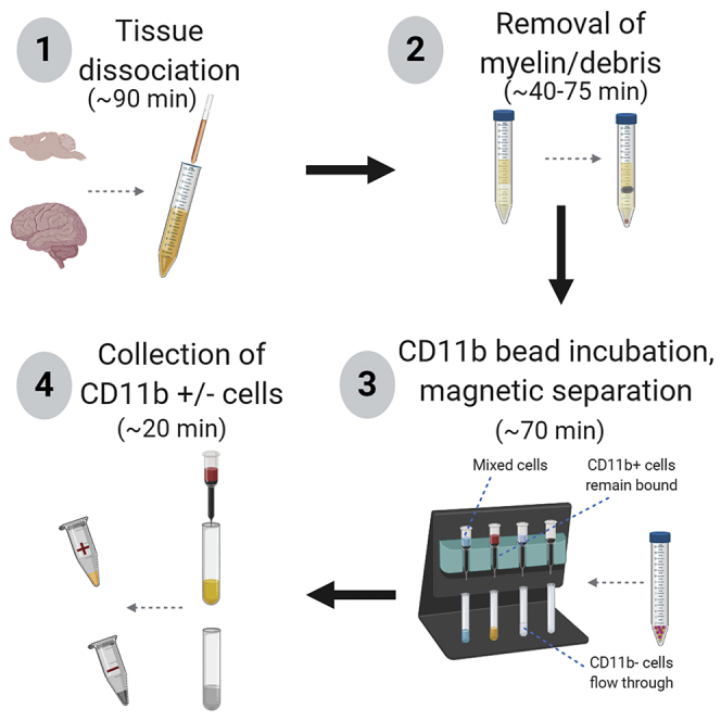
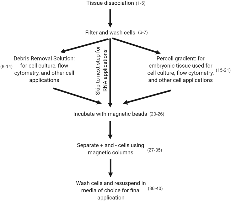
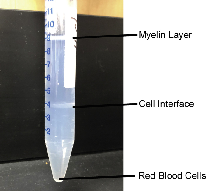
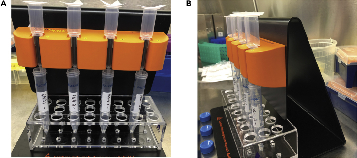
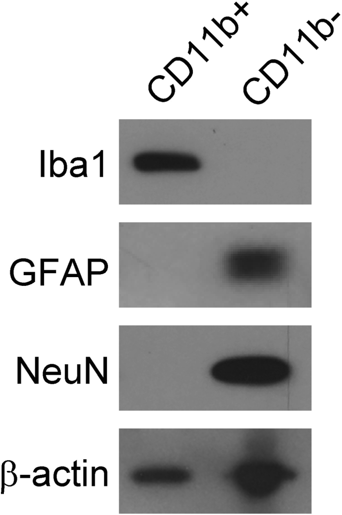
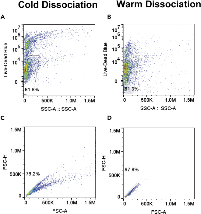
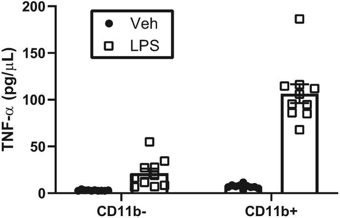
Microglia are the innate immune cells of the central nervous system. Although numerous methods have been developed to isolate microglia from the brain, the method of dissociation and isolation can have a profound effect on the function of these highly dynamic cells. Here, we present an optimized protocol to isolate CD11b+ cells (microglia) from mouse or human brain tissue using magnetic bead columns. Isolated microglia can be used to model diseases with neuroinflammatory components for potential therapeutic discoveries. For complete details on the use and execution of this protocol, please refer to Hanamsagar et al. (2017), Rivera et al. (2019), and Edlow et al. (2019).


Conflict of interest statement

DECLARATION OF INTERESTS The authors declare no competing interests.

Figures

None
Graphical abstract
Figure 1
Figure 1
Representative Images for #1, #2, and #3 Pasteur Pipets
Figure 2
Figure 2
Workflow of Isolation Protocol with Corresponding Steps in Parentheses
Figure 3
Figure 3
Dissociation of Tissue to a Single-Cell Suspension Appearance of tissue (A) prior to and (B) after dissociation steps
Figure 4
Figure 4
Debris Removal Solution Gradients Photographs of example gradients (A) before and (B) after centrifugation.
Figure 5
Figure 5
Post-centrifugation Percoll Gradients Note that myelin will separate at the top of the liquid, cells will separate out at the interface of 70% Percoll/30% Percoll, and red blood cells will pellet at the bottom of the conical tube.
Figure 6
Figure 6
Setup of LS Columns on MACS MultiStand. (A) LS Columns are placed in the MACS MultiStand over opened 15 mL tubes. The LS Columns are topped with fresh nylon filters. (B) Ensure that LS Columns are placed tightly in the MACS MultiStand.
Figure 7
Figure 7
Cell Yields Cell counts were obtained manually on a hemocytometer for CD11b+ cells isolated from adult forebrain, adult prefrontal cortex (PFC), adult hippocampus (HP), embryonic mouse forebrain, and embryonic mouse placenta.
Figure 8
Figure 8
CD11b+ Magnetic Bead Isolation Pulls down Brain Microglia (A and B) Gating strategy for (A) CD11b+ and CD45hi/lo and (B) Cx3cr1+ CD45hi/lo cells. Cells were isolated using CD11b+ magnetic bead isolation method presented in this protocol, and assessed for infiltration of non-parenchymal myeloid cells as assessed by increase in CD45 levels. (C) Quantification of CD11b+CD45lo, CD11b+CD45hi, Cx3cr1+CD45lo, and Cx3cr1+CD45hi cells obtained using this protocol.
Figure 9
Figure 9
Lysates from Human CD11b+ or CD11b- Cells Were Run on Immunoblot and Probed for Iba1 (Microglia), GFAP (Astrocytes), and NeuN (Neurons) CD11b+ cells were positive for Iba1 and negative for GFAP and NeuN, whereas CD11b- cells were negative for Iba1 and positive for GFAP and NeuN.
Figure 10
Figure 10
Cold Dissociation Results in Lowered Cell Viability and Fewer Single Cells than Does Warm Dissociation (A and B) Gating strategy for Live-Dead Blue staining from (A) cold dissociation isolated CD11b+ cells and (B) warm dissociation isolated CD11b+ cells. (C and D) Gating strategy for determining single cells from (C) cold dissociation isolated CD11b+ cells and (D) warm dissociation isolated CD11b+ cells.
Figure 11
Figure 11
Low Level of Basal Cytokine Release by Placental CD11b+ Cells Protein concentrations for TNF-α released into culture media by CD11b- or CD11b+ cells following 2 hr lipopolysachharide (LPS) or vehicle (Veh) stimulation were determined by ELISA.

References

    1. Ayata P., Badimon A., Strasburger H.J., Duff M.K., Montgomery S.E., Loh Y.E., Ebert A., Pimenova A.A., Ramirez B.R., Chan A.T. Epigenetic regulation of brain region-specific microglia clearance activity. Nat. Neurosci. 2018;21:1049–1060. - PMC - PubMed
    1. Bell A.W., Deutsch E.W., Au C.E., Kearney R.E., Beavis R., Sechi S., Nilsson T., Bergeron J.J. A HUPO test sample study reveals common problems in mass spectrometry-based proteomics. Nat. Methods. 2009;6:423–430. - PMC - PubMed
    1. Bohlen C.J., Bennett F.C., Tucker A.F., Collins H.Y., Mulinyawe S.B., Barres B.A. Diverse requirements for microglial survival, specification, and function revealed by defined-medium cultures. Neuron. 2017;94:759–773.e8. - PMC - PubMed
    1. Bohlen C.J., Bennett F.C., Bennett M.L. Isolation and culture of microglia. Curr. Protoc. Immunol. 2019;125:e70. - PMC - PubMed
    1. Edlow A.G., Glass R.M., Smith C.J., Tran P.K., James K., Bilbo S. Placental macrophages: a window into fetal microglial function in maternal obesity. Int. J. Dev. Neurosci. 2019;77:60–68. - PMC - PubMed

Publication types