. 2020 Nov;57(11):4720-4734.
doi: 10.1007/s12035-020-02044-8. Epub 2020 Aug 11.

Inhibition of Aquaporin 4 Decreases Amyloid Aβ40 Drainage Around Cerebral Vessels

Affiliations

Inhibition of Aquaporin 4 Decreases Amyloid Aβ40 Drainage Around Cerebral Vessels

Gabriela-Camelia Rosu et al. Mol Neurobiol. 2020 Nov.

Abstract

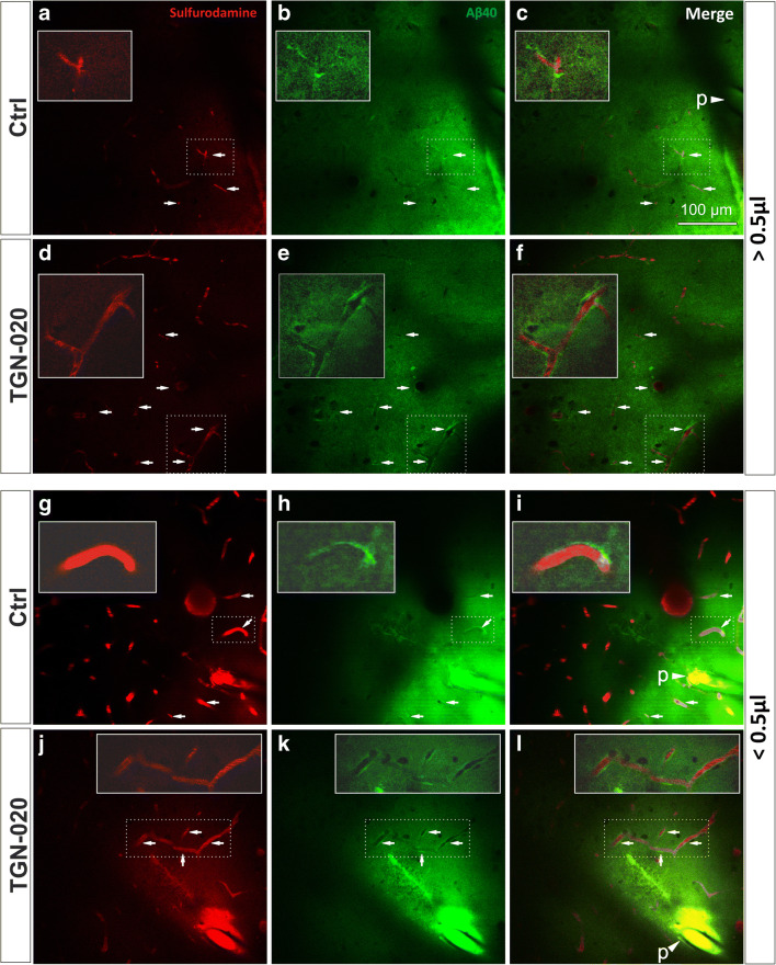
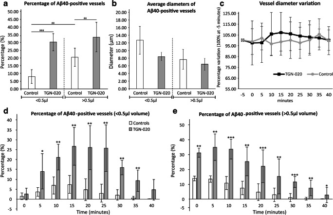
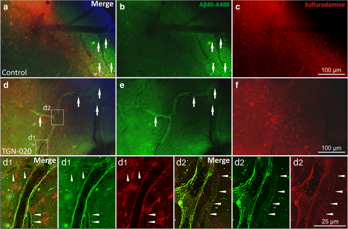
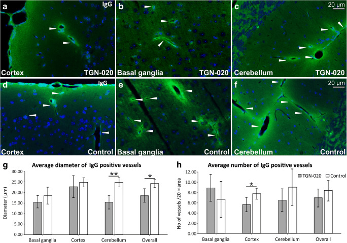
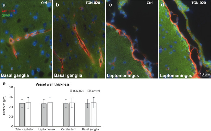
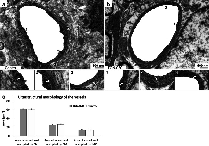
Aquaporin-4 (AQP4) is located mainly in the astrocytic end-feet around cerebral blood vessels and regulates ion and water homeostasis in the brain. While deletion of AQP4 is shown to reduce amyloid-β (Aβ) clearance and exacerbate Aβ peptide accumulation in plaques and vessels of Alzheimer's disease mouse models, the mechanism and clearing pathways involved are debated. Here, we investigated how inhibiting the function of AQP4 in healthy male C57BL/6 J mice impacts clearance of Aβ40, the more soluble Aβ isoform. Using two-photon in vivo imaging and visualizing vessels with Sulfurodamine 101 (SR101), we first showed that Aβ40 injected as a ≤ 0.5-μl volume in the cerebral cortex diffused rapidly in parenchyma and accumulated around blood vessels. In animals treated with the AQP4 inhibitor TGN-020, the perivascular Aβ40 accumulation was significantly (P < 0.001) intensified by involving four times more vessels, thus suggesting a generalized clearance defect associated with vessels. Increasing the injecting volume to ≥ 0.5 ≤ 1 μl decreased the difference of Aβ40-positive vessels observed in non-treated and AQP4 inhibitor-treated animals, although the difference was still significant (P = 0.001), suggesting that larger injection volumes could overwhelm intramural vascular clearance mechanisms. While both small and large vessels accumulated Aβ40, for the ≤ 0.5-μl volume group, the average diameter of the Aβ40-positive vessels tended to be larger in control animals compared with TGN-020-treated animals, although the difference was non-significant (P = 0.066). Using histopathology and ultrastructural microscopy, no vascular structural change was observed after a single massive dose of TGN-020. These data suggest that AQP4 deficiency is directly involved in impaired Aβ brain clearance via the peri-/para-vascular routes, and AQP4-mediated vascular clearance might counteract blood-brain barrier abnormalities and age-related vascular amyloidopathy.

Keywords: Alzheimer’s disease; Amyloid; Aquaporin 4; Aβ40; Perivascular drainage; TGN-020.


Conflict of interest statement

The authors declare that they have no competing interests.

Figures

Fig. 1
Fig. 1
Representative two-photon microscopy images of anesthetized mice, 10 min after Aβ40-A488 was injected in the upper layers of the right somatosensory cortex. Injecting a large volume floods the water drainage system, leading to some Aβ40 deposits around blood vessels in control animals (ac). The number of vessels surrounded by Aβ40 is greater when blocking AQP4 function (df). Injecting small volumes of Aβ40 leads to a limited number of vessels exhibiting Aβ40 deposits in control animals (gi); however, blocking AQP4 channels generates multiple Aβ40 accumulation around vessels (jl). In each image, insets represent magnified exemplary areas demonstrating abluminal Aβ accumulation. Arrows point to regions of interest with clear-cut Aβ40 being retained around the vascular lumens; arrow heads indicating a “p” denominate the position of the injection pipette tip, filled with the fluorescent Aβ40
Fig. 2
Fig. 2
Systemic administration of AQP4 inhibitor reveals a higher number of blood vessels showing Aβ40 deposits. a The total volume injected impacts the drainage system; however, regardless of the injected volume, blocking AQP4 channels leads to an increase frequency of Aβ40 deposits around vessels, compared with controls, predominantly for vessels (b) with a smaller diameter. c When analyzing the variation over time of the blood vessel diameters, no change could be observed. Investigating the dynamic of Aβ40 drainage around the injected site, we could see differences for both small (d) and large (e) injected volumes. The average number of elements per animal was considered for analysis in all instances. Mean ± SD; P < 0.05, ∗∗P < 0.01, ∗∗∗P < 0.001; N = 9 control animals (4 for < 0.5 μl; 5 for > 0.5 μl) and 9 TGN-020 animals (4 for < 0.5 μl; 5 for > 0.5 μl). For a, an average of n = 51 ± 9.16/43 ± 11.83 vessels have been considered for control group (< 0.5 μl/> 0.5 μl), and respectively n = 46.83 ± 9.57/53.83 ± 16.84 vessels have been considered for the TGN-020 group (< 0.5 μl/> 0.5 μl)
Fig. 3
Fig. 3
Ex vivo fluorescence imaging of Aβ40 and Sulfurodamine 101 in control (ac) and TGN-020-treated animals (df, d1, d2 insets). Colocalization of the two signals is still visible after tissue processing in more vessels for treated animals (arrows) and also in more numerous smaller vessels (arrowheads)
Fig. 4
Fig. 4
Post-fixed tissues immunostained for endogenous murine immunoglobulins (af) show only a regional decrease in IgG permeability for TGN-020-treated animals, both as the average diameter of positive vessels (g) and as the number of positive vessels (h). The average value per animal was considered for analysis. Mean ± SD; P < 0.05, ∗∗P < 0.01. N = 5 animals for the TGN-020 group and 3 animals for the control group
Fig. 5
Fig. 5
Post-fixed tissues immunostained for laminin and GFAP show no obvious differences between the basement membranes’ thickness utilizing light microscopy, nor any gliotic reaction, between the control and TGN-020-treated animals (ae). The average value per animal was considered for analysis. Mean ± SD. N = 5 animals for the TGN-020 group and 3 animals for the control group
Fig. 6
Fig. 6
Inhibition of AQP4 by TGN-020 does not alter the structural appearance of the capillary vascular wall in the grey matter. There was no difference in the ultrastructure of cerebral capillaries from mice treated with TGN-020 (b) when compared with control mice (a). Endothelial tight junctions (a1 and b1), intramural cells (a2 and b2), and basement membranes (a3 and b3) appeared normal. The average value per animal was considered for analysis. Mean ± SD. N = 3 animals each of the control and TGN-020 groups

References

    1. Hardy J, Selkoe DJ. The amyloid hypothesis of Alzheimer’s disease: progress and problems on the road to therapeutics. Science. 2002;297(5580):353–356. doi: 10.1126/science.1072994. - DOI - PubMed
    1. Zlokovic BV. Neurovascular pathways to neurodegeneration in Alzheimer’s disease and other disorders. Nat Rev Neurosci. 2011;12(12):723–738. doi: 10.1038/nrn3114. - DOI - PMC - PubMed
    1. Piaceri I, Nacmias B, Sorbi S. Genetics of familial and sporadic Alzheimer’s disease. Front Biosci (Elite Ed) 2013;5:167–177. doi: 10.2741/E605. - DOI - PubMed
    1. Dorszewska J, Prendecki M, Oczkowska A, Dezor M, Kozubski W. Molecular basis of familial and sporadic Alzheimer’s disease. Curr Alzheimer Res. 2016;13(9):952–963. doi: 10.2174/1567205013666160314150501. - DOI - PubMed
    1. Ritchie K, Carriere I, Ritchie CW, Berr C, Artero S, Ancelin ML. Designing prevention programmes to reduce incidence of dementia: prospective cohort study of modifiable risk factors. BMJ (Clinical research ed) 2010;341:c3885. doi: 10.1136/bmj.c3885. - DOI - PMC - PubMed

MeSH terms

LinkOut - more resources