. 2021 Apr 1;529(5):1066-1080.
doi: 10.1002/cne.25004. Epub 2020 Aug 31.

The SNARE regulator Complexin3 is a target of the cone circadian clock

Affiliations

The SNARE regulator Complexin3 is a target of the cone circadian clock

Jacob D Bhoi et al. J Comp Neurol. .

Abstract

BMAL1 is a core component of the mammalian circadian clockwork. Removal of BMAL1 from the retina significantly affects visual information processing in both rod and cone pathways. To identify potential pathways and/or molecules through which BMAL1 alters signal transmission at the cone pedicle, we performed an RNA-seq differential expression analysis between cone-specific Bmal1 knockout cones (cone-Bmal1-/- ) and wild-type (WT) cones. We found 88 genes differentially expressed. Among these, Complexin3 (Cplx3), a SNARE regulator at ribbon synapses, was downregulated fivefold in the mutant cones. The purpose of this work was to determine whether BMAL1 and/or the cone clock controls CPLX3 protein expression at cone pedicles. We found that CPLX3 expression level was decreased twofold in cone-Bmal1-/- cones. Furthermore, CPLX3 expression was downregulated at night compared to the day in WT cones but remained constitutively low in mutant cones both day and night. The transcript and protein expression levels of Cplx4-the other complexin expressed in cones-were similar in WT and mutant cones; in WT cones, CPLX4 protein level did not change with the time of day. In silico analysis revealed four potential BMAL1:CLOCK binding sites upstream from exon one of Cplx3 and none upstream of exon one of Cplx4. Our results suggest that CPLX3 expression is regulated at the transcriptional level by the cone clock. The modulation of CPLX3 may be a mechanism by which the clock controls the cone synaptic transfer function to second-order cells and thereby impacts retinal signal processing during the day/night cycle.

Keywords: Bmal1; Cplx3; Cplx4; SNARE proteins; circadian clock; cones; retina; ribbon synapses.


Conflict of interest statement

CONFLICT OF INTEREST

The authors declare no conflict of interest.

Figures
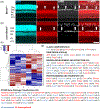
FIGURE 1
FIGURE 1
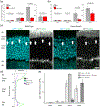
RNA-seq and analysis in cone-Bmal1−/− cones. (a) Wild-type retina section reacted with an antibody against BMAL1. Note the presence of BMAL1 expression in the inner retina (INL and GCL) and cone somas (white arrows). (b) Cone-Bmal1−/− retina section reacted with an antibody against BMAL1. Note the normal expression of BMAL1 in the inner retina and the absence of BMAL1 immunoreactivity in the cones (white arrows). Cones were labeled with an antibody against cone-arrestin [cARR]). GCL, ganglion cell layer; INL, inner nuclear layer; IPL, inner plexiform layer; ONL, outer nuclear layer; OPL, outer plexiform layer. DAPI (cyan) stains the cell nuclei. Bar is 50 μm (applies to all). (c) Heat map showing the expression pattern of 88 protein-coding gene transcripts between wild-type mice (C1, C2, C3) and cone-Bmal1−/− mutant mice (M1, M2, M3) and within triplicates. Red indicates upregulation and blue downregulation. Tissue was collected in the middle of the light phase (ZT 05-07). One triplicate = 3 × 1 animals (two retinas/animal). (d) Biological processes overrepresented in transcripts upregulated or downregulated in cones from cone-Bmal1−/− retinas (x fold increase in mutant cones)
FIGURE 2
FIGURE 2
Cplx mRNA levels in wild-type (WT) and cone and rod specific BMAL1 knockouts. (a) Cplx3 is the only member of the Cplx family which is differentially expressed in WT and cone-Bmal1−/− cones. Cones also show high Cplx4 expression in both WT and cone-Bmal1−/− cones. (b) Cplx3 is the only member of the Cplx family which is differentially expressed in WT and rod-Bmal1−/−. A small difference was found between WT and mutant rods for Cplx2 but the levels were overall very low. Rods show a very high expression of Cplx4, consistent with previous research indicating that it is the primary Cplx found in rods. Error bars are SEM. Dots show individual data points. n = 1 animal (two retinas)/point, three animals/group. Tissue was collected in the middle of the light phase (ZT05-07). p-Values: unpaired t-test, two-tailed. Note the difference in y-axis scale for Cplx4 in rods and cones. (c) RNAscope in situ hybridization (ISH) followed by immunocytochemistry (ICC) revealed that Cplx3 mRNA (green dots) is found primarily in the outer layer of the outer nuclear layer (ONL), where the somas of cones primarily stratify, in WT retinas. Cplx3 mRNA is primarily surrounding cone somas (white), designated by white arrows. In cone-Bmal1−/− retinas, there is less Cplx3 mRNA in the outer ONL. DAPI (cyan) stains the cell nuclei. Scale bar: 25 μm (applies to all). (d) Vertical intensity plot of Cplx3 mRNA in WT (green) and cone-Bmal1−/− (purple). Cplx3 mRNA was higher in the upper ONL where the somas of cones stratify. (e) Expression of Cplx mRNA in cone photoreceptors, rod bipolar cells, and rod photoreceptors based on published single-cell RNA-seq data (Shekhar et al., 2016). Cplx1 and Cplx2 are not highly expressed in any of these cell types. Cplx3 mRNA is primarily in cones and rod bipolar cells. Cplx4 is primarily expressed in rods and cones. Cones (n = 14); rod bipolar cells (n = 7,784); and rods (n = 32). Error bars show SEM
FIGURE 3
FIGURE 3
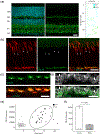
CPLX3 is expressed in cone pedicles. (a) CPLX3 labeling (green) is found in both plexiform layers of mouse retina. Note intensity plot of CPLX3 and DAPI stain vertically through retinal section to the right. DAPI (cyan) stains the cell nuclei. Scale bar: 25 μm. (b) Labeling cones with cArr (red) reveals strong colocalization of CPLX3 at cone pedicles (asterisks). Also note the CPLX3 hotspots at the axon initial segment of some cones (arrowheads). DAPI (cyan) stains the cell nuclei. Scale bar: 25 μm. (c) Detail of (b). CPLX3 (green) fills the cone pedicles. (d) PSD95 (white) labels rod and cone terminals which can be distinguished based on their size. CPLX3 (green) strongly labels cone pedicles (asterisks) and there is slight CPLX3 in rod spherules (arrows). All images from retinas collected in the middle of the day subjective day (CT 05-07). Scale bar: 10 μm. (e) Rods (n = 94 rods from three animals) and cones (n = 34 cones from three animals) form two clusters based on area and CPLX3 intensity (Dunn index = 0.007) (f) mean CPLX3 intensity for rods (n = 94 rods from three animals) and cones (n = 34 cones from three animals). p-Value: unpaired t-test, two-tailed
FIGURE 4
FIGURE 4
CPLX4 is expressed in both rods and cones. (a) CPLX4 (red) is expressed in the outer plexiform layer (OPL) and the inner plexiform layer (IPL). Note the intensity plot of CPLX4 and DAPI stain vertically through retinal section to the right. DAPI (cyan) stains the cell nuclei. Scale bar: 25 μm. (b) An antibody against PSD95 labels rod and cone terminals (white), which can be differentiated based on their relative sizes (i.e., big ones: cone pedicles and small one: rod spherules). CPLX4 is expressed in both synaptic terminals but to a lower degree in cones (asterisks). White arrows indicate rod spherules. All images from retinas collected in the middle of the subjective day (CT 05-07). Scale bar: 5 μm. (c) Rods (n = 144 rods from five animals) and cones (n = 45 cones from five animals) form two clusters based on area and CPLX4 intensity (Dunn index = 0.18). (e) Mean CPLX4 intensity for rods (n = 144 rods from five animals) and cones (n = 45 cones from five animals). p-Values: unpaired t-test, two-tailed
FIGURE 5
FIGURE 5
CPLX3 expression is controlled by the cone clock. (a–c) Wild-type (WT) retinal section collected during the subjective day (a). Cone-Bmal1−/− retinal section collected during the subjective day (b). Wild-type retinal section collected during the subjective night (c). Note that CPLX3 immunolabeling is dramatically decreased in the outer plexiform layer (OPL) in cone-Bmal1−/− retina but is normal in the IPL, compared to the wild-type. In wild-type, CPLX3 is decrease in both OPL and IPL during subjective night, compared to subjective daytime. Scale bar: 25 μm, applies to (a–c). (d–e) High magnification micrographs showing that during the subjective day, cone pedicles (white asterisk) of WT animals (d) have significant higher CPLX3 immunoreactivity than cone pedicles of cone-Bmal1−/− animals (e). PSD95 (white) labels photoreceptor terminals. DAPI (cyan) stains the cell nuclei. Arrows point to rod spherules. Scale bar: 5 μm
FIGURE 6
FIGURE 6
Quantification of CPLX3 and CPLX4. (a) Quantification of CPLX3 in cone pedicles in the wild-type (WT) and mutant cones in the middle of the day under room light (ZT 05-07, yellow) or subjective day (CT 05-07, gray) showed that CPLX3 expression is decreased in cone-Bmal1−/− cones and there is no effect of light adaptation. (b) Quantification of CPLX3 in cone pedicles during the subjective day (CT 05-07, light gray) or during the subjective night (CT 07–19, dark gray) shows that CPLX3 is downregulated at night in WT cones and is constitutively low in cone-Bmal1−/− cones. (c,d) Quantification of CPLX4 in the outer plexiform layer (OPL) showed that there were no differences in CPLX4 expression in the OPL under the conditions depicted in (a,b). n is shown on the figures and represents number of animals (5–18 cones per animal). Error bars are SEM. Dots show individual data points. p-Values: unpaired t-test, two-tailed
FIGURE 7
FIGURE 7
Cplx3 and Cplx4 DNA promotor region analysis. (a) The Cplx3 DNA promotor region contains three E-boxes and one ROR element. (b) The Cplx4 DNA promotor region contains no E- or D-boxes and no ROR or CRE elements. The promotor region was designated as the 600 bp upstream of Exon 1. Gene sequences were downloaded from ENSEMBL

References

    1. Anders S, Pyl PT, & Huber W (2015). HTSeq—A Python framework to work with high-throughput sequencing data. Bioinformatics, 31(2), 166–169. 10.1093/bioinformatics/btu638 - DOI - PMC - PubMed
    1. Baba K, Piano I, Lyuboslavsky P, Chrenek MA, Sellers JT, Zhang S, … Iuvone PM (2018). Removal of clock gene Bmal1 from the retina affects retinal development and accelerates cone photoreceptor degeneration during aging. Proceedings of the National Academy of Sciences of the United States of America, 115(51), 13099–13104. 10.1073/pnas.1808137115 - DOI - PMC - PubMed
    1. Baba K, Ribelayga CP, Michael Iuvone P, & Tosini G (2018). The retinal circadian clock and photoreceptor viability. In Ash JD, Anderson RE, LaVail MM, Bowes Rickman C, Hollyfield JG, & Grimm C (Eds.), Retinal degenerative diseases (Vol. 1074, pp. 345–350). New York: Springer International Publishing. 10.1007/978-3-319-75402-4_42 - DOI - PMC - PubMed
    1. Babai N, Sendelbeck A, Regus-Leidig H, Fuchs M, Mertins J, Reim K, … Brandstätter JH (2016). Functional roles of Complexin 3 and Complexin 4 at mouse photoreceptor ribbon synapses. The Journal of Neuroscience, 36(25), 6651–6667. 10.1523/JNEUROSCI.4335-15.2016 - DOI - PMC - PubMed
    1. Besharse JC, & McMahon DG (2016). The retina and other light-sensitive ocular clocks. Journal of Biological Rhythms, 31(3), 223–243. 10.1177/0748730416642657 - DOI - PMC - PubMed

Publication types

MeSH terms