. 2021 Jan;73(1):89-99.
doi: 10.1002/art.41486. Epub 2020 Nov 17.

Synergistic Roles of Macrophages and Neutrophils in Osteoarthritis Progression

Affiliations

Synergistic Roles of Macrophages and Neutrophils in Osteoarthritis Progression

Ming-Feng Hsueh et al. Arthritis Rheumatol. 2021 Jan.

Abstract

Objective: To evaluate the role of immune cells and their effector cytokines in the pathogenesis and progression of knee osteoarthritis (OA) in matched OA synovial fluid (SF) and synovial tissue samples.

Methods: Cells from matched samples of synovial tissue and SF acquired from individuals undergoing total knee replacement for OA (n = 39) were characterized for immune cell-associated surface markers and intracellular cytokine expression using polychromatic flow cytometry. Additional individuals with radiographic knee OA (Kellgren/Lawrence severity grades ≥1) who had available etarfolatide (inflammatory cell) imaging (n = 26) or baseline and 3-year data on progression of radiographic knee OA (n = 85) were also assessed. SF cytokine concentrations in all cohorts were evaluated for associations with synovial tissue and SF cell phenotypes and severity of radiographic knee OA.

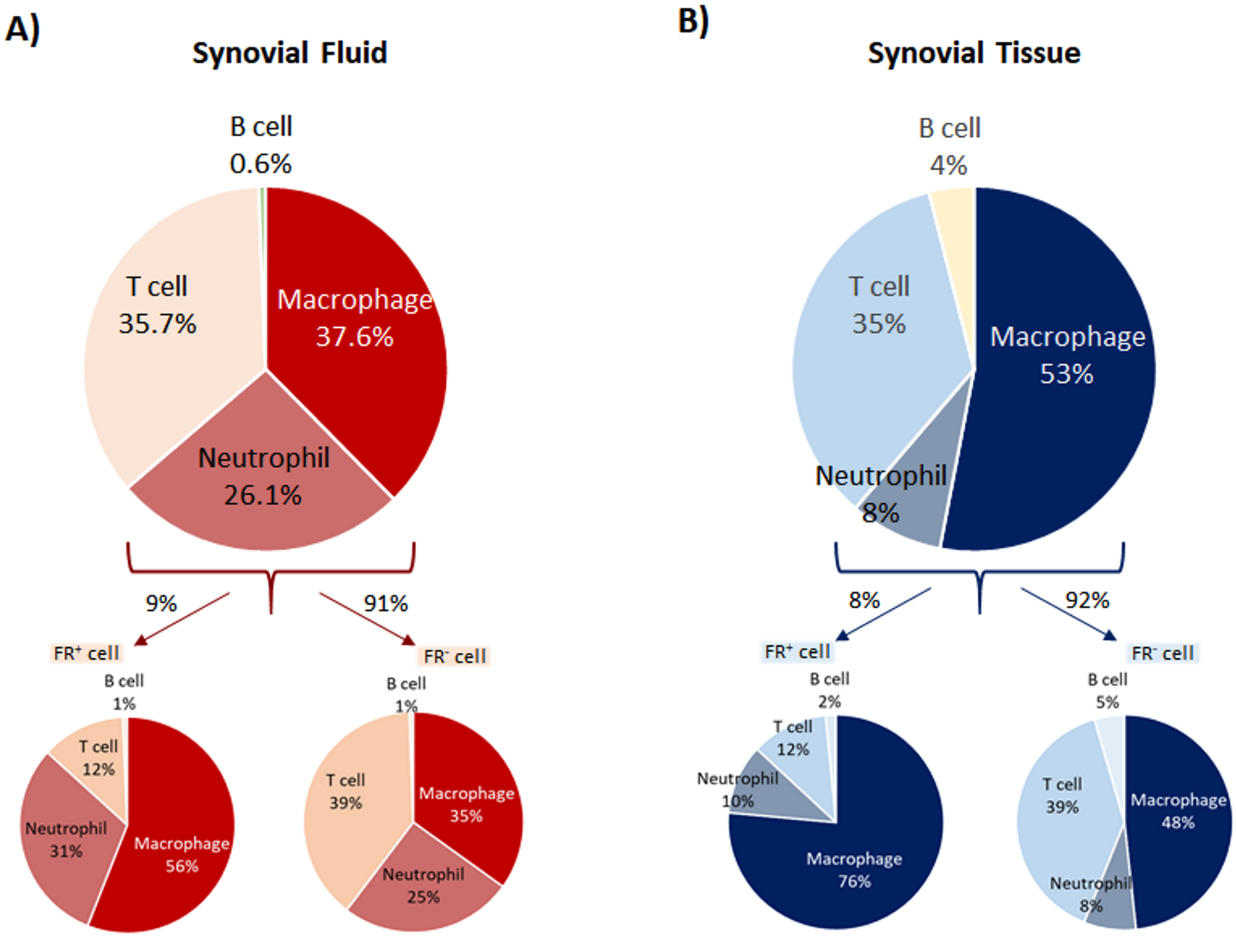
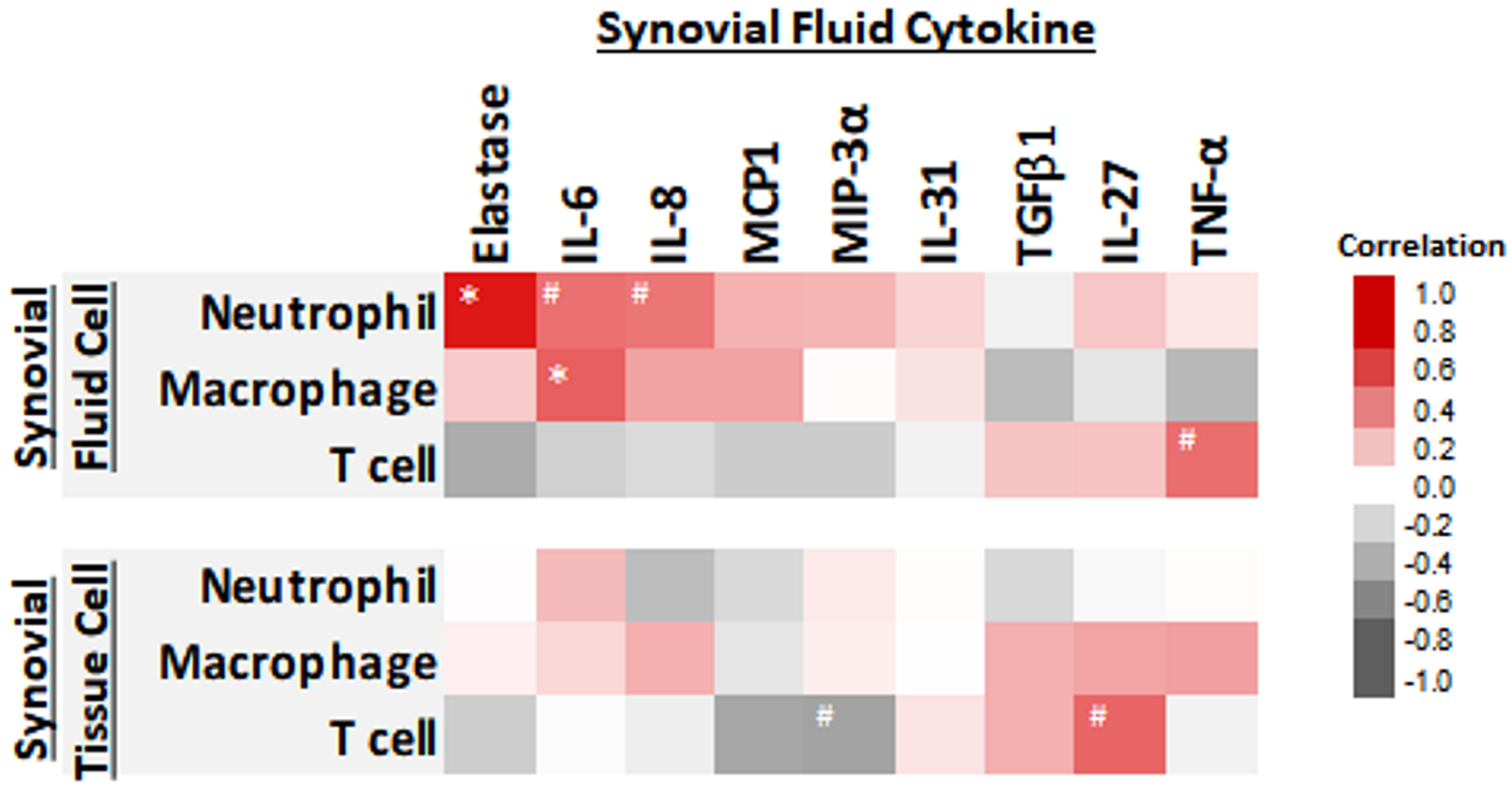
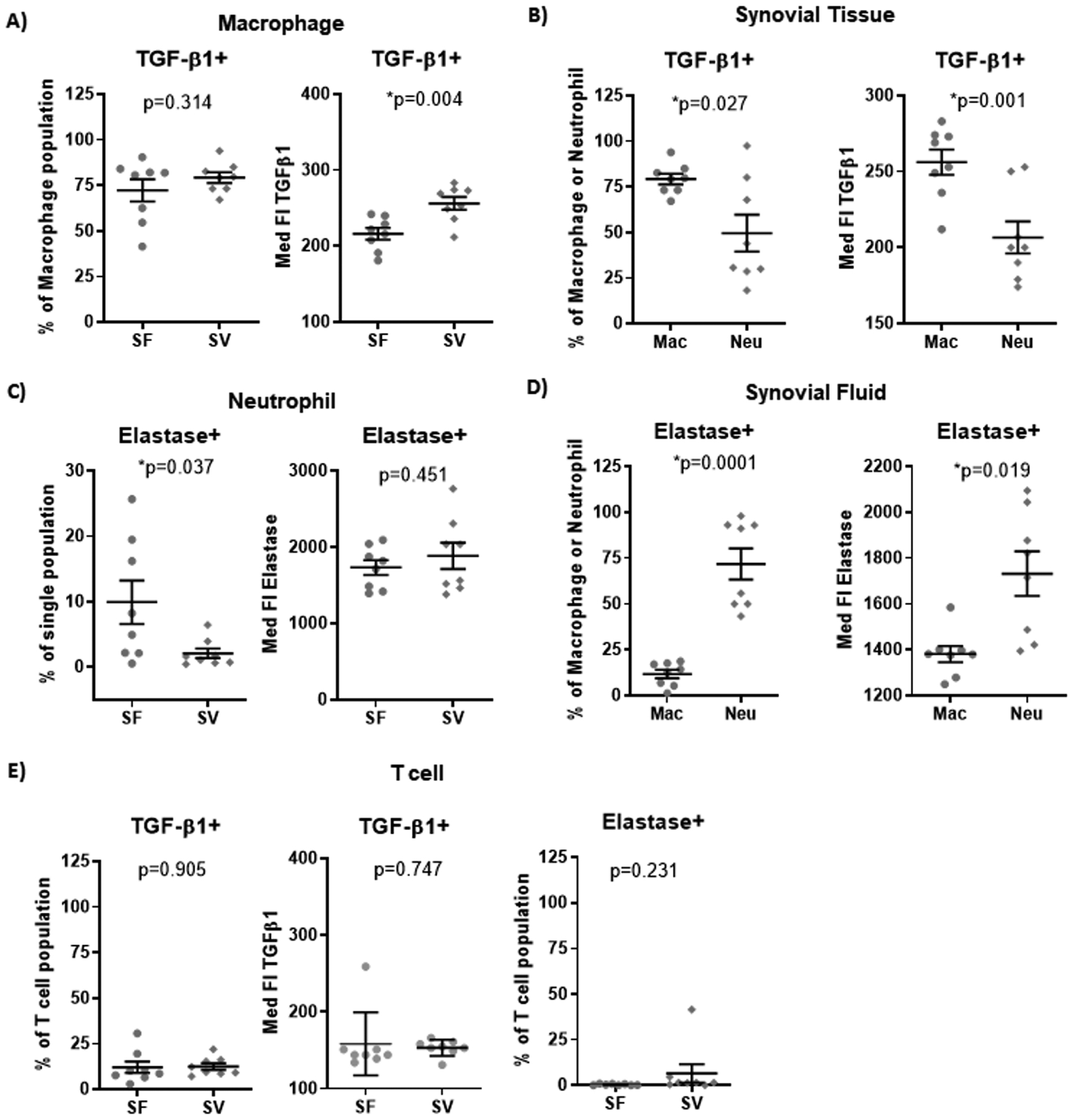
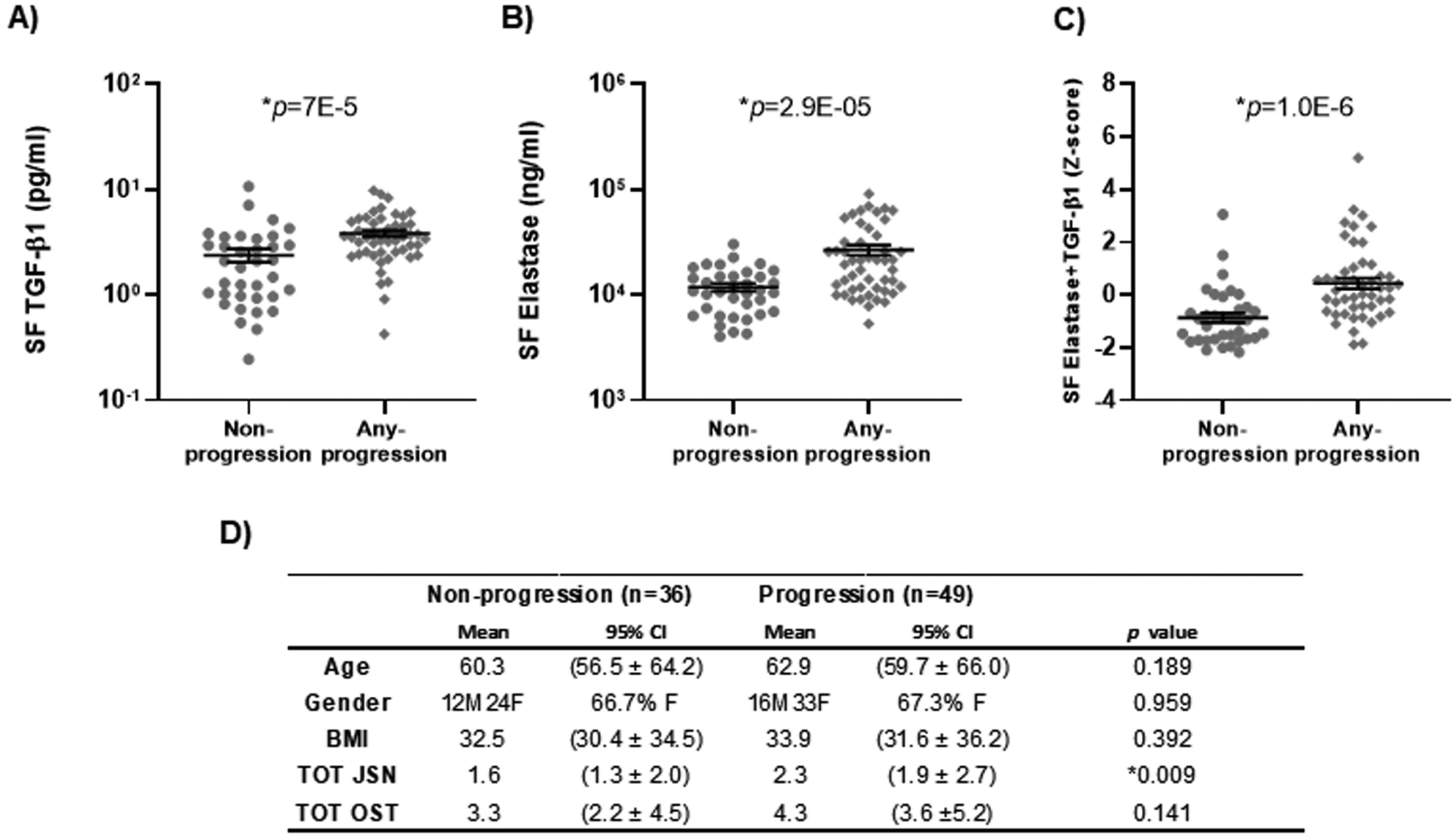
Results: Macrophages (predominant in the synovial tissue, 53% of total cells) and neutrophils (predominant in the SF, 26% of total cells) were the major immune cell populations identified in the OA knee joints, exhibiting expression of or association with transforming growth factor β1 (TGFβ1) and elastase, respectively, in the SF. Expression levels of TGFβ1 and elastase were significantly associated with severity of radiographic knee OA. Baseline SF concentrations of TGFβ1 and elastase along with radiographic knee OA severity scores were predictive of knee OA progression, with areas under the receiver operating characteristic curves of 0.810 (for TGFβ1), 0.806 (for elastase), and 0.846 (for both TGFβ1 and elastase combined), with greater stability of prediction when both markers were utilized.

Conclusion: Our findings demonstrate the hitherto underappreciated role of neutrophils in the sterile inflammatory process and progression of OA. Two soluble mediators, SF elastase and TGFβ1, are strong predictors of knee OA progression, reflecting a synergistic role of neutrophil and macrophage populations in the pathogenesis and worsening of OA that could potentially be utilized to identify patients who may have a greater risk of more rapid disease progression.


Conflict of interest statement

Competing interest statement

The authors have no conflict of interest of any kind with regard to this work.

Figures

Figure 1.
Figure 1.
Phenotypic characterization of immune cells determined by flow cytometric analyses. A&B) Macrophages (CD14+/CD11c+/HLADR+/CD11b+ /CD16low), neutrophils (CD14/CD11c/HLADR/CD11b+/CD16high), and T cells (CD45+/CD3+) are major immune cell populations in SF and SV (n=17 each group). A higher mean percentage of neutrophils was present in SF compared to SV. In contrast, a higher mean percentage of macrophages was present in SV compared to SF. Overall, a mean 8–9% of the SV and SF cells were FR+. By flow cytometric analysis, macrophages and neutrophils constituted the major FR+ immune cell populations in both SV and SF. The FR+ cell subtype was enriched for macrophages and neutrophils but had fewer T cells compared to the FR majority cell population
Figure 2.
Figure 2.
Heat map representing the associations of synovial fluid (SF) and synovial tissue (SV) immune cells and SF cytokines. Elastase, IL-6, IL-8, MCP1 and MIP-3α were positively associated with the total number of SF neutrophils. TGF-β1, TNF-α, and IL-27 were positively associated with the total number of SV macrophages, and the number of T cells in both SF and SV. #= p-value less than 0.1. *= p-value less than 0.05. N=17 patients.
Figure 3.
Figure 3.
Intracellular cytokine production by macrophages and neutrophils in SF and SV based on flow cytometric analyses. Although the percentage of total macrophages (CD3/CD14+/CD16low)(A) producing TGF-β1 was similar in SF and SV, the amount of TGF-β1 produced on average per cell (based on MFI) was significantly greater for macrophages in SV compared to SF. (B) In SV, the percentage of TGF-β1 producing macrophages was significantly higher than the percentage of TGF-β1 producing neutrophils; the amount of TGF-β1 produced on average per cell was also significantly higher in macrophages compared to neutrophils. In contrast, elastase was produced by a greater number of total neutrophils (CD3/CD14/CD16high) (C) in SF compared to SV although the amount of elastase produced on average per cell (based on MFI) was similar for neutrophils in SF compared to SV. (D) In SF, elastase+ neutrophils were significantly more abundant compared to the basal level of elastase+ macrophages; the amount of elastase produced on average per cell was also significantly higher in neutrophils compared to macrophages. (E) The number of TGF-β1+ T cells was much lower than TGF-β1+ macrophages and no or very few elastase+ T cells were detected. The amount of TGF-β1 produced per T cell was lower than the amount produced by macrophages in SF and SV. Med FI: Median Fluorescent Intensity. Mac: macrophages. Neu: neutrophils. N=8 patients per group.
Figure 4.
Figure 4.
Prediction of osteoarthritis progression by baseline synovial fluid cytokines. Knee OA progression was defined as “non-progression” (n=36) or “any progression” (n=49) based on either 1 unit increase in OST, 1 unit increase in JSN or total knee replacement (TKR) during a 3-year follow-up interval from the POP cohort. Scatter plots by knee OA progression status demonstrate: (A) significantly higher mean SF TGF-β1 of 3.8 pg/ml (95% confidence interval [95% CI] 3.3 to 4.4 pg/ml) in the any progression group compared to 2.4 pg/ml (95% CI 1.7 to 3.1 pg/ml) in the non-progression group; (B) significantly higher mean SF elastase of 26770 ng/ml (95% CI 20845 to 32695 ng/ml) compared to 11850 ng/ml (95% CI 9852 to 13847 ng/ml) in the non-progression group; and (C) significantly higher mean concentration of the combination of the two in the any progression group with Z score 0.4 (95% CI 0.03 to 0.84) compared to Z score −0.88 (95% CI −1.24 to −0.51) in the non-progression group. (D) The demographic information and radiographic feature score separated by progression group and non-progression group.

References

    1. Berenbaum F Osteoarthritis as an inflammatory disease (osteoarthritis is not osteoarthrosis!). Osteoarthritis Cartilage 2013;21(1):16–21. - PubMed
    1. Muley MM, Reid AR, Botz B, Bolcskei K, Helyes Z, McDougall JJ. Neutrophil elastase induces inflammation and pain in mouse knee joints via activation of proteinase-activated receptor-2. Br J Pharmacol 2016;173(4):766–77. - PMC - PubMed
    1. de Lange-Brokaar BJ, Ioan-Facsinay A, van Osch GJ, Zuurmond AM, Schoones J, Toes RE, Huizinga TW, Kloppenburg M. Synovial inflammation, immune cells and their cytokines in osteoarthritis: a review. Osteoarthritis Cartilage 2012;20(12):1484–99. - PubMed
    1. Tarhan S, Unlu Z. Magnetic resonance imaging and ultrasonographic evaluation of the patients with knee osteoarthritis: a comparative study. Clin Rheumatol 2003;22(3):181–8. - PubMed
    1. Song IH, Althoff CE, Hermann KG, Scheel AK, Knetsch T, Schoenharting M, Werner C, Burmester GR, Backhaus M. Knee osteoarthritis. Efficacy of a new method of contrast-enhanced musculoskeletal ultrasonography in detection of synovitis in patients with knee osteoarthritis in comparison with magnetic resonance imaging. Ann Rheum Dis 2008;67(1):19–25. - PubMed

Publication types

MeSH terms