

Neurofluid Dynamics and the Glymphatic System: A Neuroimaging Perspective
- PMID: 32783417
- PMCID: PMC7462760
- DOI: 10.3348/kjr.2020.0042
Neurofluid Dynamics and the Glymphatic System: A Neuroimaging Perspective
Abstract
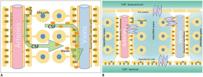
The glymphatic system hypothesis is a concept describing the clearance of waste products from the brain. The term "glymphatic system" combines the glial and lymphatic systems and is typically described as follows. The perivascular space functions as a conduit that drains cerebrospinal fluid (CSF) into the brain parenchyma. CSF guided to the perivascular space around the arteries enters the interstitium of brain tissue via aquaporin-4 water channels to clear waste proteins into the perivascular space around the veins before being drained from the brain. In this review, we introduce the glymphatic system hypothesis and its association with fluid dynamics, sleep, and disease. We also discuss imaging methods to evaluate the glymphatic system.
Keywords: Cerebrospinal fluid; Contrast media; Glymphatic system; Interstitial fluid; Magnetic resonance imaging, Diffusion imaging.
Copyright © 2020 The Korean Society of Radiology.
Conflict of interest statement
Department of Innovative Biomedical Visualization (iBMV), Nagoya University is supported by CANON MEDICAL SYSTEMS CORPORATION.
Figures
Similar articles
-
The Glymphatic System: A Review of the Challenges in Visualizing its Structure and Function with MR Imaging.Magn Reson Med Sci. 2022 Mar 1;21(1):182-194. doi: 10.2463/mrms.rev.2020-0122. Epub 2020 Nov 27. Magn Reson Med Sci. 2022. PMID: 33250472 Free PMC article. Review.
-
Current Concepts in Intracranial Interstitial Fluid Transport and the Glymphatic System: Part II-Imaging Techniques and Clinical Applications.Radiology. 2021 Dec;301(3):516-532. doi: 10.1148/radiol.2021204088. Epub 2021 Oct 26. Radiology. 2021. PMID: 34698564 Review.
-
Glymphatic imaging using MRI.J Magn Reson Imaging. 2020 Jan;51(1):11-24. doi: 10.1002/jmri.26892. Epub 2019 Aug 18. J Magn Reson Imaging. 2020. PMID: 31423710 Review.
-
The perivascular space is a conduit for cerebrospinal fluid flow in humans: A proof-of-principle report.Proc Natl Acad Sci U S A. 2024 Oct 15;121(42):e2407246121. doi: 10.1073/pnas.2407246121. Epub 2024 Oct 7. Proc Natl Acad Sci U S A. 2024. PMID: 39374384 Free PMC article.
-
Gadolinium-based Contrast Media, Cerebrospinal Fluid and the Glymphatic System: Possible Mechanisms for the Deposition of Gadolinium in the Brain.Magn Reson Med Sci. 2018 Apr 10;17(2):111-119. doi: 10.2463/mrms.rev.2017-0116. Epub 2018 Jan 25. Magn Reson Med Sci. 2018. PMID: 29367513 Free PMC article.
Cited by
-
Dynamic changes in glymphatic function in reversible cerebral vasoconstriction syndrome.J Headache Pain. 2024 Feb 5;25(1):17. doi: 10.1186/s10194-024-01726-1. J Headache Pain. 2024. PMID: 38317074 Free PMC article.
-
Giant Tumefactive Perivascular Space: Advanced Fusion MR Imaging and Tractography Study-A Case Report and a Systematic Review.Diagnostics (Basel). 2023 Apr 30;13(9):1602. doi: 10.3390/diagnostics13091602. Diagnostics (Basel). 2023. PMID: 37174993 Free PMC article.
-
Reproducibility of diffusion tensor image analysis along the perivascular space (DTI-ALPS) for evaluating interstitial fluid diffusivity and glymphatic function: CHanges in Alps index on Multiple conditiON acquIsition eXperiment (CHAMONIX) study.Jpn J Radiol. 2022 Feb;40(2):147-158. doi: 10.1007/s11604-021-01187-5. Epub 2021 Aug 14. Jpn J Radiol. 2022. PMID: 34390452 Free PMC article.
-
Repeat and single dose administration of gadodiamide to rats to investigate concentration and location of gadolinium and the cell ultrastructure.Sci Rep. 2021 Jul 6;11(1):13950. doi: 10.1038/s41598-021-93147-2. Sci Rep. 2021. PMID: 34230532 Free PMC article.
-
The Glymphatic System: A Review of the Challenges in Visualizing its Structure and Function with MR Imaging.Magn Reson Med Sci. 2022 Mar 1;21(1):182-194. doi: 10.2463/mrms.rev.2020-0122. Epub 2020 Nov 27. Magn Reson Med Sci. 2022. PMID: 33250472 Free PMC article. Review.
References
-
- Agarwal N, Contarino C, Toro EF. Neurofluids: a holistic approach to their physiology, interactive dynamics and clinical implications for neurological diseases. Veins and Lymphatics. 2019;8:49–58.
-
- Taoka T, Naganawa S. Glymphatic imaging using MRI. J Magn Reson Imaging. 2020;51:11–24. - PubMed
-
- Cushing H. Cameron lecture. Lancet. 1925;206:851–857.
Publication types
MeSH terms
Substances
LinkOut - more resources
Full Text Sources
Other Literature Sources
