. 2020 Sep 1;124(3):815-821.
doi: 10.1152/jn.00339.2020. Epub 2020 Aug 12.

Early life stress facilitates synapse premature unsilencing to enhance AMPA receptor function in the developing hippocampus

Affiliations

Early life stress facilitates synapse premature unsilencing to enhance AMPA receptor function in the developing hippocampus

Aycheh Al-Chami et al. J Neurophysiol. .

Abstract

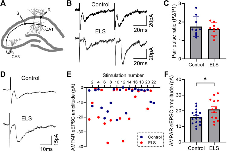
Chronic early life stress (ELS) increases vulnerability to psychopathologies and cognitive deficits in adulthood by disrupting the function of related neural circuits. However, whether this disruption emerges early in the developing brain remains largely unexplored. In the current study, using an established limited-bedding and nesting model of ELS in postnatal day (P)2-10 mice, we provide direct evidence that ELS caused early modification of hippocampal glutamatergic synapses in the developing brain. We demonstrated that ELS induced rapid enhancement of AMPA receptor function in hippocampal CA1 pyramidal neurons through a postsynaptic mechanism, and importantly, this was associated with premature unsilencing of NMDA receptor-only silent hippocampal synapses. These results suggest that potentiation of AMPAR function may represent an early mediator of ELS-induced alterations of neural networks in the developing brain and may potentially contribute to subsequent cognitive impairments later in life.NEW & NOTEWORTHY Early life stress (ELS) is known to increase the risk of later life cognitive deficits by disrupting neural circuit function. However, whether this disruption emerges early in the developing brain remains largely unexplored. The current study presents direct evidence that ELS prematurely unsilences hippocampal synapses to enhance AMPA receptor functions in a limited-bedding and nesting model, revealing an early mediator of ELS-induced neural circuit reorganizations.

Keywords: AMPA receptor; brain development; critical period; early life stress; silent synapse.


Conflict of interest statement

No conflicts of interest, financial or otherwise, are declared by the authors.

Figures

Fig. 1.
Fig. 1.
Early life stress (ELS) induces early enhancement of AMPA receptor (AMPAR) function in CA1 pyramidal neurons. A: schematic of limited-bedding and nesting (LBN) procedure. Electrophysiological (Ephys) recordings were performed at postnatal day (P)10–11. B: photographs show typical setups of control (top) and LBN (bottom) cages. Note that the LBN cage has a fitted plastic-coated mesh platform with half nestlet provided. C: body weight of control and LBN mice at P10–11; n = 14, 10; **P < 0.001 by unpaired t test. D: representative AMPAR-mediated spontaneous excitatory postsynaptic currents (sEPSCs) in CA1 pyramidal neurons from P10 control (top traces) and ELS (bottom traces) mice. E and F: group data of AMPAR-mediated sEPSC rise time (E) and decay time (F) in CA1 pyramidal neurons from control and ELS mice; n = 24, 12. Error bars indicate SE. G: cumulative distribution shows significantly higher amplitude of AMPAR sEPSCs in neurons from ELS mice compared with control. ***P < 0.001 by Kolmogorov-Smirnov test. H: group data of AMPAR-mediated sEPSC amplitude in CA1 pyramidal neurons from control and ELS mice; n = 24, 12; ***P < 0.001 by unpaired t test. Error bars indicate SE. I: cumulative distribution for AMPAR sEPSC interevent intervals in neurons from control and ELS mice. P > 0.05 by Kolmogorov-Smirnov test. J: group data of AMPAR-mediated sEPSC frequency in CA1 pyramidal neurons from control and ELS mice; n = 24, 12. Error bars indicate SE.
Fig. 2.
Fig. 2.
Early life stress (ELS) potentiates minimally evoked AMPA receptor (AMPAR)-mediated evoked excitatory postsynaptic currents (eEPSCs) with no significant alterations in paired-pulse facilitation in CA1 pyramidal neurons. A: schematic of stimulation (S) and recording (R) sites in the ex vivo hippocampal slice. B: representative traces of paired-pulse evoked AMPAR EPSCs at a holding potential of −60 mV in CA1 pyramidal neurons by stimulating Schaffer collateral pathway in hippocampal slices from control and ELS mice. The pulse interval was 50 ms. C: group data of paired-pulse facilitation in CA1 pyramidal neurons from control and ELS mice; n = 9, 9; unpaired t test was used. Error bars indicate SE. D: representative minimally evoked AMPAR EPSCs in CA1 pyramidal neurons from control and ELS mice. E: amplitudes for 20 consecutive minimally eEPSC responses at a holding potential of −60 mV in CA1 pyramidal neurons from control and ELS mice. F: group data of minimally eEPSC amplitude in CA1 pyramidal neurons from control and ELS mice; n = 16, 13; *P < 0.05 by unpaired t test. Error bars indicate SE.
Fig. 3.
Fig. 3.
Early life stress (ELS) diminishes hippocampal NMDA receptor (NMDAR)-only silent synapses in the developing brain. A: representative minimally evoked excitatory postsynaptic currents (eEPSCs) recordings at holding potentials of +40 (top trace) and −60 mV (bottom trace) in CA1 pyramidal neurons from a control mouse. B: successes (blue dots) and failures (gray dots) of individual eEPSCs are shown. C: representative minimally eEPSC recordings at holding potentials of +40 (top trace) and −60 mV (bottom trace) in CA1 pyramidal neurons from an ELS mouse. D: successes (red dots) and failures (gray dots) of individual evoked EPSCs are shown. E: group data of eEPSC failure rates at −60 and +40 mV holding potentials in CA1 pyramidal neurons from control mice; n = 16; **P < 0.001 by paired t test. Error bars indicate SE. F: group data of eEPSC failure rates at −60 and +40 mV holding potentials in CA1 pyramidal neurons from ELS mice; n = 13; *P < 0.05 by paired t test. Error bars indicate SE. G: group data of eEPSC failure rate difference between −60 and +40 mV holding potentials in CA1 pyramidal neurons from control and ELS mice; n = 16, 13; ***P < 0.001 by unpaired t test. Error bars indicate SE. H: group data of the calculated fraction of silent synapses in CA1 pyramidal neurons from control and ELS mice; n = 16, 13; ***P < 0.001 by unpaired t test. Error bars indicate SE.

References

    1. Ali I, Salzberg MR, French C, Jones NC. Electrophysiological insights into the enduring effects of early life stress on the brain. Psychopharmacology (Berl) 214: 155–173, 2011. doi:10.1007/s00213-010-2125-z. - DOI - PubMed
    1. Bath KG, Nitenson AS, Lichtman E, Lopez C, Chen W, Gallo M, Goodwill H, Manzano-Nieves G. Early life stress leads to developmental and sex selective effects on performance in a novel object placement task. Neurobiol Stress 7: 57–67, 2017. doi:10.1016/j.ynstr.2017.04.001. - DOI - PMC - PubMed
    1. Brunson KL, Kramár E, Lin B, Chen Y, Colgin LL, Yanagihara TK, Lynch G, Baram TZ. Mechanisms of late-onset cognitive decline after early-life stress. J Neurosci 25: 9328–9338, 2005. doi:10.1523/JNEUROSCI.2281-05.2005. - DOI - PMC - PubMed
    1. Chandrasekaran AR, Shah RD, Crair MC. Developmental homeostasis of mouse retinocollicular synapses. J Neurosci 27: 1746–1755, 2007. doi:10.1523/JNEUROSCI.4383-06.2007. - DOI - PMC - PubMed
    1. Chaouloff F, Groc L. Temporal modulation of hippocampal excitatory transmission by corticosteroids and stress. Front Neuroendocrinol 32: 25–42, 2011. doi:10.1016/j.yfrne.2010.07.004. - DOI - PubMed

Publication types

Substances

Grants and funding