. 2020 Nov-Dec:49:107263.
doi: 10.1016/j.carpath.2020.107263. Epub 2020 Jul 16.

Analysis of cardiopulmonary findings in COVID-19 fatalities: High incidence of pulmonary artery thrombi and acute suppurative bronchopneumonia

Affiliations

Analysis of cardiopulmonary findings in COVID-19 fatalities: High incidence of pulmonary artery thrombi and acute suppurative bronchopneumonia

Claudia Grosse et al. Cardiovasc Pathol. 2020 Nov-Dec.

Abstract

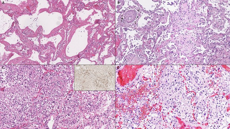
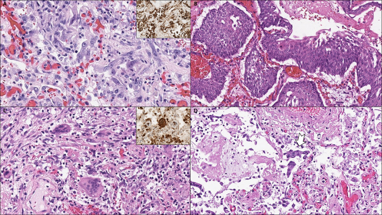
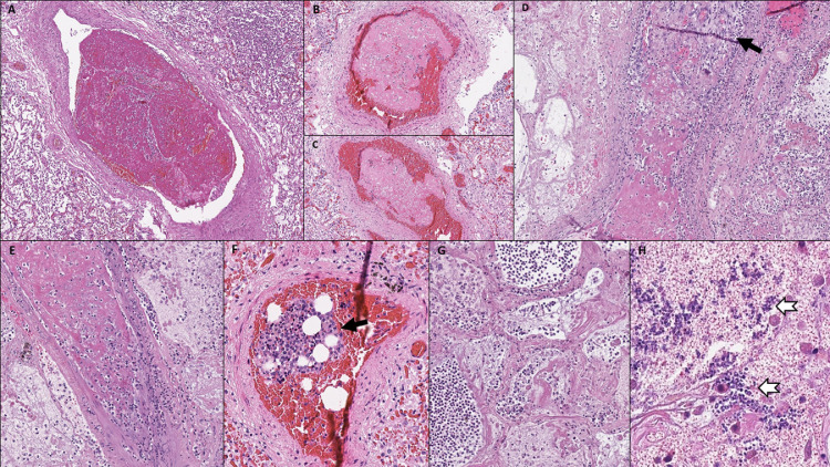
Since its recognition in December 2019, coronavirus disease 2019 (COVID-19) caused by SARS-CoV-2 has rapidly spread globally causing a pandemic that represents the greatest medical challenge in decades. The aim of the study was to evaluate the spectrum of cardiopulmonary pathology of COVID-19 based on (non-minimal invasive) autopsies performed on 14 COVID-19 decedents. Bilateral diffuse alveolar damage (DAD) was found in all patients. Superimposed acute bronchopneumonia was present in 11 of 14 (78.6%) patients and was considered the major cause of death in 2 patients. A key finding was the presence of thrombotic/thromboembolic vascular occlusions. We classified 5 types of pulmonary thrombi: 1. capillary microthrombi (11/14, 78.6%); 2. partially organized thrombi in mid-sized pulmonary arteries with complete vessel occlusion; 3. non-organized thrombi in mid-sized pulmonary arteries that did not completely fill out the vessel lumen and probably represented thromboemboli rather than thrombosis; 4. bone marrow emboli (1/14, 7.1%); and 5. septic pulmonary thromboemboli (1/14, 7.1%). Pulmonary thrombi in mid-sized arteries were noted in 5 of 14 (35.7%) patients, causing pulmonary infarction and/or pulmonary hemorrhage. All patients had evidence of chronic cardiac disease, including myocardial hypertrophy (13/14, 92.9%), mild to marked coronary artery atherosclerosis (14/14, 100%) and focal myocardial fibrosis (3/14, 21.4%). Acute myocardial infarction was found as concurrent cause of death in 3 (21.4%) patients, and significant cardiac hypertrophy (heart weight 750 g) was present in 1 (7.1%) patient with ATTR-positive cardiac amyloidosis. The autopsy findings confirm that COVID-19 is a systemic disease, with major involvement of the lungs, that increases the risk of cardiac and vascular complications including acute myocardial injury and thrombotic/thromboembolic events. Secondary acute bronchopneumonia is a common complication in patients with COVID-19 and may be the major cause of death.

Keywords: Autopsy; COVID-19; Cardiopulmonary pathology; Coronavirus; Diffuse alveolar damage; SARS-CoV-2.


Figures

Fig 1
Fig. 1
(A) Acute-phase diffuse alveolar damage with hyaline membranes, highlighted by (B) pancytokeratin and (C) Elastica van Gieson stain.
Fig 2
Fig. 2
(A) Acute-phase diffuse alveolar damage with hyaline membranes. (B-D) Chronic-phase diffuse alveolar damage with (B) interstitial and airspace fibroblastic proliferation, (C) lymphocytic infiltrate, mainly composed of CD3-positive T-lymphocytes (inset, CD3), and (D) focal pulmonary hemorrhage.
Fig 3
Fig. 3
(A) Hyperplastic type II pneumocytes with some degree of cytological atypia, positive for pancytokeratin showing epithelial lineage of atypical cells (inset). (B) Squamous metaplasia. (C) Multinucleated cells in chronic-phase diffuse alveolar damage, positive for CD68 suggestive of histiocytic cell origin (inset). (D) Syncytical cells of pneumocytic origin (arrow).
Fig 4
Fig. 4
(A) Interstitial fibroblastic proliferation and hyaline membranes. (B) Organizing diffuse alveolar damage (arrowhead) is seen next to features of acute pneumonia (arrow). (C) Acute bronchopneumonia with dense aggregates of neutrophile granulocytes within the airspaces. (D) A foreign particle (arrow) in a bronchial lumen indicative of terminal aspiration.
Fig 5
Fig. 5
(A-C) Thrombi in mid-sized pulmonary arteries with incomplete occlusion of the vessel lumen. (D, E) Partially organized thrombi (arrow in D) in mid-sized pulmonary arteries, completely occluding the vessel lumen. (F) Bone marrow embolus. (G) Pulmonary infarction caused by thrombi in mid-sized pulmonary arteries. (H) Fungal superinfection (arrows) in infarcted lung tissue.
Fig 6
Fig. 6
Pulmonary septicoemboli in a patient with fungal sepsis. (A) Low power magnification shows massively dilated mid-sized pulmonary arteries with intraluminal fibrin deposits and entrapment of neutrophils. (Lower image) High power magnification shows intraluminal fibrin deposits with neutrophilic entrapment and incomplete occlusion of the vessel lumen. (B) Septic embolus consisting of intraluminal fibrin deposits admixed with fungal structures on periodic acid-Schiff stain (right image, high power magnification). (C) Mid-sized pulmonary artery with septic embolus, perivascular hemorrhage and transmural neutrophilic infiltration of the vessel wall (arrows). (D) Septic embolus with transmural neutrophilic infiltration of the vessel wall and involvement of the adjacent lung tissue (arrows).
Fig 7
Fig. 7
Extrapulmonary manifestations of COVID-19. (A) Myocardium with few CD3-positive interstitial T-lymphocytes. (B) Spleen with increased neutrophil granulocytes in the perifollicular and marginal zone (splenitis). (C) Spleen with white pulp atrophy. (D) Liver with mild portal lymphocytic infiltration. (E) Kidney with acute tubular damage.

Comment in

References

    1. Park SE. Epidemiology, virology, and clinical features of severe acute respiratory syndrome-coronavirus-2 (SARS-CoV-2; Coronavirus Disease-19) Clin Exp Pediatr. 2020;63:119–124. doi: 10.3345/cep.2020.00493. - DOI - PMC - PubMed
    1. Lu H, Stratton CW, Tang YW. Outbreak of pneumonia of unknown etiology in Wuhan, China: The mystery and the miracle. J Med Virol. 2020;92:401–402. doi: 10.1002/jmv.25678. - DOI - PMC - PubMed
    1. Johns Hopkins University&Medicine. COVID-19 dashboard by the Center for Systems Science and Engineering (CSSE). Available at: https://coronavirus.jhu.edu/map.html; Accessed July 6, 2020.
    1. Xu Z, Shi L, Wang Y, Zhang J, Huang L, Zhang C. Pathological findings of COVID-19 associated with acute respiratory distress syndrome. Lancet Respir Med. 2020;8:420–422. doi: 10.1016/S2213-2600(20)30076-X. - DOI - PMC - PubMed
    1. Tian S, Xiong Y, Liu H, Niu L, Guo J, Liao M. Pathological study of the 2019 novel coronavirus disease (COVID-19) through postmortem core biopsies. Mod Pathol. 2020 doi: 10.1038/s41379-020-0536-x. - DOI - PMC - PubMed

MeSH terms