. 2020 Aug 8;12(8):867.
doi: 10.3390/v12080867.

Mapping Attenuation Determinants in Enterovirus-D68

Affiliations

Mapping Attenuation Determinants in Enterovirus-D68

Ming Te Yeh et al. Viruses. .

Abstract

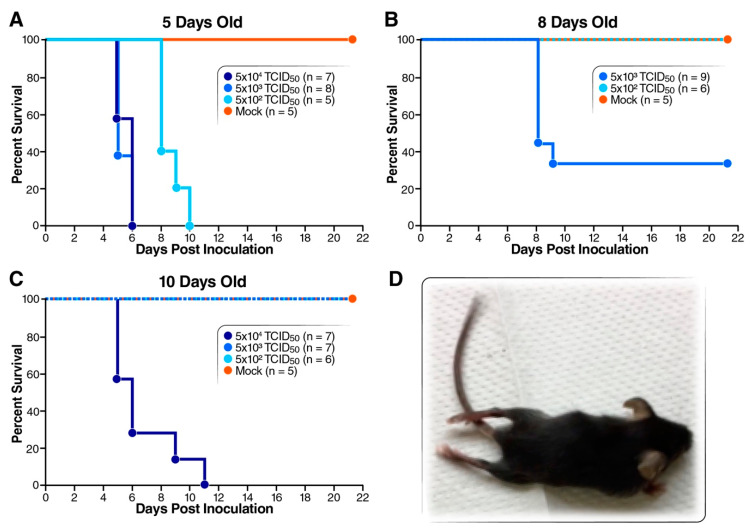
Enterovirus (EV)-D68 has been associated with epidemics in the United Sates in 2014, 2016 and 2018. This study aims to identify potential viral virulence determinants. We found that neonatal type I interferon receptor knockout mice are susceptible to EV-D68 infection via intraperitoneal inoculation and were able to recapitulate the paralysis process observed in human disease. Among the EV-D68 strains tested, strain US/MO-14-18949 caused no observable disease in this mouse model, whereas the other strains caused paralysis and death. Sequence analysis revealed several conserved genetic changes among these virus strains: nucleotide positions 107 and 648 in the 5'-untranslated region (UTR); amino acid position 88 in VP3; 1, 148, 282 and 283 in VP1; 22 in 2A; 47 in 3A. A series of chimeric and point-mutated infectious clones were constructed to identify viral elements responsible for the distinct virulence. A single amino acid change from isoleucine to valine at position 88 in VP3 attenuated neurovirulence by reducing virus replication in the brain and spinal cord of infected mice.

Keywords: VP3; enterovirus; enterovirus-D68; infectious clones; mouse model; paralysis; virulence determinant.


Conflict of interest statement

The authors declare no conflict of interest.

Figures

Figure 1
Figure 1
Susceptibility of type I interferon receptor deficient mice to EV-D68 infection. EV-D68 49129 used in this experiment was from cell culture. (AC) Mice of 5, 8 and 10 days old were i.p. inoculated with 5 × 10²–5 × 10⁴ TCID50 of EV-D68 strain 49129 (US/MO/14-18947) and monitored for survival for 21 days. Mice in the control group were injected with 100 µL of viral medium. (D) Paralysis on both posterior limbs was observed after i.p. inoculation of strain 49129.
Figure 2
Figure 2
Characteristics of various EV-D68 strains. (A) Virulence test for various EV-D68 strains. Seven-day-old mice were i.p. inoculated with 100 uL of virus delivering 1 × 10⁴ TCID50 of EV-D68 strain NR-49129, NR-49130, NR-49131 or CA/14-4231. Survival of the mice was monitored for 21 days. Asterisk (*) represents significantly different survival curve, comparing to that of 49131-infected mice (log-rank test, p < 0.05). (B) Replication in RD cells. RD cells were infected with EV-D68 strains tested in Figure 2A at m.o.i. of 0.01 and harvested at 0, 24, 48 and 72 h for TCID50 to determine virus titers. Results of triplicates are shown as mean ± S.D. Asterisks represent significant differences in virus titers compared with 49131 (Student’s t test, p < 0.05).
Figure 3
Figure 3
Virulence test for 5′-UTR-modified EV-D68 viruses. (A) Schematic presentation of the gene-swapped and point-mutated viruses. Strains 49131 and 49130 were used as backbone to generate these 5′-UTR modified viruses. Strain 30-5UTR-49131 is 49131 carrying 5′-UTR from 49130. Nucleotide changes U107C and G648C were individually introduced into 49131. (B) Replication analysis for the engineered EV-D68 viruses was performed with RD cells, as described in Figure 1B. Results of triplicates are shown as mean ± S.D. No significant difference in virus replication ability compared with strain 49131 was detected by Student’s t test (p < 0.05). (C) Seven-day-old Tg21/IFNR-ko mice were i.p. inoculated with 1 × 10⁵ TCID50 of virus and monitored for 21 days to compare the virulence of the 5′-UTR-modified EV-D68 viruses. Viruses 49131, 30-5UTR (30-5UTR-49131), 49131-5UTR-U107C and 49131-5UTR-G648C were tested with 2 litters of pups. Asterisk represents significantly different survival curve comparing with that of 49131-infected mice detected by using log-rank test (p < 0.05).
Figure 4
Figure 4
Validation of genetic changes in the non-structural genes on EV-D68 virulence with strain 49131 as a backbone. (A) Schematic of the gene-swapped and point-mutated viruses. Strain 49131 was used as backbone to generate these recombinant viruses. Amino acid changes 2A-T22A and 3C-H47R were introduced into 49131 individually. (B) Replication analysis for the engineered EV-D68 viruses carrying mutations in the non-structural region (NSR) was performed with RD cells, as described in Figure 1B. Results of triplicates are shown as mean ± S.D. Asterisk: significantly lower virus titer of 49130 at 72 h post infection comparing with 49131 was detected by Student’s t test (p < 0.05). (C) Seven-day-old mice were i.p. inoculated with 1 × 10⁵ TCID50 of viruses carrying 2A-T22A or 3C-H47R and monitored for 21 days. 49131, 2A-T22A, and 3C-H47R were tested in 2 litters of pups. Asterisks represent survival curves which, compared with those of 49131-infected mice, were determined to be significantly different by using log-rank test (p < 0.05).
Figure 5
Figure 5
Validation of the conserved amino acid changes in structural proteins on replication and virulence of EV-D68. Asterisks represent significant difference as compared with 49131. (A) Representation of the gene composition for viruses carrying swapped VP1 or P1 gene and amino acid substitutions in the structural gene. Strain 49131 was used as backbone to generate these recombinant viruses. Amino acid changes VP3-I88V, VP1-L1P and VP1-V148A were introduced into 49131 individually and VP1-K282R and VP1-G283E were changed together. (B) Replication analysis for VP1 or P1 swapped viruses, as shown in Figure 1B. (C) Seven-day-old mice were inoculated i.p. with 1 × 10⁵ TCID50 of strain 49131 carrying VP1 or P1 gene from 49130 and monitored for 21 days. (D) Replication analysis for viruses carrying mutations in the structural genes, as shown in Figure 1B. (E) Virulence test for viruses carrying mutations in the structural genes were performed by i.p. injecting 7-day-old mice with 1 × 10⁵ TCID50 of virus and monitored for 21 days. In this experiment, 49131, VP3-I88V and VP1-L1P were tested with 2 litters of pups. (B,D) Results of triplicates are shown as mean ± S.D. Asterisks represent significantly different virus titers of mutant viruses compared with 49131 detected by Student’s t test (p < 0.05). (C,E) Asterisks represent significantly different survival curves, compared with that of 49131-infected mice, as detected by log-rank test (p < 0.05).
Figure 6
Figure 6
Neurovirulence test for 49131-VP3-I88V mutant. Five-day-old mice were i.c. inoculated with 10 µL of inoculum delivering 1 × 10³ or 1 × 10⁴ TCID50 of 49131 or 49131-VP3-I88V virus. Mice in control group were injected with 10 µL of viral medium. Survival was monitored for 21 days. Asterisk represents significantly different survival curves of VP3-I88V-infected mice, compared with that of 49131 (lower dose)-infected mice, as detected by log-rank test (p < 0.05).
Figure 7
Figure 7
Tissue distribution of EV-D68 virus in Tg21/IFNR-ko mice. Virus titers were determined by a standard TCID50 assay and shown as log TCID50 per milligram of tissue. Results are shown as mean ± S.E.M. (A) Mice of seven days old were i.p. inoculated with 1 × 10⁵ TCID50 of 49131 (shown as black) or 49131-VP3-I88V (shown as blue). At days 1, 3 and 5, tissues of three mice from each group were harvested to determine virus titers. (B) Mice of 5 days old were i.c. inoculated with 1 × 10⁴ TCID50 of 49131 or 49131-VP3-I88V. At days 1, 3 and 5, tissues of three mice from each group were harvested to determine virus titers.
Figure 8
Figure 8
Replication analysis for 49131 and 49131-VP3-I88V in cell lines. SK-N-SH (A), RD (B) and Neuro-2A (C) cells were infected with 49131 or VP3-I88V at an m.o.i. of 0.01 in triplicate and harvested at indicated time points for TCID50 assay to determine virus titers. Results of triplicates are shown as mean ± S.D.
Figure 9
Figure 9
Comparison of the simulated VP3-I88 and VP3-I88V structures. (A) VP1 (blue), VP2 (green) and VP3 (red) of EV-D68 are represented in new cartoon format. I88 is represented in van der Waals sphere format. The inset reports on the position of I88 within the pentamer with respect to VP1, VP2 and VP3. (B) Representation of the VP3 residues within 12 Å of I88 in new cartoon format. Residues interacting with I88 or showing relevant changes between VP3-I88 and VP3-I88V simulated structures are displayed in licorice format. (C) Comparison between the VP3-I88 (red) and VP3-I88V (pink) simulated structures extracted from the last frame of the simulations. In all panels, residues are colored according to the atoms: oxygen in red, carbon in cyan, nitrogen in blue; hydrogens are omitted for clarity.

References

    1. Blomqvist S., Savolainen C., Raman L., Roivainen M., Hovi T. Human rhinovirus 87 and enterovirus 68 represent a unique serotype with rhinovirus and enterovirus features. J. Clin. Microbiol. 2002;40:4218–4223. doi: 10.1128/JCM.40.11.4218-4223.2002. - DOI - PMC - PubMed
    1. Furuse Y., Chaimongkol N., Okamoto M., Oshitani H. Evolutionary and Functional Diversity of the 5′ Untranslated Region of Enterovirus D68: Increased Activity of the Internal Ribosome Entry Site of Viral Strains during the 2010s. Viruses. 2019;11:626. doi: 10.3390/v11070626. - DOI - PMC - PubMed
    1. Schieble J.H., Fox V.L., Lennette E.H. A probable new human picornavirus associated with respiratory diseases. Am. J. Epidemiol. 1967;85:297–310. doi: 10.1093/oxfordjournals.aje.a120693. - DOI - PubMed
    1. Meijer A., Van der Sanden S., Snijders B.E., Jaramillo-Gutierrez G., Bont L., Van der Ent C.K., Overduin P., Jenny S.L., Jusic E., Van der Avoort H.G., et al. Emergence and epidemic occurrence of enterovirus 68 respiratory infections in The Netherlands in 2010. Virology. 2012;423:49–57. doi: 10.1016/j.virol.2011.11.021. - DOI - PubMed
    1. Imamura T., Fuji N., Suzuki A., Tamaki R., Saito M., Aniceto R., Galang H., Sombrero L., Lupisan S., Oshitani H. Enterovirus 68 among children with severe acute respiratory infection, the Philippines. Emerg. Infect. Dis. 2011;17:1430–1435. doi: 10.3201/eid1708.101328. - DOI - PMC - PubMed

Publication types

MeSH terms

Substances