

Preliminary Post-Mortem COVID-19 Evidence of Endothelial Injury and Factor VIII Hyperexpression
- PMID: 32784826
- PMCID: PMC7460315
- DOI: 10.3390/diagnostics10080575
Preliminary Post-Mortem COVID-19 Evidence of Endothelial Injury and Factor VIII Hyperexpression
Abstract
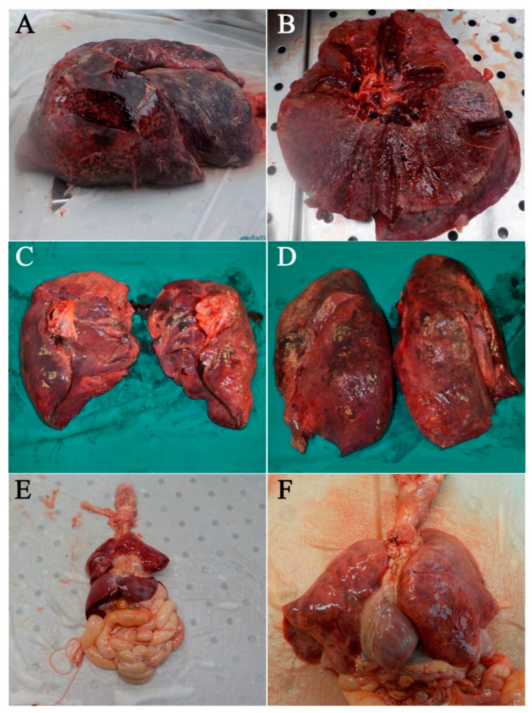
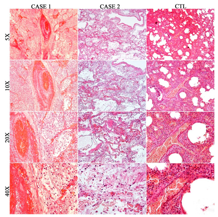
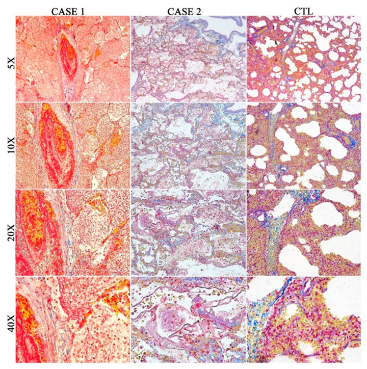
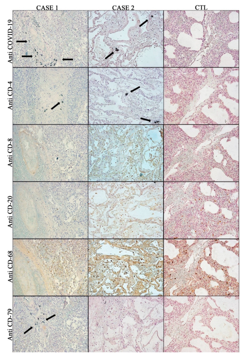
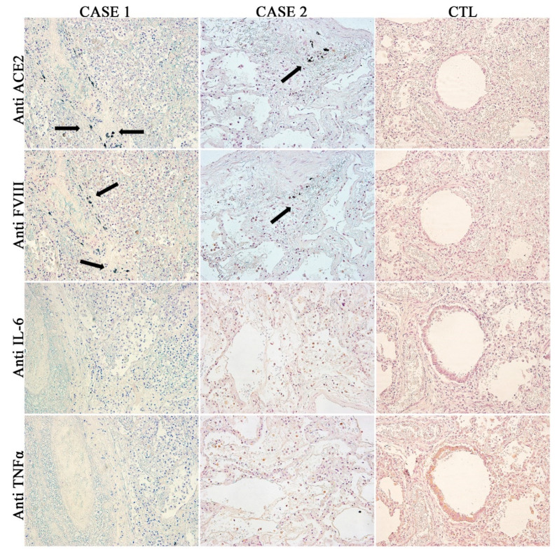
(1) Background: The current outbreak of COVID-19 infection is an ongoing challenge and a major threat to public health that requires surveillance, prompt diagnosis, as well as research efforts to understand the viral pathogenesis. Despite this, to date, very few studies have been performed concerning autoptic specimens. Therefore, this study aimed: (i) to reiterate the importance of the autoptic examination, the only method able to precisely define the cause of death; (ii) to provide a complete post-mortem histological and immunohistochemical investigation pattern capable of diagnosing death from COVID-19 infection. (2) Methods: In this paper, the lung examination of two subjects who died from COVID-19 are discussed, comparing the obtained data with those of the control, a newborn who died from pneumonia in the same pandemic period. (3) Results: The results of the present study suggest that COVID-19 infection can cause different forms of acute respiratory distress syndrome (ARDS), due to diffuse alveolar damage and diffuse endothelial damage. Nevertheless, different patterns of cellular and cytokine expression are associated with anti-COVID-19 antibody positivity, compared to the control case. Moreover, in both case studies, it is interesting to note that COVID-19, ACE2 and FVIII positivity was detected in the same fields. (4) Conclusions: COVID-19 infection has been initially classified as exclusively interstitial pneumonia with varying degrees of severity. Subsequently, vascular biomarkers showed that it can also be considered a vascular disease. The data on Factor VIII discussed in this paper, although preliminary and limited in number, seem to suggest that the thrombogenicity of Sars-CoV2 infection might be linked to widespread endothelial damage. In this way, it would be very important to investigate the pro-coagulative substrate both in all subjects who died and in COVID-19 survivors. This is because it may be hypothesized that the different patterns with which the pathology is expressed could depend on different individual susceptibility to infection or a different personal genetic-clinical background. In light of these findings, it would be important to perform more post-mortem investigations in order to clarify all aspects of the vascular hypothesis in the COVID-19 infection.
Keywords: COVID-19; COVID-19 diagnostic; autopsy; forensic pathology; forensic science; immunohistochemistry; post-mortem examination.
Conflict of interest statement
The authors declare no conflict of interest.
Figures
References
-
- Chan J.F.W., Yuan S., Kok K.H., To K.K.W., Chu H., Yang J., Xing F., Liu J., Yip C.C.Y., Poon R.W.S., et al. A familial cluster of pneumonia associated with the 2019 novel coronavirus indicating person-to-person transmission: A study of a family cluster. Lancet. 2020;395:514–523. doi: 10.1016/S0140-6736(20)30154-9. - DOI - PMC - PubMed
-
- Fineschi V., Aprile A., Aquila I., Arcangeli M., Asmundo A., Bacci M., Cingolani M., Cipolloni L., D’Errico S., De Casamassimi I., et al. Management of the corpse with suspect, probable or confirmed COVID-19 respiratory infection—Italian interim recommendations for personnel potentially exposed to material from corpses, including body fluids, in morgue structures, during autopsy practice. Pathol. J. Ital. Soc. Anat. Pathol. Diagn. Cytopathol. 2020;112:64–77. - PMC - PubMed
-
- WHO Interm Guidance Infection Prevention and Control for the safe management of a dead body in the context of COVID-19. J. Hosp. Infect. 2020;104:246–251. - PubMed
-
- Salthouse T.N. Histopathologic Technic and Practical Histochemistry. Bull. Soc. Pharmacol. Environ. Pathol. 1977;5:15. doi: 10.1177/019262337700500405. - DOI
LinkOut - more resources
Full Text Sources
Miscellaneous
