Review
. 2020 Aug 9;21(16):5707.
doi: 10.3390/ijms21165707.

SARS-CoV-2 Entry Inhibitors: Small Molecules and Peptides Targeting Virus or Host Cells

Affiliations
Review

SARS-CoV-2 Entry Inhibitors: Small Molecules and Peptides Targeting Virus or Host Cells

Rolando Cannalire et al. Int J Mol Sci. .

Abstract

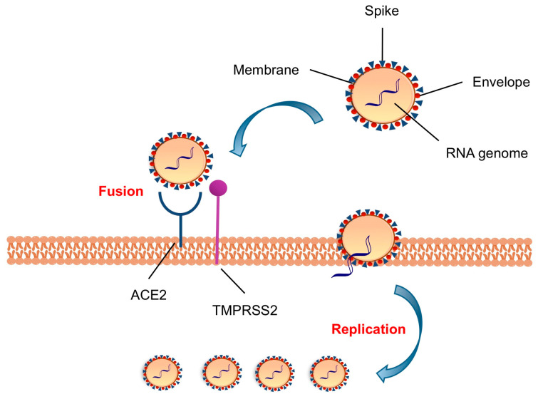
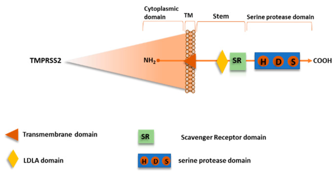
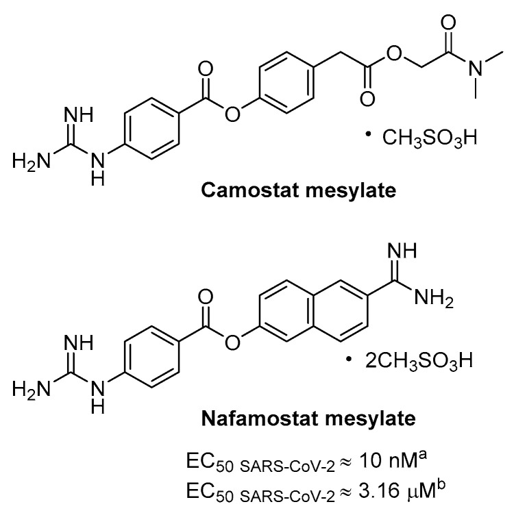
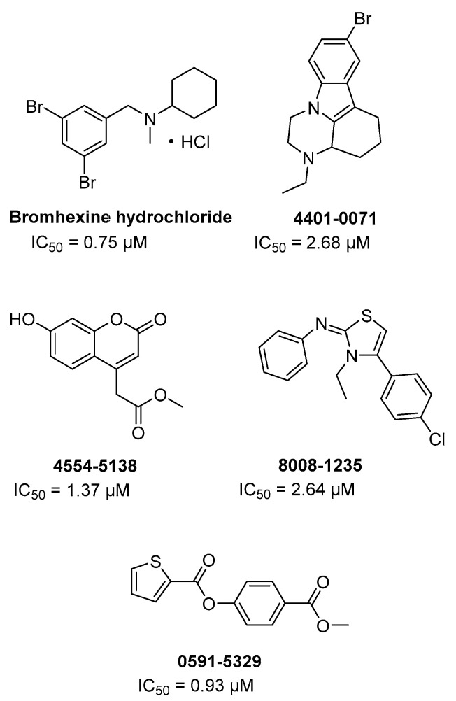
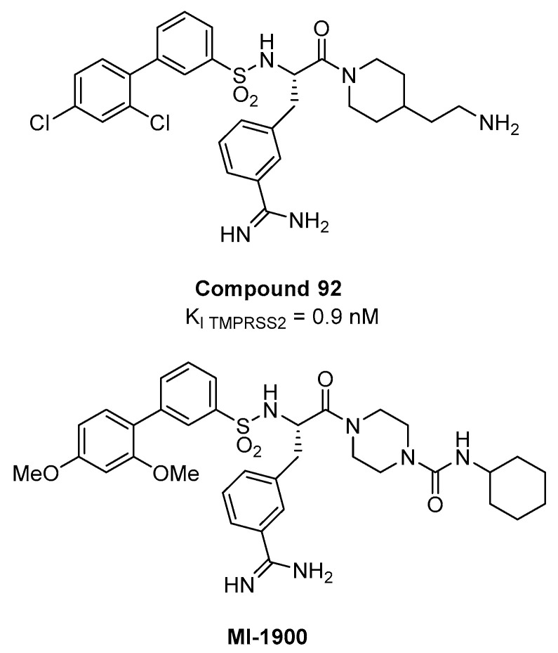
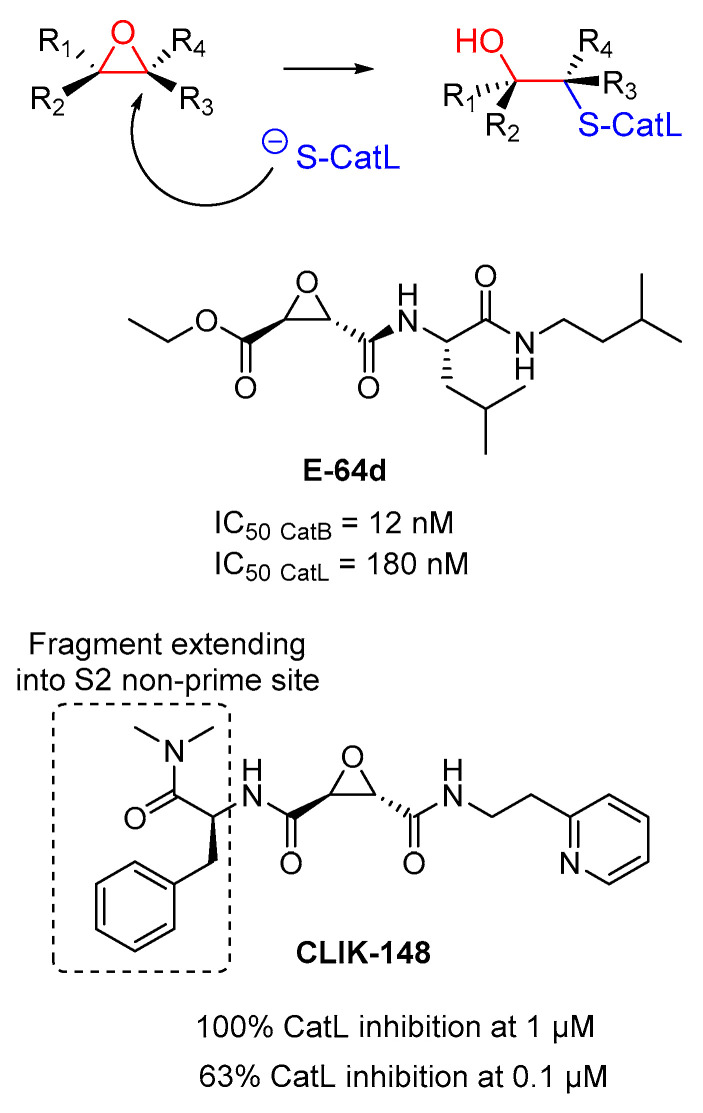
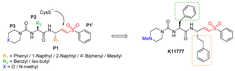
The pandemic evolution of SARS-CoV-2 infection is forcing the scientific community to unprecedented efforts to explore all possible approaches against COVID-19. In this context, targeting virus entry is a promising antiviral strategy for controlling viral infections. The main strategies pursued to inhibit the viral entry are considering both the virus and the host factors involved in the process. Primarily, direct-acting antivirals rely on inhibition of the interaction between ACE2 and the receptor binding domain (RBD) of the Spike (S) protein or targeting the more conserved heptad repeats (HRs), involved in the membrane fusion process. The inhibition of host TMPRSS2 and cathepsins B/L may represent a complementary strategy to be investigated. In this review, we discuss the development entry inhibitors targeting the S protein, as well as the most promising host targeting strategies involving TMPRSS2 and CatB/L, which have been exploited so far against CoVs and other related viruses.

Keywords: COVID-19; SARS-CoV-2 entry inhibitors; TMPRSS2; cathepsins; coronavirus; peptides inhibitors; small molecules inhibitors; spike.


Conflict of interest statement

The authors declare no conflict of interest

Figures

Figure 1
Figure 1
Schematic representation of SARS-CoV-2 virion and viral entry into host cell.
Figure 2
Figure 2
Receptor binding domain (RBD) “down” and “up” conformations of SARS-CoV-2. (A) Single protomer of SARS-CoV-2 with the RBD in the down conformation, displayed as cartoon (PDB ID: 6VSB). RBD is colored cyan, the N-terminal domain (NTD) is pale green, subdomains 1 and 2 (SD1 and SD2) are yellow, the S2 domain is white, with HR1 colored salmon and FP hotpink. (B) Single protomer of SARS-CoV-2 in the RBD up conformation next to (C) a protomer of SARS-CoV (colored gray) in the RBD up conformation (PDB: 6CRZ).
Figure 3
Figure 3
Structural details of the interface between SARS-CoV and SARS-CoV-2 RBDs and ACE2. (A) Structural overlay of SARS-CoV-2 (cyan, PDB ID: 6M0J) and SARS-CoV (light pink, PDB ID: 2AJF) RBDs bound to ACE2 (wheat), displayed as cartoon. The four disulfide bonds in SARS-CoV-2 RBD are shown as sticks. The region enclosed by the black dashed lines, encompassing the interface between RBD and ACE2 is illustrated in detail in panel (B). Overlay of the RBD interface residues of SARS-CoV-2 (cyan sticks) and SARS-CoV (light pink sticks). Q493 is shown in two alternate positions.
Figure 4
Figure 4
Comparisons of the interactions formed at the interfaces between SARS-CoV and SARS-CoV-2 RBDs and ACE2. Overall structure of (A) SARS-CoV (light pink, PDB ID: 2AJF) and (B) SARS-CoV-2 (cyan, PDB ID: 6M0J) RBDs bound to ACE2 (wheat). Key residues involved in SARS-CoV RBD/ACE2 (C) and SARS-CoV-2/ACE2 (D) complex formation are shown as sticks and labeled. ACE2 residues are underlined for clarity. Hydrogen bonds and salt bridges are displayed as dashed red lines.
Figure 5
Figure 5
Structure of 6-HB fusion core in SARS-CoV-2. (A) The HR1 domain of SARS-CoV-2 is depicted as electrostatic surface, with hydrophobic residues in white, basic in blue, and acidic in red. The HR2 domain is shown in cartoon representation, with the hydrophobic residues in the central fusion core region shown as sticks and labeled. (B) The superposition of 6-HB structures of SARS-CoV-2 (PDB ID: 6LXT) and SARS-CoV (PDB ID: 1WYY), shown as ribbon. The HR1 and HR2 domains are colored salmon and slate for SARS-CoV-2, raspberry, and deep teal for SARS-CoV, respectively.
Figure 6
Figure 6
Comparisons of 6-HB fusion core in SARS-CoV-2 and SARS-CoV. (A) Sequence alignment between SARS-CoV-2 and SARS-CoV S proteins for the HR1 and HR2 regions, indicated with boxes colored salmon and raspberry for HR1 and slate and deepteal for HR2, respectively. Top view of the 6-HB fusion core structure of (B) SARS-CoV-2 (PDB ID: 6LXT) and (C) SARS-CoV (PDB ID: 1WYY) displayed as cartoon. The HR1 and HR2 domains are colored as in (A) and labeled. Side view of 6-HB of (D) SARS-CoV-2 and (E) SARS-CoV. A zoomed view of the interactions between HR1 and HR2, mediating fusion core formation, is shown on the right side. Key residues are displayed as sticks and labeled; hydrogen bonds and salt bridges are displayed as dashed red lines.
Figure 7
Figure 7
The interactions of the broad-spectrum peptide inhibitor EK1 with HR1 residues of different HCoVs. EK1 and HR1 residues connected with dashed gray lines locate to the same layers on the 3HR1 triple helix. Burying EK1 residues are highlighted in yellow, and ridge-packing EK1 residues are highlighted in green. HR1 residues that mediate assembly of the 3HR1 cores are highlighted in yellow, while those involved in ridge packing are highlighted in green. HR1 residues forming conserved side chain-to-side chain and side chain-to-main chain hydrophilic interactions with EK1 residues are indicated with boxes colored blue and purple, respectively. Adapted from ref. [46].
Figure 8
Figure 8
Amino acids sequences of EK-1 and its lipopeptide derivatives.
Figure 9
Figure 9
SARS-CoV-2 HR2-derived peptides.
Figure 10
Figure 10
Schematic representation of serine protease TMPRSS2.
Figure 11
Figure 11
Chemical structures of phenyl-4-guanidinobenzoate derivatives known drugs as serine protease and antiviral activity of nafamostat agaisnt SARS-CoV-2 in Calu-3 cells measured by CPE; a pre-treatment; b added after virus inoculation.
Figure 12
Figure 12
Chemical structures of Bromhexine hydrochloride and additional four hits identified by a biochemical HTS as TMPRSS2 inhibitors, with their inhibitory activity.
Figure 13
Figure 13
Substrate analogues containing 3-amidinophenylalanine as P1 residue as TMPRSS2 inhibitors compound 92 and MI-1900 showing antiviral activity against SARS-CoV-2.
Figure 14
Figure 14
Chemical structures of Benzoselenoxanthene analogues, TMPRSS2 G-quadruplex stabilizers.
Figure 15
Figure 15
Structures and inhibitory activities of epoxysuccinates E-64d and CLIK-148 against human cathepins.
Figure 16
Figure 16
Peptidyl aryl vinylsulfone SAR (left) and the best compound K11777 as irreversible inhibitors of cathepsins and SARS-CoV replication in Vero 76 cells.
Figure 17
Figure 17
Chemical structure and biological activity of aldehyde MDL28170 as potent covalent reversible inhibitor of CatL and anti-CoV agents; a virus yield reduction assay (biological data from Ref. [108]).
Figure 18
Figure 18
Structures and biological activities of tetrahydroquinoline oxocarbaze CID 23631927 and its analog CID 16725315 covalent reversible inhibitors of human CatL and anti-SARS-CoV-2 agents; a Preincubation for 4 h; b Cathepsin L/B selectivity ratio.

References

    1. WHO . Ten Threats to Global Health in 2019. WHO; Geneva, Switzerland: 2019. [(accessed on 8 June 2020)]. Available online: https://www.who.int/emergencies/ten-threats-to-global-health-in-2019.
    1. WHO . Prioritizing Diseases for Research and Development in Emergency Contexts. WHO; Geneva, Switzerland: 2020. [(accessed on 8 June 2020)]. Available online: Https://www.who.int/activities/prioritizing-diseases-for-research-anddev....
    1. Wu F., Zhao S., Yu B., Chen Y.M., Wang W., Song Z.G., Hu Y., Tao Z.W., Tian J.H., Pei Y.Y., et al. A new coronavirus associated with human respiratory disease in China. Nature. 2020;579:265–269. doi: 10.1038/s41586-020-2008-3. - DOI - PMC - PubMed
    1. Zhu N., Zhang D., Wang W., Li X., Yang B., Song J., Zhao X., Huang B., Shi W., Lu R., et al. A novel coronavirus from patients with pneumonia in China, 2019. N. Engl. J. Med. 2020;382:727–733. doi: 10.1056/NEJMoa2001017. - DOI - PMC - PubMed
    1. Rolling Updates on Coronavirus Disease (COVID-19) [(accessed on 8 June 2020)]; Available online: Https://www.who.int/emergencies/diseases/novel-coronavirus-2019/events-a....

MeSH terms

Substances

Grants and funding

LinkOut - more resources