. 2020 Aug 9;21(16):5709.
doi: 10.3390/ijms21165709.

The Evolution of Duplicated Genes of the Cpi-17/Phi-1 (ppp1r14) Family of Protein Phosphatase 1 Inhibitors in Teleosts

Affiliations

The Evolution of Duplicated Genes of the Cpi-17/Phi-1 (ppp1r14) Family of Protein Phosphatase 1 Inhibitors in Teleosts

Irene Lang et al. Int J Mol Sci. .

Abstract

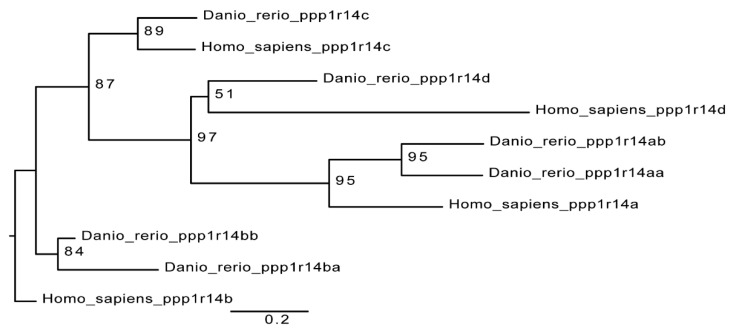
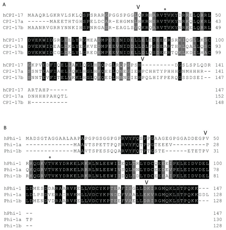
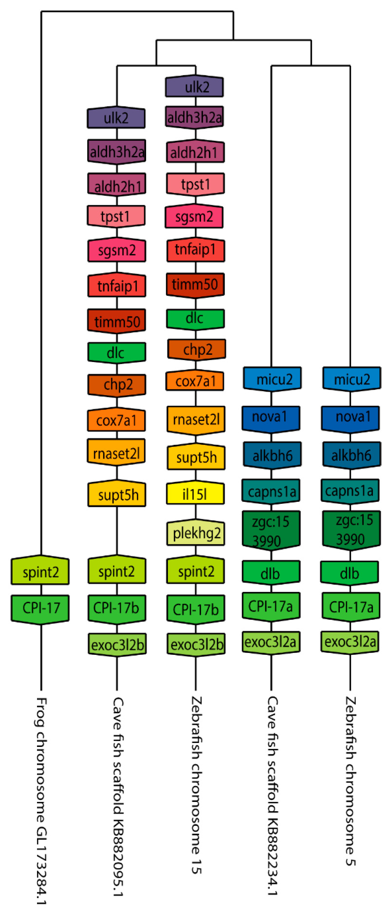
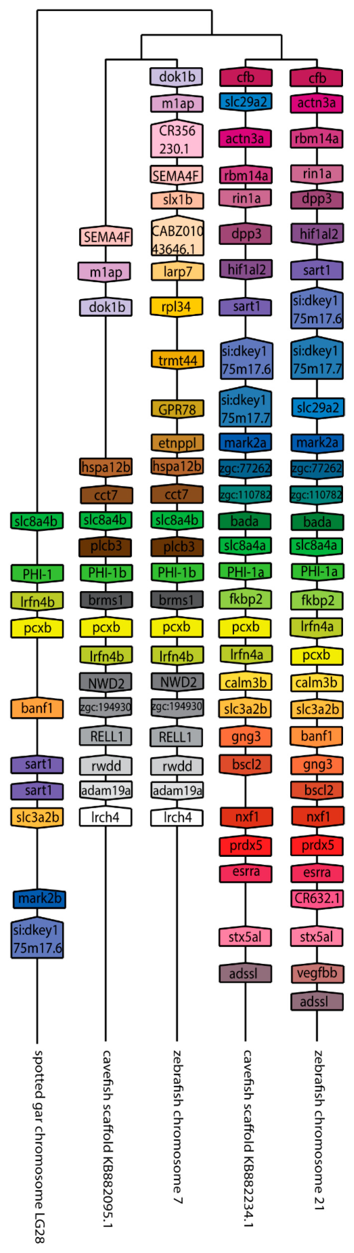
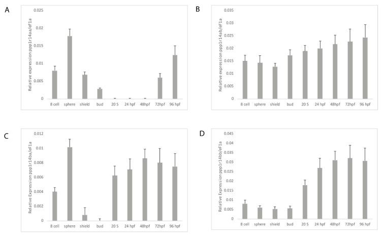
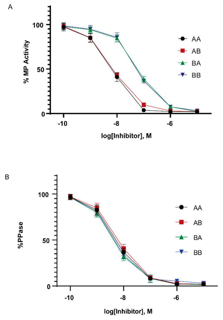
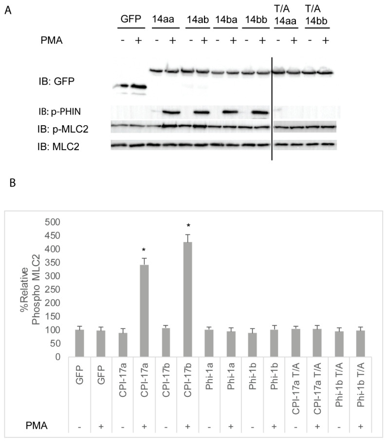
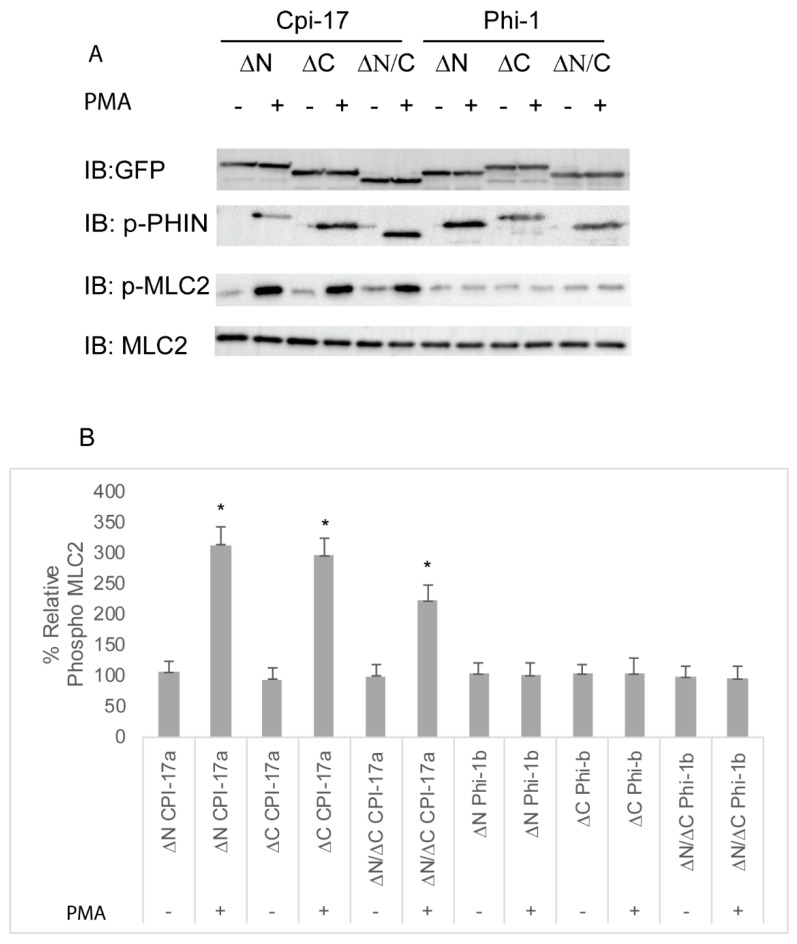
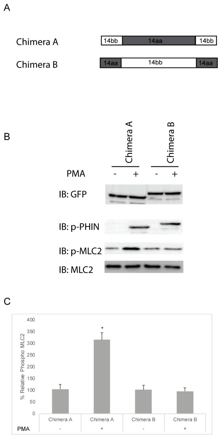
The Cpi-17 (ppp1r14) gene family is an evolutionarily conserved, vertebrate specific group of protein phosphatase 1 (PP1) inhibitors. When phosphorylated, Cpi-17 is a potent inhibitor of myosin phosphatase (MP), a holoenzyme complex of the regulatory subunit Mypt1 and the catalytic subunit PP1. Myosin phosphatase dephosphorylates the regulatory myosin light chain (Mlc2) and promotes actomyosin relaxation, which in turn, regulates numerous cellular processes including smooth muscle contraction, cytokinesis, cell motility, and tumor cell invasion. We analyzed zebrafish homologs of the Cpi-17 family, to better understand the mechanisms of myosin phosphatase regulation. We found single homologs of both Kepi (ppp1r14c) and Gbpi (ppp1r14d) in silico, but we detected no expression of these genes during early embryonic development. Cpi-17 (ppp1r14a) and Phi-1 (ppp1r14b) each had two duplicate paralogs, (ppp1r14aa and ppp1r14ab) and (ppp1r14ba and ppp1r14bb), which were each expressed during early development. The spatial expression pattern of these genes has diverged, with ppp1r14aa and ppp1r14bb expressed primarily in smooth muscle and skeletal muscle, respectively, while ppp1r14ab and ppp1r14ba are primarily expressed in neural tissue. We observed that, in in vitro and heterologous cellular systems, the Cpi-17 paralogs both acted as potent myosin phosphatase inhibitors, and were indistinguishable from one another. In contrast, the two Phi-1 paralogs displayed weak myosin phosphatase inhibitory activity in vitro, and did not alter myosin phosphorylation in cells. Through deletion and chimeric analysis, we identified that the difference in specificity for myosin phosphatase between Cpi-17 and Phi-1 was encoded by the highly conserved PHIN (phosphatase holoenzyme inhibitory) domain, and not the more divergent N- and C- termini. We also showed that either Cpi-17 paralog can rescue the knockdown phenotype, but neither Phi-1 paralog could do so. Thus, we provide new evidence about the biochemical and developmental distinctions of the zebrafish Cpi-17 protein family.

Keywords: Cpi-17 (ppp1r14a); Danio rerio; Mypt1; PP1; Phi-1 (ppp1r14b); genome duplication.


Conflict of interest statement

The authors declare no conflict of interest. The funders had no role in the design of the study; in the collection, analyses, or interpretation of data; in the writing of the manuscript, or in the decision to publish the results.

Figures

Figure 1
Figure 1
Cpi-17 gene family in zebrafish. The Cpi-17 family in humans consists of ppp1r14a, ppp1r14b, ppp1r14c, and ppp1r14d. The Cpi-17 family in zebrafish consists of two paralogs of both ppp1r14a and ppp1r14b, and a single copy of ppp1r14c and ppp1r14d. Ppp1r14c and ppp1r14d were not expressed in zebrafish early embryonic development.
Figure 2
Figure 2
Protein sequences of zebrafish Cpi-17 and Phi-1 paralogs aligned to their human orthologs. (A). Protein alignment of zebrafish Cpi-17a and Cpi-17b with human Cpi-17. (B). Protein alignments of zebrafish Phi-1a and Phi-1b with human Phi-1. The conserved 86-residue long conserved PHIN domain is marked with flanking V, with inhibitory phosphorylation sites T31 (Cpi-17a), T37 (Cpi-17b), T35 (Phi-1a), and T38 (Phi-1b) indicated by *.
Figure 3
Figure 3
The genomic organization of the Cpi-17 (ppp1r14a) paralogs in zebrafish. Genomicus was used to analyze Cpi-17 (ppp1r14a) synteny between Danio rerio (Zebrafish), and the most closely related sequenced relative, Astyanax mexicanus (Cavefish). Xenopus laevis (African clawed frog) was used as an outgroup. Homologous genes are shown with matching coloration, and the gene direction on the chromosome is indicated by the direction of the boxed arrow. Species and chromosome numbers are noted.
Figure 4
Figure 4
The genomic organization of the ppp1r14b paralogs in zebrafish. Genomicus was used to analyze ppp1r14b synteny between Danio rerio (zebrafish), and the most closely related sequenced relative, Astyanax mexicanus (cavefish). Lepisosteus oculatus (spotted gar) was used as an outgroup. Homologous genes are shown with matching coloration, and the gene direction on the chromosome is indicated by the direction of the boxed arrow. Species and chromosome numbers are noted.
Figure 5
Figure 5
Expression of ppp1r14a and ppp1r14b paralogs during early embryonic development in zebrafish. Embryos were collected shortly after fertilization (8 cell), at sphere stage (4 hpf), shield stage (6 hpf), bud stage (10 hpf), 20 somite stage, Prim-5 stage (24 hpf), long-pec stage (48 hpf), protruding-month stage (72 hpf), and day 4 (96 hpf). Amplification of eF1a and total RNA without addition of reverse transcriptase were used as positive and negative controls, respectively. (A) Gene specific primers were used to detect ppp1r14aa (A), ppp1r14ab (B), ppp1r14ba (C), or ppp1r14bb (D). Values are means of three biological replicates performed in duplicate. Error bars represent standard deviation. All values are reported as expression relative to ef1a.
Figure 6
Figure 6
In vitro inhibition of myosin phosphatase by Cpi-17 and Phi-1 paralogs. (A) The myosin phosphatase holoenzyme was isolated from HEK cells and used to dephosphorylate recombinant Mlc2. Increasing concentrations of thiophosphorylated ppp1r14aa (AA, circle), ppp1r14ab (AB, red square), ppp1r14ba (BA, green triangle), or ppp1r14bb (BB, blue triangle) proteins were added, and the extent of Mlc2 dephosphorylation is reported as percent phosphatase activity. (B) Isolated PP1catalytic subunit was used to dephosphorylate Mlc2. Increasing concentrations of thiophosphorylated ppp1r14aa (AA, circle), ppp1r14ab (AB, red square), ppp1r14ba (BA, green triangle), or ppp1r14bb (BB, blue triangle) proteins were added, and the extent of Mlc2 dephosphorylation is reported as percent phosphatase activity. Each experiment was repeated three times in duplicate, and error bars indicate standard error.
Figure 7
Figure 7
Differential inhibition of the myosin phosphatase by Cpi-17 family members in HeLa cells. (A) HeLa cells were transfected with either GFP alone, ppp1r14aa, ppp1r14ab, ppp1r14ba, ppp1r14bb, mutant ppp1r14aa lacking the regulatory phosphorylation site, or mutant ppp1r14bb lacking the regulatory phosphorylation site. Moreover, 15 h post transfection cells were treated with either 1 µM PMA or 0.1% DMSO for 3 h, and lysates were blotted with anti-GFP, anti-phospho-PHIN, anti-Mlc2, and anti phospho-Mlc2. (B) Mlc2 phosphorylation was normalized to total Mlc2 content, and is shown as fold change compared with cells expressing the empty GFP vector. Error bars indicate standard error, and an * indicates a statistically significant difference from control. Statistical significance was calculated using a one-factor ANOVA with Holm-Šídák post-hoc analysis, and is defined as p < 0.05 from at least 4 biological replicates.
Figure 8
Figure 8
Truncation mutants of Cpi-17 family members highlight the critical role of the PHIN domain in myosin phosphatase inhibition. (A) HeLa cells were transfected with either ppp1r14aa lacking the N-terminus (ΔN), ppp1r14aa lacking the C terminus (ΔC), ppp1r14aa lacking both the N and C termini (ΔN/ΔC), ppp1r14bb lacking the N-terminus (ΔN), ppp1r14bb lacking the C-terminus (ΔC), or ppp1r14bb lacking both the N- and C-termini (ΔN/ΔC). Notably, 15 h post transfection cells were treated with either 1 µM PMA or 0.1% DMSO for 3 h, and lysates were blotted with anti-GFP, anti-phospho-PHN, anti-Mlc2, or anti phospho-Mlc2. (B) Mlc2 phosphorylation was normalized to total Mlc2 content, and is shown as fold change compared with cells expressing the empty GFP-vector. Error bars indicate standard error, and an * indicates a statistically significant difference from control. Statistical significance was calculated using a one-factor ANOVA with Holm–Šídák post-hoc analysis and is defined as p < 0.05 from at least four biological replicates.
Figure 9
Figure 9
Cpi-17/Phi-1 chimeric proteins indicate that the PHIN domain is required for Myosin Phosphatase specificity. (A,B) HeLa cells were transfected with either chimera A, which contains the PHIN domain of ppp1r14aa and the N and C-termini of ppp1r14bb, or chimera B, which contains the PHIN domain of ppp1r14bb and the N and C-termini of ppp1r14aa. Notably, 15 h post transfection cells were treated with either 1 µM PMA or 0.1% DMSO for 3 h, and lysates were blotted with anti-GFP, anti-phospho-PHIN, anti-Mlc2, and anti phospho-Mlc2. (C) Mlc2 phosphorylation was normalized to total Mlc2 content and is shown as fold change compared with cells expressing the empty GFP-vector. Error bars indicate standard error, and an * indicates a statistically significant difference from control. Statistical significance was calculated using a one-factor ANOVA with Holm–Šídák post-hoc analysis and is defined as p < 0.05 from at least four biological replicates.
Figure 10
Figure 10
Expression of Cpi-17 paralogs, but not Phi-1 paralogs, can rescue ppp1r14ab loss-of-function. Embryos at bud stage stained with hgg1 (prechordal plate), shh (midline), pax2.1 (midbrain-hindbrain boundary), and dlx3 (neural plate) after injection of 200 pg GFP (A,B) or 200 pg ppp1r14aa mRNA (C,D). (EM) embryos were collected at 10.5 hpf and stained with fgf3 after injection of 4ng of control MO (E), 4 ng of ppp1r14ab MO (F), 50pg of ppp1r14aa mRNA + 4 ng of ppp1r14ab MO (G), 50pg of ppp1r14ab mRNA + 4 ng of ppp1r14ab MO (H), 50pg of ppp1r14bb mRNA + 4 ng of ppp1r14ab MO (I), 50pg of ppp1r14aa mRNA + 4 ng of ppp1r14ab MO (J), 50pg of ppp1r14aa T38A mRNA + 4 ng of ppp1r14ab MO (K), 50 pg chimera A mRNA + 4 ng of ppp1r14ab MO (L), or 50ng chimera B mRNA 4 ng of ppp1r14ab MO (M).

References

    1. Ravi V., Venkatesh B. The Divergent Genomes of Teleosts. Annu. Rev. Anim. Biosci. 2018 doi: 10.1146/annurev-animal-030117-014821. - DOI - PubMed
    1. Hoegg S., Brinkmann H., Taylor J.S., Meyer A. Phylogenetic timing of the fish-specific genome duplication correlates with the diversification of teleost fish. J. Mol. Evol. 2004 doi: 10.1007/s00239-004-2613-z. - DOI - PubMed
    1. Amores A., Catchen J., Ferrara A., Fontenot Q., Postlethwait J.H. Genome evolution and meiotic maps by massively parallel DNA sequencing: Spotted gar, an outgroup for the teleost genome duplication. Genetics. 2011 doi: 10.1534/genetics.111.127324. - DOI - PMC - PubMed
    1. Meyer A., Schartl M. Gene and genome duplications in vertebrates: The one-to-four (-to-eight in fish) rule and the evolution of novel gene functions. Curr. Opin. Cell Biol. 1999;11:699–704. doi: 10.1016/S0955-0674(99)00039-3. - DOI - PubMed
    1. Meyer A., Van De Peer Y. From 2R to 3R: Evidence for a fish-specific genome duplication (FSGD) BioEssays. 2005;27:937–945. doi: 10.1002/bies.20293. - DOI - PubMed

MeSH terms

LinkOut - more resources