. 2020 Aug 10;9(8):726.
doi: 10.3390/antiox9080726.

In Vitro Antioxidant, Antiinflammation, and Anticancer Activities and Anthraquinone Content from Rumex crispus Root Extract and Fractions

Affiliations

In Vitro Antioxidant, Antiinflammation, and Anticancer Activities and Anthraquinone Content from Rumex crispus Root Extract and Fractions

Taekil Eom et al. Antioxidants (Basel). .

Abstract

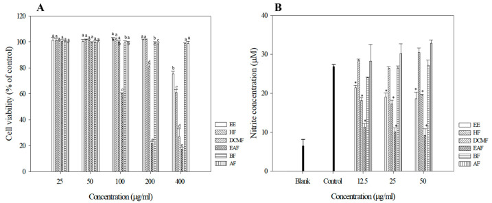
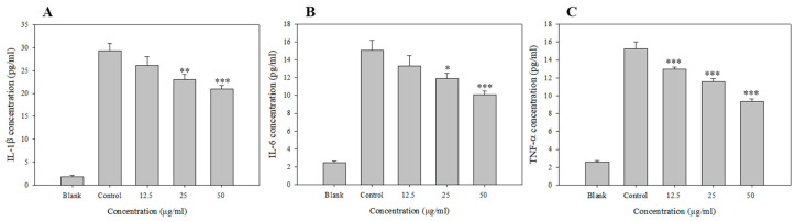
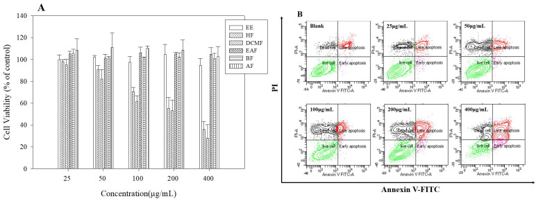
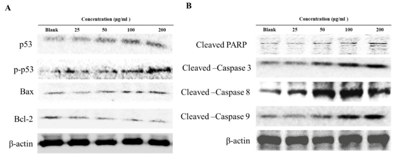
Rumex crispus is a perennial plant that grows in humid environments across Korea. Its roots are used in traditional Korean medicine to treat several diseases, including diseases of the spleen and skin and several inflammatory pathologies. In this study, different solvent fractions (n-hexane, dichloromethane, ethyl acetate, n-butanol, and aqueous fractions) from an ethanol extract of R. crispus roots were evaluated for the presence and composition of anthraquinone compounds and antioxidants by checking for such things as free radical scavenging activity, and electron and proton atom donating ability. In addition, anti-inflammatory activity was measured by NO scavenging activity and inflammatory cytokine production; furthermore, anti-cancer activity was measured by apoptosis-inducing ability. Polyphenolic and flavonoid compounds were shown to be abundant in the dichloromethane and ethyl acetate fractions, which also exhibited strong antioxidant activity, including free radical scavenging and positive results in FRAP, TEAC, and ORAC assays. HPLC analysis revealed that the dichloromethane fractions had higher anthraquinone contents than the other fractions; the major anthraquinone compounds included chrysophanol, emodin, and physcione. In addition, results of the anti-inflammatory assays showed that the ethyl acetate fraction showed appreciable reductions in the levels of nitric oxide and inflammatory cytokines (TNF-α, IL-1β, and IL-6) in Raw 264.7 cells. Furthermore, the anthraquinone-rich dichloromethane fraction displayed the highest anticancer activity when evaluated in a human hepatoma cancer cell line (HepG2), in which it induced increased apoptosis mediated by p53 and caspase activation.

Keywords: Rumex crispus; anthraquinone; apoptosis; free radical scavenging; inflammatory cytokines.


Conflict of interest statement

The authors have no conflicts of interest to declare.

Figures

Figure 1
Figure 1
Cell viabilities of R crispus extracts and solvent fractions. (A) NO production (B) in Raw 264.7 cells. RAW 264.7 cells were treated with various concentrations (25, 50, 100, 200, 400 μg/mL) of R. crispus extracts and fractions for 24 h. Cell viability was measured by MTT assay. RAW 264.7 cells were pre-incubated with 12.5, 25, and 50 μg/mL of extracts and fractions for 1 h and then treated with 1 μg/mL of LPS for 24 h. The NO production was measured by the Griess reagent system. Data are represented as means ± SEMs. The different superscripts are significantly different at p < 0.05. * Statistical significance of the difference between LPS and LPS + sample treatment groups: * p < 0.05.
Figure 2
Figure 2
Inhibition of LPS induced IL-1b, IL-6 and TNF-a in the EAF. RAW 264.7 cells were preincubated with 12.5 or 25 μg/mL of EAF for 1 h and then treated with 1 μg/mL of LPS for 24 h. The IL-1β, IL-6, and TNF-α production was measured by ELISA, as described in Materials and Methods. Data are represented as means ± SEMs. * Statistical significance of the difference between LPS and LPS + sample treatment groups: * p < 0.05, ** p < 0.01, *** p < 0.001.
Figure 3
Figure 3
Cell viability of R crispus extracts and solvent fractions. (A) apoptosis induced (B) in HepG2 cells. HepG2 cells were treated with various concentrations (25, 50, 100, 200, 400 μg/mL) of R. crispus extracts and fractions for 24 h. Cell viability was measured by MTT assay. Flow cytometry analysis of apoptosis after exposure to various concentrations (25, 50, 100, 200, 400 μg/mL) of DCMF for 24 h, using annexin V-FITC/PI. The lower right indicates the percentage of early apoptotic cells; the upper right indicates the percentage of necrotic and late apoptotic cells.
Figure 4
Figure 4
Effects of DCMF on the Bcl-2 family and p53 (A) and caspase family (B) protein expression in HepG2 cells. HepG2 were treated with the indicated concentrations of DCMF for 24 h. The equal amounts of cellular proteins were probed with the indicated antibodies, and the proteins were visualized using an ECL detection system. Actin was used as an internal control.

References

    1. Finkel T. Signal transduction by reactive oxygen species. J. Cell Biol. 2011;194:7–15. doi: 10.1083/jcb.201102095. - DOI - PMC - PubMed
    1. Valko M., Leibfritz D., Moncol J., Cronin M.T., Mazur M., Telser J. Free radicals and antioxidants in normal physiological functions and human disease. Int. J. Biochem. Cell Biol. 2007;39:44–84. doi: 10.1016/j.biocel.2006.07.001. - DOI - PubMed
    1. Morgan M.J., Liu Z.G. Crosstalk of reactive oxygen species and NF-kappaB signaling. Cell Res. 2011;21:103–115. doi: 10.1038/cr.2010.178. - DOI - PMC - PubMed
    1. Mittal M., Siddiqui M.R., Tran K., Reddy S.P., Malik A.B. Reactive oxygen species in inflammation and tissue injury. Antioxid. Redox Signal. 2014;20:1126–1167. doi: 10.1089/ars.2012.5149. - DOI - PMC - PubMed
    1. Grivennikov S.I., Greten F.R., Karin M. Immunity, inflammation, and cancer. Cell. 2010;140:883–899. doi: 10.1016/j.cell.2010.01.025. - DOI - PMC - PubMed