. 2020 Aug 12;15(8):e0237300.
doi: 10.1371/journal.pone.0237300. eCollection 2020.

Structural insight into the role of novel SARS-CoV-2 E protein: A potential target for vaccine development and other therapeutic strategies

Affiliations

Structural insight into the role of novel SARS-CoV-2 E protein: A potential target for vaccine development and other therapeutic strategies

Manish Sarkar et al. PLoS One. .

Abstract

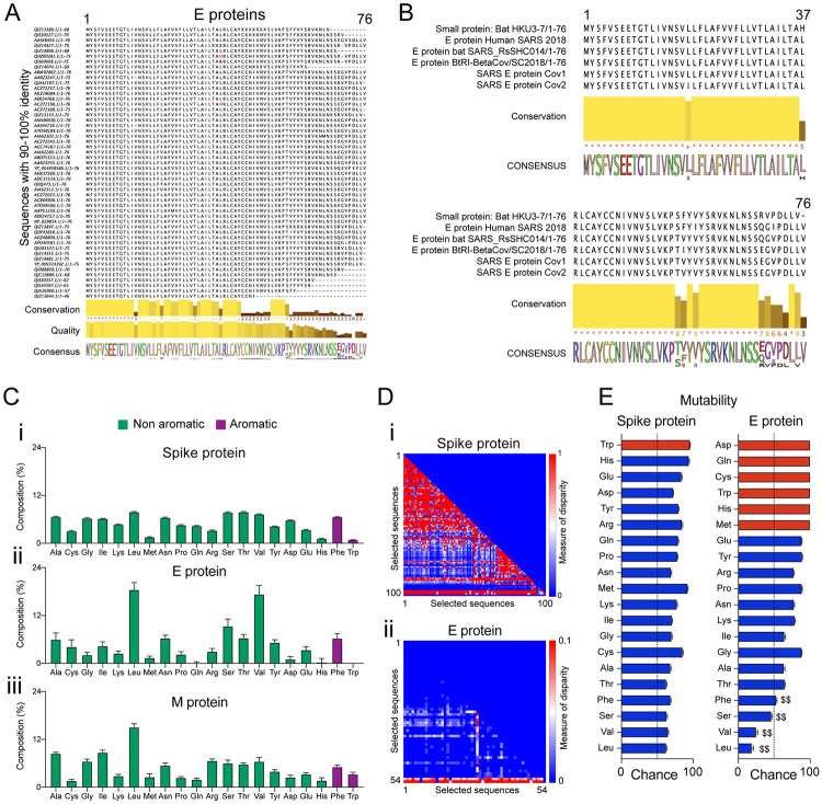
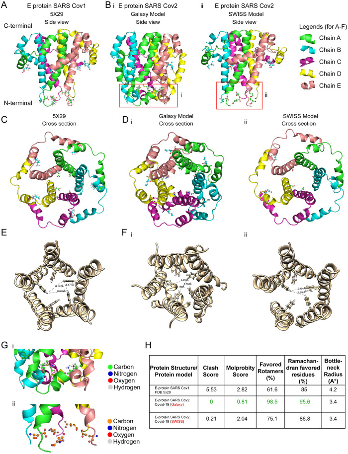
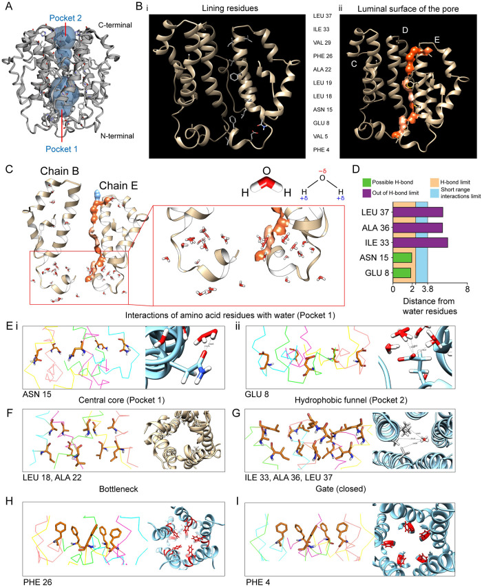
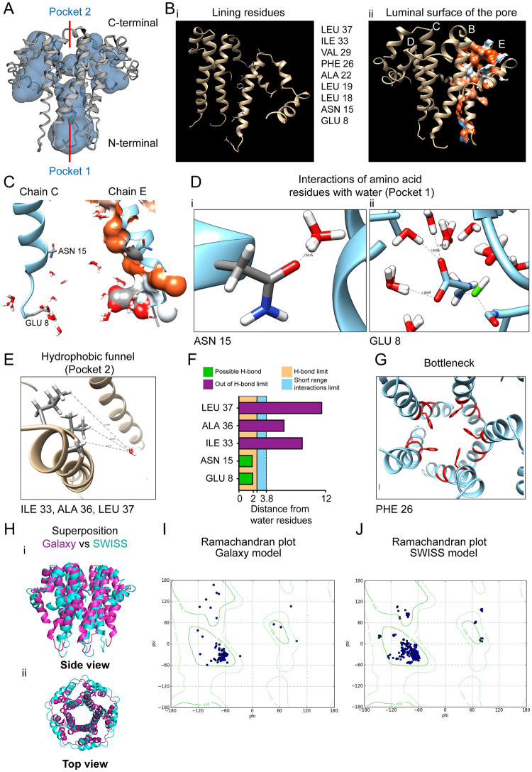
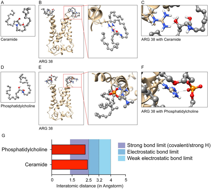
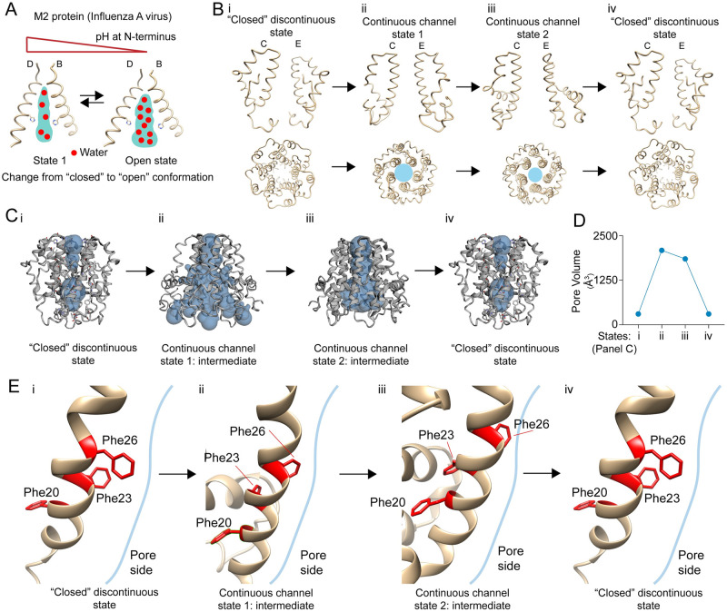
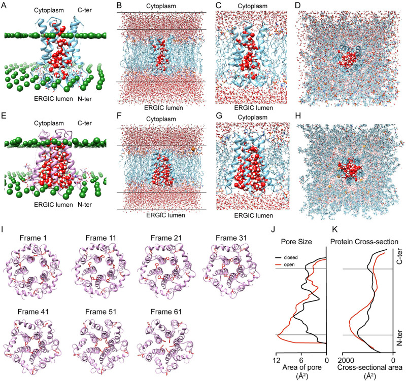
The outbreak of COVID-19 across the world has posed unprecedented and global challenges on multiple fronts. Most of the vaccine and drug development has focused on the spike proteins and viral RNA-polymerases and main protease for viral replication. Using the bioinformatics and structural modelling approach, we modelled the structure of the envelope (E)-protein of novel SARS-CoV-2. The E-protein of this virus shares sequence similarity with that of SARS- CoV-1, and is highly conserved in the N-terminus regions. Incidentally, compared to spike proteins, E proteins demonstrate lower disparity and mutability among the isolated sequences. Using homology modelling, we found that the most favorable structure could function as a gated ion channel conducting H+ ions. Combining pocket estimation and docking with water, we determined that GLU 8 and ASN 15 in the N-terminal region were in close proximity to form H-bonds which was further validated by insertion of the E protein in an ERGIC-mimic membrane. Additionally, two distinct "core" structures were visible, the hydrophobic core and the central core, which may regulate the opening/closing of the channel. We propose this as a mechanism of viral ion channeling activity which plays a critical role in viral infection and pathogenesis. In addition, it provides a structural basis and additional avenues for vaccine development and generating therapeutic interventions against the virus.


Conflict of interest statement

The authors have declared that no competing interests exist.

Figures

Fig 1
Fig 1. Sequence alignment of SARS CoV E proteins, disparity index and mutability.
(A) Sequence alignment of CoV E-proteins performed by Clustal Omega after a BLASTp search against human SARS-CoV-2 E protein (Accession number: QII57162.1). The selected aligned sequences were based on the criteria of 90–100% similarity. The conserved sequences, quality of sequence alignment, and consensus sequence are shown below. (B) Sequence alignment of CoV E-proteins from the bat (HKU3-7), human SARS 2018, bat SARS_RsSHC014, BtRI-BetaCoV, SARS-CoV-1 and SARS-CoV-2 showing highly conserved regions in the N-terminal region. The consensus sequence is shown below. (C) Percentage composition of each amino acid computed from the aligned selected sequences for (i) Spike protein, (ii) E-protein, and (iii) M-protein. (D) Lower triangular heatmap representation of the disparity index computed from the sequence alignment for (i) spike protein, and (ii) E-protein. Scale: 0–1 (for i), and 0–0.1 (for ii). (E) Mutability (probability of amino acid change) calculated for spike protein and E-protein. The dotted line indicates a 50% probability chance, while red bars indicate amino acids absent/single-site presence on the sequences analyzed. $$ indicates residues with lower mutability in E-protein compared to spike protein.
Fig 2
Fig 2. Pentameric homology model of the E protein of SARS-CoV-2.
(A) NMR structure of pentameric E protein (PDB id: 5X29) of SARS-CoV-1. (B) Pentameric model of E protein of SARS-CoV-2 (COVID-19) (i) generated in GALAXY (E-put), and (ii) generated in SWISS-MODEL. A magnified figure for each is provided in (red box). (C) Top view of pentameric E protein SARS-CoV-1 (5X29). (D) Top view of pentameric E protein SARS-CoV-2 (i) generated in GALAXY (E-put), and (ii) generated in SWISS-MODEL. (E) Bottleneck radius measurement of C (5X29). (F) Bottleneck radii measurement of D (i) GALAXY (E-put) and (ii) SWISS-MODEL. (G) N-terminal residues of (i) GALAXY model (E-put) (as in B-i) and (ii) SWISS-MODEL model (as in B-ii). (H) Structure validation parameters and pore radii of 5X29, GALAXY model (E-put), and SWISS-MODEL model.
Fig 3
Fig 3. Pore volume estimation of E put protein by GALAXY-WEB and docking with water molecules.
(A) Estimation of the pore volume of the GALAXY-WEB E-put protein using the CASTp 3.0 server. (B) (i) Residues lining the luminal side of the E-put protein core and (ii) the luminal surface of the pore determined by the CASTp 3.0 server. (C) Docking of water to the E-put protein by SWISS-DOCK showing the chains B and E of the pentamer and magnification of the region of interaction. (D) Distances of water molecules from different lining residues and the limit of H-bond. (E) Residues’ orientation of the E-put protein generated from PYMOL and interaction of water with residues showing the distance between water and (i) ASN 15 and (ii) GLU 8 by CHIMERA. (F) Residues’ orientation generated from PYMOL (side view) and CHIMERA (top view) lining the Central core of the E-put protein. (G) Residues’ orientation generated from PYMOL and interaction of water with the lining residues of the Hydrophobic funnel of the E-put protein by CHIMERA. (H) Residues’ orientation generated from PYMOL (side view) and CHIMERA (top view) showing the Gate of the E-put protein in the closed conformation. (I) Residues’ orientation generated from PYMOL (side view) and CHIMERA (top view) forming the bottleneck of the E-put protein.
Fig 4
Fig 4. Pore volume estimation of E put protein by SWISS MODEL and docking with water molecules.
(A) Estimation of the pore volume of the SWISS-MODEL E-put protein using the CASTp 3.0. (B) (i) Residues lining the luminal side of the SWISS-MODEL protein core and (ii) the luminal surface of the pore determined by the CASTp 3.0 server. (C) Docking of water to the SWISS-MODEL protein by SWISS-DOCK showing the chains C and E of the pentamer. (D) Residues’ interaction of water with residues showing the distance between water and (i) ASN 15 and (ii) GLU 8 by CHIMERA. (E) Residues’ interaction of water with the lining residues of the hydrophobic funnel of the SWISS-MODEL protein by CHIMERA. (F) Distances of water molecules from different lining residues and the limit of H-bond. (G) Residues’ orientation in CHIMERA forming the bottleneck of the SWISS-MODEL protein. (H) Superposition of the E-put and the SWISS-MODEL protein (purple: E-put; cyan: SWISS-MODEL) as (i) side view and (ii) top view. (I) Ramachandran plot of the E-put protein (Galaxy model). (J) Ramachandran plot of the SWISS-MODEL protein.
Fig 5
Fig 5. E-put protein interacts with the lipid molecule components of ERGIC membrane.
(A) Ceramide: Sphingolipid generated in OpenBabel and CHIMERA. (B) Docking of Ceramide to the N-terminus of the E-put protein in CHIMERA-VINA and magnification of the region of interaction. (C) Interaction of ARG 38 in the C terminal with the docked ligand showing the distances between the charged groups of ARG 38 and the ligand. (D) Phosphatidylcholine: Glycerophospholipid generated in OpenBabel and CHIMERA. (E) Docking of Phosphatidylcholine to the N-terminus of the E-put protein in CHIMERA and Autodock Vina and magnification of the region of interaction. (F) Interaction of ARG 38 in the C terminal of E-put with the docked ligand showing the distances between the charged groups of ARG 38 and the ligand in CHIMERA. (G) Distances of the ligands from ARG 38 showing the bond limits of different interactions.
Fig 6
Fig 6. Proposed mechanism of proton chaneling activity in E-put protein.
(A) Proton channeling by water hopping mechanism in M2 viroporin of influenza A virus in a pH-dependent fashion generated with CHIMERA. (B) Different conformational states of E-put protein in SARS-CoV-2 by CHIMERA showing its (i) Closed Discontinuous state (ii) Continuous Channel state 1 (iii) Continuous Channel state 2 (iv) Closed Discontinuous state generated with CHIMERA. (C) Pore volumes of the E-put protein (i) Closed Discontinuous state (ii) Continuous Channel state 1 (iii) Continuous Channel state 2 (iv) Closed Discontinuous state generated with CHIMERA and CASTp. (D) Representation of the change of pore volume as in C- i, ii, iii, iv. (E) The orientation of the bottleneck PHE 26 in different conformational states as in C- i, ii, iii, iv.
Fig 7
Fig 7. Membrane insertion of E-put protein and structural morphing from open to closed state.
(A) Membrane insertion outline of the closed discontinuous state of E put TM with pore water (in red and white spheres) inside the pore structure and packed by lipid-like pseudo atoms (green spheres). Only pore water molecules remain after pore water generation through high-temperature dynamics. (B) The closed discontinuous state of E put TM inserted in a 1.5 lipid bilayer (POPC, POPE, POPI, POPS, Cholesterol, and SM) having 194 lipid components (94 in the upper leaflet and 100 in the lower leaflet) and modified with a TIP3P water model. The water molecules are seen in the cytoplasmic and the ERGIC luminal side. (C) A magnified side view of the model water molecules in the pore structure of the closed discontinuous state of the E put TM inserted in the ERGIC membrane mimic. (D) Top view of the model water molecules in the pore structure of the E put TM inserted in the closed discontinuous state within the ERGIC membrane mimic. (E) Membrane insertion outline of the continuous channel state 1 of E put TM with pore water (in red and white spheres) inside the pore structure and packed by lipid-like pseudo atoms (green spheres). Only pore water molecules remain after pore water generation through high-temperature dynamics. (F) The continuous channel state 1 of E put TM inserted in a 1.5 lipid bilayer (POPC, POPE, POPI, POPS, Cholesterol, and SM) having 194 lipid components (94 in the upper leaflet and 100 in the lower leaflet) and modified with a TIP3P water model. The water molecules are seen in the cytoplasmic and the ERGIC luminal side. (G) A magnified side view of the model water molecules in the pore structure of the continuous channel state 1 of the E put TM inserted in the ERGIC membrane mimic. (H) Top view of the model water molecules in the pore structure of the E put TM inserted in the continuous channel state 1 within the ERGIC membrane mimic. (I) Snapshots of the morphed protein models between open and closed state intermediate of the E put at a 10-frame interval (from frame 1–61). (J) Cross-sectional pore size profile of the membrane-inserted E put TM along the Z-axis in the closed discontinuous state (black) and continuous channel state 1 (red). (K) Cross-sectional pore area profile of the membrane-inserted E put TM along the Z-axis in the closed discontinuous state (close in black) and continuous channel state 1 (open in red).

References

    1. Wu F, Zhao S, Yu B, Chen Y-M, Wang W, Song Z-G, et al. A new coronavirus associated with human respiratory disease in China. Nature. 2020;579(7798):265–9. 10.1038/s41586-020-2008-3 - DOI - PMC - PubMed
    1. W.H.O. T. Coronavirus disease (COVID-2019) situation reports. https://www.who.int/emergencies/diseases/novel-coronavirus-2019/situatio...
    1. Worldometer. Coronavirus Pandemic 2019 Worldometer. https://www.worldometers.info/coronavirus/
    1. Su S, Wong G, Shi W, Liu J, Lai ACK, Zhou J, et al. Epidemiology, Genetic Recombination, and Pathogenesis of Coronaviruses. Trends Microbiol. 2016;24(6):490–502. 10.1016/j.tim.2016.03.003 - DOI - PMC - PubMed
    1. Gordon DE, Jang GM, Bouhaddou M, Xu J, Obernier K, White KM, et al. A SARS-CoV-2 protein interaction map reveals targets for drug repurposing. Nature. 2020. April 30; - PMC - PubMed

MeSH terms