Review
. 2020 Aug;42(4):431-450.
doi: 10.1007/s00281-020-00808-x. Epub 2020 Aug 12.

Novel pathways of inflammation in human fetal membranes associated with preterm birth and preterm pre-labor rupture of the membranes

Affiliations
Review

Novel pathways of inflammation in human fetal membranes associated with preterm birth and preterm pre-labor rupture of the membranes

Ramkumar Menon et al. Semin Immunopathol. 2020 Aug.

Abstract

Spontaneous preterm birth (PTB) and preterm pre-labor rupture of the membranes (pPROM) are major pregnancy complications. Although PTB and pPROM have common etiologies, they arise from distinct pathophysiologic pathways. Inflammation is a common underlying mechanism in both conditions. Balanced inflammation is required for fetoplacental growth; however, overwhelming inflammation (physiologic at term and pathologic at preterm) can lead to term and preterm parturition. A lack of effective strategies to control inflammation and reduce the risk of PTB and pPROM suggests that there are several modes of the generation of inflammation which may be dependent on the type of uterine tissue. The avascular fetal membrane (amniochorion), which provides structure, support, and protection to the intrauterine cavity, is one of the key contributors of inflammation. Localized membrane inflammation helps tissue remodeling during pregnancy. Two unique mechanisms that generate balanced inflammation are the progressive development of senescence (aging) and cyclic cellular transitions: epithelial to mesenchymal (EMT) and mesenchymal to epithelial (MET). The intrauterine build-up of oxidative stress at term or in response to risk factors (preterm) can accelerate senescence and promote a terminal state of EMT, resulting in the accumulation of inflammation. Inflammation degrades the matrix and destabilizes membrane function. Inflammatory mediators from damaged membranes are propagated via extracellular vesicles (EV) to maternal uterine tissues and transition quiescent maternal uterine tissues into an active state of labor. Membrane inflammation and its propagation are fetal signals that may promote parturition. This review summarizes the mechanisms of fetal membrane cellular senescence, transitions, and the generation of inflammation that contributes to term and preterm parturitions.

Keywords: Amniochorion; Cytokines; EMT; Exosome; Fetal membranes; Inflammation; Mesenchymal cells; Progesterone; p38MAPK.


Conflict of interest statement

Conflicts of interest: Authors report no conflict of interest

Figures

Figure 1:
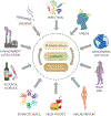
Figure 1:. Oxidative stress induces p38MAPK mediated activation of senescence and inflammation in fetal membranes at term.
Various factors as shown in the figure can contribute to excessive reactive oxygen species (ROS) build-up in the intra-amniotic cavity. This ROS can accelerate fetal membrane senescence and senescence-associated secretory phenotype (SASP). Senescence and SASP factors, in a feedback loop, can cause further damage to non-senescent and neighboring tissues to cause further enhanced inflammation. This is a natural and physiological process during normal parturition.
Figure 2:
Figure 2:. Pregnancy risk factors increase oxidative stress and cause increased activation of p38MAPK, senescence and inflammation in fetal membranes in preterm pregnancies.
Multiple risk factors can cause an increase in ROS in the intra-amniotic cavity. Pathways of ROS generation, characteristics of oxidative stress and p38MAPK activation may not be the same for all risk factors. Regardless, many of these factors can increase p38MAPK activation pathologically prior to term. Senescence and SASP factors can cause preterm labor and or pPROM. Premature activation of p38MAPK is the pathological activation of senescence leading to preterm parturition.
Figure 3:
Figure 3:. Schematics of changes to membrane structure during normal gestation and parturition.
Top panel: Membranes during gestation: Fetal membranes, specifically amnion layer undergoes a cyclic transition of EMT←→MET that maintains membrane homeostasis and a 10:1 ratio between AEC and AMC. Middle panel: During gestation, the TGF-b/TAB/p38MAPK-mediated pathway forces the EMT to shed AECs which are transformed into AMC. Since the accumulation of AMC is an unstable state, these cells are transitioned back to AMC by the P4/PGRMC2/c-MYC pathway. Bottom panel: At term, ROS buildup and p38MAPK activation (see Figure 1), can lead to a terminal state of EMT with the accumulation of AMC and no MET to balance the cell ratio between AEC and AMC. This is an unstable state of inflammation and cause local inflammatory build-up, matrix degradation and membrane weakening.
Figure 4:
Figure 4:. Two distinct mechanisms of inflammatory activation in fetal membranes and fetal inflammatory signaling for parturition.
Physiologic (Fig 1) or pathologic (Fig 2) signals increase ROS in the intra-amniotic cavity and cause non-canonical activation of p38MAPK in human fetal membrane cells. p38MAPK activation can lead to: 1. Senescence of the fetal membrane cells, the production of SASP and the generation of DAMPS. 2. p38MAPK forces a terminal state of EMT and the accumulation of AMCs, causing membrane matrix damage and weakening along with increased localized inflammation. Inflammatory mediators generated are packaged into extracellular vesicles released by fetal membrane cells – fetal signals – that can reach the myometrium and decidua and cause their activation (inflammation) which can transition these tissues from their quiescent state to an active state of labor.

References

    1. Abrahams VM, Potter JA, Bhat G, Peltier MR, Saade G, & Menon R (2013). Bacterial modulation of human fetal membrane Toll-like receptor expression. Am J Reprod Immunol, 69(1), 33–40. doi:10.1111/aji.12016 - DOI - PMC - PubMed
    1. Adams Waldorf KM, Singh N, Mohan AR, Young RC, Ngo L, Das A, … Johnson MR (2015). Uterine overdistention induces preterm labor mediated by inflammation: observations in pregnant women and nonhuman primates. Am J Obstet Gynecol, 213(6), 830.e831–830.e819. doi:10.1016/j.ajog.2015.08.028 - DOI - PMC - PubMed
    1. Agarwal A, Gupta S, & Sharma RK (2005). Role of oxidative stress in female reproduction. Reprod Biol Endocrinol, 3, 28. doi:10.1186/1477-7827-3-28 - DOI - PMC - PubMed
    1. Agrawal V, & Hirsch E (2012). Intrauterine infection and preterm labor. Semin Fetal Neonatal Med, 17(1), 12–19. doi:10.1016/j.siny.2011.09.001 - DOI - PMC - PubMed
    1. Alijotas-Reig J, Llurba E, & Gris JM (2014). Potentiating maternal immune tolerance in pregnancy: a new challenging role for regulatory T cells. Placenta, 35(4), 241–248. doi:10.1016/j.placenta.2014.02.004 - DOI - PubMed

Publication types

Substances

LinkOut - more resources