. 2020 Aug 12;11(1):92.
doi: 10.1186/s13244-020-00901-7.

Structured reporting of chest CT in COVID-19 pneumonia: a consensus proposal

Affiliations

Structured reporting of chest CT in COVID-19 pneumonia: a consensus proposal

E Neri et al. Insights Imaging. .

Abstract

Objectives: The need of a standardized reporting scheme and language, in imaging of COVID-19 pneumonia, has been welcomed by major scientific societies. The aim of the study was to build the reporting scheme of chest CT in COVID-19 pneumonia.

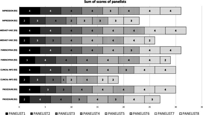
Methods: A team of experts, of the Italian Society of Medical and Interventional Radiology (SIRM), has been recruited to compose a consensus panel. They used a modified Delphi process to build a reporting scheme and expressed a level of agreement for each section of the report. To measure the internal consistency of the panelist ratings for each section of the report, a quality analysis based on the average inter-item correlation was performed with Cronbach's alpha (Cα) correlation coefficient.

Results: The overall mean score of the experts and the sum of score were 3.1 (std.dev. ± 0.11) and 122 in the second round, and improved to 3.75 (std.dev. ± 0.40) and 154 in the third round. The Cronbach's alpha (Cα) correlation coefficient was 0.741 (acceptable) in the second round and improved to 0.789 in the third round. The final report was built in the management of radiology report template (MRRT) and includes n = 4 items in the procedure information, n = 5 items in the clinical information, n = 16 in the findings, and n = 3 in the impression, with overall 28 items.

Conclusions: The proposed structured report could be of help both for expert radiologists and for the less experienced who are faced with the management of these patients. The structured report is conceived as a guideline, to recommend the key items/findings of chest CT in COVID-19 pneumonia.

Keywords: COVID-19; Computed tomography; Structured reporting.


Conflict of interest statement

The authors declare that they have no competing interests.

Figures

Fig. 1
Fig. 1
Flow chart of the Delphi consensus
Fig. 2
Fig. 2
Sum of scores of each section of the structured report in round 2 (R2) and round 3 (R3)
Fig. 3
Fig. 3
Structured report displayed in the MMRT format (www.radreport.org). a Sections “Procedure Information” and “Clinical Information”. b Section “Parenchyma”. c Sections “Mediastinum and Vascular findings”, and “Impression.” The subsection classification is based on the paper by Simpson et al. [13], as follows: Typical—peripheral, bilateral, GGO with or without consolidation or visible intralobular lines (crazy-paving). Multifocal GGO of rounded morphology with or without consolidation or visible intralobular lines (crazy-paving). Reverse halo sign or other findings of organizing pneumonia (seen later in the disease). Atypical—absence of typical or indeterminate features and presence of: isolated lobar or segmental consolidation without GGO. Discrete small nodules (centrilobular, tree in-bud). Lung cavitation. Smooth interlobular septal thickening with pleural effusion. Indeterminate—absence of typical features and presence of: multifocal, diffuse, perihilar, or unilateral GGO with or without consolidation lacking a specific distribution and are non-rounded or non-peripheral. Few very small GGO with a non-rounded and non-peripheral distribution. Negative—no CT features to suggest pneumonia. The subsection “Select CO-RADS category” is based on the paper by Prokop et al. [10], as follows: CO-RADS 0 not interpretable: scan technically insufficient for assigning a score; CO-RADS 1 very low: normal or non-infections; CO-RADS 2 low: typical for other infection but not COVID-19. CO-RADS 3 equivocal/unsure: features compatible with COVID-19, but also other diseases. CO-RADS 4 high: suspicious for COVID-19. CO-RADS 5 very high: typical for COVID-19. CO-RADS 6 proven: RT-PCR positive for SARS-CoV-2

References

    1. Fanelli D, Piazza F. Analysis and forecast of COVID-19 spreading in China, Italy and France. Chaos, Solitons & Fractals. 2020;134:109761. doi: 10.1016/j.chaos.2020.109761. - DOI - PMC - PubMed
    1. Goletti O, Castoldi M, Bombardieri E (2020) Keep or release: experience on management of COVID-19 during maximum emergency in Bergamo and impact on patient outcomes. Eur J Emerg Med 27:309. 10.1097/MEJ.0000000000000720 - PubMed
    1. COVID-19 Map. In: Johns Hopkins Coronavirus Resource Center. https://coronavirus.jhu.edu/map.html. Accessed 8 Jul 2020
    1. Huang Z, Zhao S, Li Z, et al (2020) The battle against Coronavirus Disease 2019 (COVID-19): emergency management and infection control in a radiology department. J Am Coll Radiol. 10.1016/j.jacr.2020.03.011 - PMC - PubMed
    1. Rubin GD, Ryerson CJ, Haramati LB, et al (2020) The role of chest imaging in patient management during the COVID-19 pandemic: a multinational consensus statement from the Fleischner society. Radiology 201365. 10.1148/radiol.2020201365 - PMC - PubMed