. 2020 Aug 12;13(1):56.
doi: 10.1186/s12284-020-00418-z.

UvAtg8-Mediated Autophagy Regulates Fungal Growth, Stress Responses, Conidiation, and Pathogenesis in Ustilaginoidea virens

Affiliations

UvAtg8-Mediated Autophagy Regulates Fungal Growth, Stress Responses, Conidiation, and Pathogenesis in Ustilaginoidea virens

Shuai Meng et al. Rice (N Y). .

Abstract

Background: Ustilaginoidea virens has become one of the most devastating rice pathogens in China, as well as other rice-growing areas. Autophagy is an important process in normal cell differentiation and development among various organisms. To date, there has been no optimized experimental system introduced for the study of autophagy in U. virens. In addition, the function of autophagy in pathogenesis remains unknown in U. virens. Therefore, the functional analyses of UvAtg8 may potentially shed some light on the regulatory mechanism and function of autophagy in U. virens.

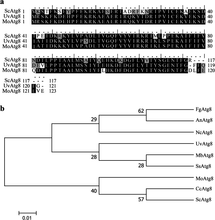
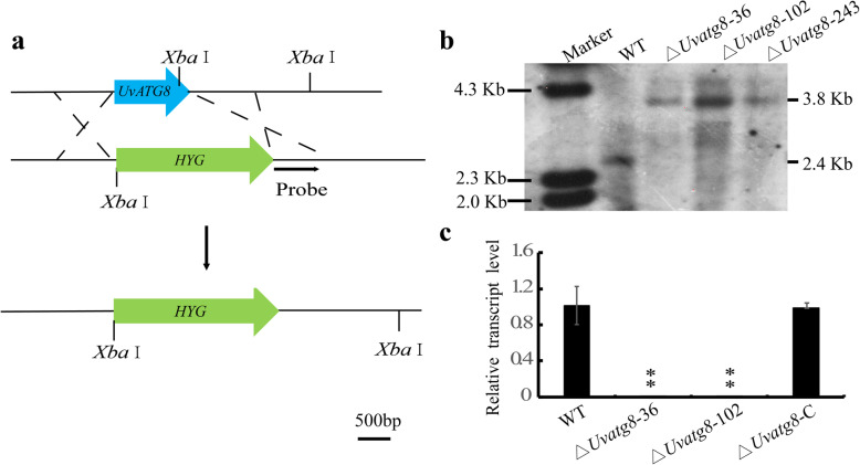
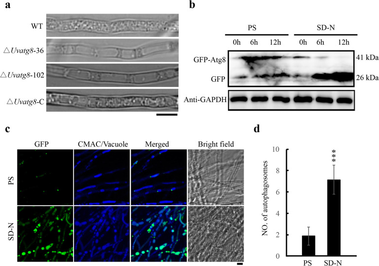
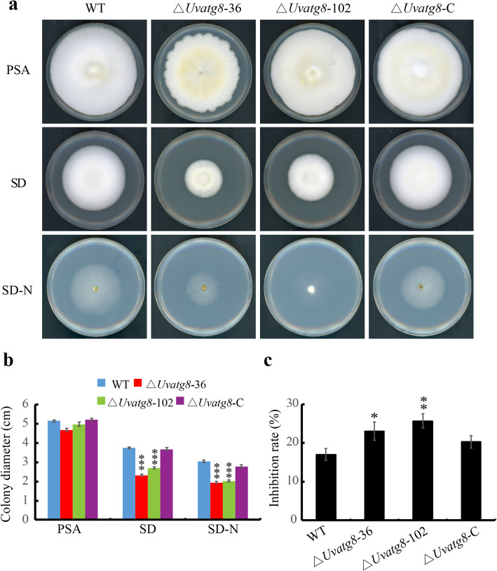
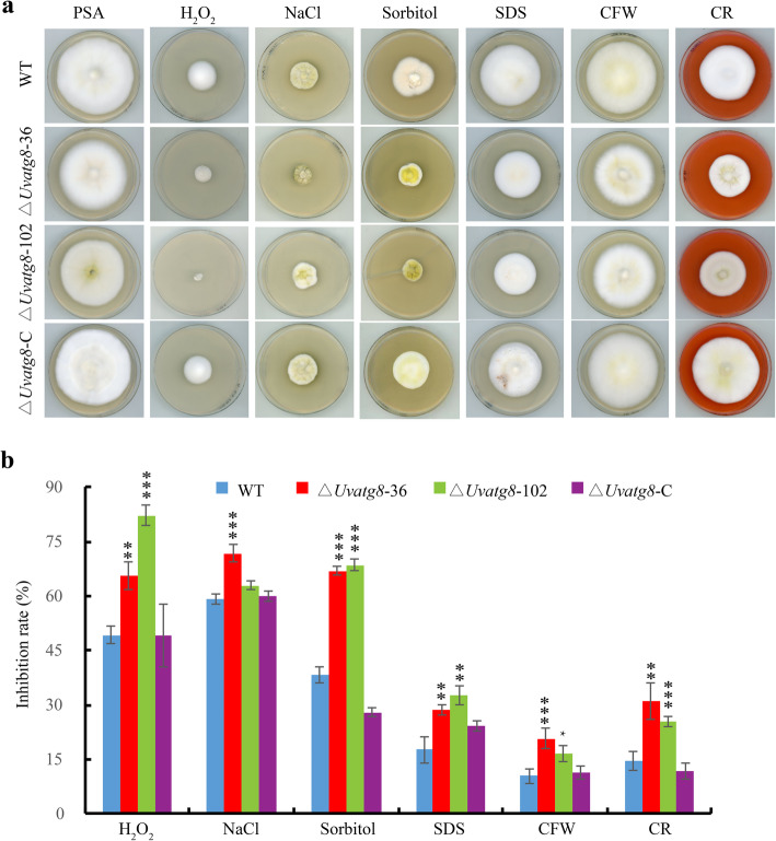
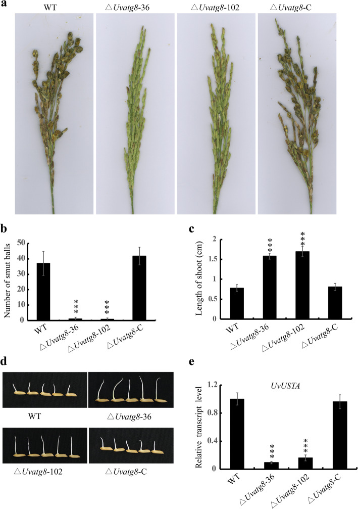
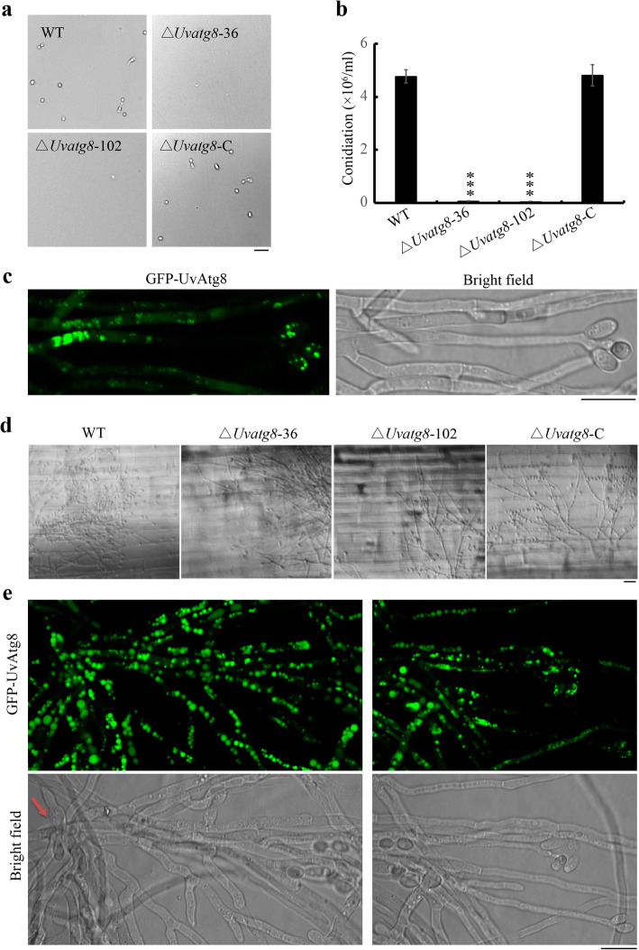
Results: In this study, we characterized the functions of UvAtg8, which is a homolog of Saccharomyces cerevisiae ScAtg8, in the rice false smut fungus U. virens. The results showed that UvATG8 is essential for autophagy in U. virens. Also, the GFP-UvATG8 strain, which could serve as an appropriate marker for monitoring autophagy in U. virens, was generated. Furthermore, this study found that the ΔUvatg8 mutant was defective in the vegetative growth, conidiation, adaption to oxidative, hyperosmotic, cell wall stresses, and production of toxic compounds. Pathogenicity assays indicated that deletion of UvATG8 resulted in significant reduction in virulence of U. virens. Further microscopic examinations of the infection processes revealed that the severe virulence defects in the ∆Uvatg8 were mainly caused by the highly reduced conidiation and secondary spore formation.

Conclusions: Our results indicated that the UvAtg8 is necessary for the fungal growth, stresses responses, conidiation, secondary spore formation, and pathogenicity of U. virens. Moreover, our research finding will potentially assist in further clarifying the molecular mechanism of U. virens infection, as well as provide a good marker for autophagy in U. virens and a good reference value for the further development of effective fungicides based on gene targeting.

Keywords: Autophagy; Pathogenicity; Rice false smut; Secondary spore; Ustilaginoidea virens; UvAtg8.


Conflict of interest statement

There is no conflict of interest.

Figures

Fig. 1
Fig. 1
Evolution analysis of UvAtg8. a, The mutiple alignment of amino acid sequences of UvAtg8, ScAtg8, and MoAtg8. The identitical amino acid were shaded in solid black, and the amino acids with 50 % similarity were highlighted in gray. b, Phylogenetic tree of Atg8 orthologs from different fungi was constructed by MEGA 5.0 using the neighbour-joining method. The sequences included FgAtg8 (Fusarium graminearum, XP_001392077.1), AnAtg8 (Aspergillus niger, XP_001392077.1), NcAtg8 (Neurospora crassa, XP_956248.1), UvAtg8 (Ustilaginoidea virens, KDB12146.1), MbAtg8 (Metarhizium brunneum, XP_014545792.1), SsAtg8 (Sclerotinia sclerotiorum, XP_001597408.1), MoAtg8 (Magnaporthe oryzae, XP_003717877.1), CcAtg8 (Coprinopsis cinerea, XP_001833603.1), and ScAtg8 (Saccharomyces cerevisiae, NP_009475.1)
Fig. 2
Fig. 2
Targeted knockout of UvATG8 and complement assay of the ∆Uvatg8 in Ustilaginoidea virens. a, Disruption strategy for the U. virens UvATG8 gene. The UvATG8 coding region was replaced with the hygromycin phosphotransferase gene cassette (HYG) by homologous recombination. b, Southern blot analysis of the WT (wild type) and ∆Uvatg8 mutant strains. The Xba I digested genomic DNA from the WT and ∆Uvatg8 strains were processed for the Southern blotting with the 1 Kb downstream of UvATG8 as the probe. c, qRT-PCR analysis of the expression of UvATG8 in the WT, ∆Uvatg8, and ∆Uvatg8-C strains. β-tubulin gene was used as the endogenous reference gene. The data represent the mean ± SD from three independent replicates. The data were subject to Duncan’s Test and the significant differences were indicated in the figure with two asterisks (**, p < 0.005)
Fig. 3
Fig. 3
UvAtg8 is essential for the autophagy in Ustilaginoidea virens. a, Autophagy was blocked in ∆Uvatg8 mutants. The strains were cultured in the PS (potato sucrose medium) medium for 3 d, following subjected to SD-N (nitrogen starvation condition) medium for 4 h to determine the autophagic affuxes in the WT and ∆Uvatg8 strain. The images were taken by bright field microscopy. b, The degradation of the GFP-UvAtg8 was observed under nitrogen starvation condition via Western blot assay with an anti-GFP. GAPDH was used as an internal reference. c, Confocal microscopy image of the GFP-UvAtg8 strain under nitrogen rich and starvation condition. The indicated strain was cultured in the PS medium for 3 d, and then shifted to an SD-N medium for 12 h. The vacuoles were stained with CAMC (7-amino-4-chloromethylcoumarin) before the observation using confocal laser scanning microscope. Scale bar = 5 μm. d, The number of autophagosomes was counted. The data were subjected to Duncan’s Test and the significant difference was indicated by asterisks (***, p < 0.001)
Fig. 4
Fig. 4
UvAtg8 plays important roles in the fungal growth. a, Radial growth of ∆Uvatg8 was reduced. Colonies of the WT, ∆Uvatg8, and ∆Uvatg8-C strains were grown on the PSA、SD and SD-N medium in dark for 15 d and then imaged. b, Statistical analysis of colonies diameters of test strains on the PSA, SD and SD-N plates. c, Statistical analysis of inhibition rate of test strains under nitrogen starvation condition by comparing the fungal growth on the SD-N and SD medium. Three independent biological experiments were performed with three replicates each time. The data were subjected to Duncan’s Test and the significant differences were shown in the figure with asterisks (*, p < 0.01; **, p < 0.005; ***, p < 0.001)
Fig. 5
Fig. 5
Defects of the ∆Uvatg8 mutants in response to different stresses. a, The represent strains were cultured on regular PSA plates or PSA plates with 0.03% H202, 0.4 M NaCl, 0.7 M sorbitol, 0.03% Sodium dodecyl sulfate (SDS), 200 μg/mL Calcofluor white (CFW), 240 μg/mL Congo red (CR) plates. Typical cultures were photographed after incubation for 15 d at 28 °C. b, Statistical analysis of inhibition rate of test strains under different stress conditions. The diameters of colonies were detected and analyzed. Similar results were obtained by three repeated experiments. The error bars represent the standard deviation and the asterisk represents the significant difference compared to the WT strain under the same conditions (*, p < 0.01; **, p < 0.005; ***, p < 0.001)
Fig. 6
Fig. 6
Virulence assays of the ∆Uvatg8 mutants. a, Disease symptoms caused by the WT, ∆Uvatg8, and ∆Uvatg8-C strains on 21 d after inoculation. b, Statistical analysis results of the average number of false smut balls per spikelet. At least three independent biological experiments were performed with 30 inoculated panicles each time. c, Culture filtrates of the ∆Uvatg8 mutants displayed less inhibitory of rice seedling germination. The shoot lengths were measured and statistically analyzed after 7 d in 28 °C illumination incubators. d, The growths of the rice shoots of the indicated strains were treated with testing culture filtrates. More than 100 rice seeds were sterilized with 0.1% potassium permanganate, then shifted to 35 mL culture filtrates at 28 °C for 7 d. e, qRT-PCR analysis of the expression of UvUSTA in the WT, ∆Uvatg8, and ∆Uvatg8-C strains. β-tubulin gene was used as the endogenous reference gene. Three independent experiments (n = 100) were carried out (mean ± SD). *** indicates p < 0.001
Fig. 7
Fig. 7
Deletion of UvATG8 resulted in decreased conidiation. a and b, Conidiation of the tested strains. The strains were cultured in 50 mL of PS medium with 170 rpm at 28 °C for 7 d prior to the observation. Three repetitions were performed, with similar results obtained. c, The expression of GFP-UvAtg8 during conidiation. d, Conidial germination of U. virens on rice sheath. 5 μL conidial droplets (1 × 106 conidia/mL) were inoculated and incubated at 28 °C for 3 d. Photographs were taken under an Olympus BX53 microscope equipped with bright field optics. e, The GFP-UvAtg8 was highly expressed, and autophagy had occurred during the secondary spore formation stage. The images were obtained using a confocal laser scanning microscope. Red arrow showed the conidial germination. Scale bar = 2.5 μm

References

    1. Chen X, Hai D, Tang JT, Liu H, Huang JB, Luo CX et al (2019) UvCom1 is an important regulator required for development and infection in the rice false smut fungus Ustilaginoidea virens. Phytopathology. 10.1094/PHYTO-05-19-0179-R - PubMed
    1. Deng YZ, Ramos-Pamplona M, Naqvi NI. Autophagy-assisted glycogen catabolism regulates asexual differentiation in Magnaporthe oryzae. Autophagy. 2009;5(1):33–43. doi: 10.4161/auto.5.1.7175. - DOI - PubMed
    1. Fan J, Du N, Li L, Li GB, Wang YQ, Zhou YF, et al. A core effector UV_1261 promotes Ustilaginoidea virens infection via spatiotemporally suppressing plant defense. Phytopathol Res. 2019;1(1):11. doi: 10.1186/s42483-019-0019-5. - DOI
    1. Fan J, Guo XY, Huang F, Li Y, Liu YF, Wang WM. Epiphytic colonization of ustilaginoidea virens on biotic and abiotic surfaces implies the widespread presence of primary inoculum for rice false smut disease. Plant Pathol. 2013;63:937–945. doi: 10.1111/ppa.12167. - DOI
    1. Fan J, Yang J, Wang YQ, Li GB, Li Y, Huang F, et al. Current understanding on Villosiclava virens, a unique flower-infecting fungus causing rice false smut disease. Mol Plant Pathol. 2016;17(9):1321–1330. doi: 10.1111/mpp.12362. - DOI - PMC - PubMed

LinkOut - more resources