. 2020 Aug 19;142(33):14295-14306.
doi: 10.1021/jacs.0c06066. Epub 2020 Aug 10.

Dismantling and Rebuilding the Trisulfide Cofactor Demonstrates Its Essential Role in Human Sulfide Quinone Oxidoreductase

Affiliations

Dismantling and Rebuilding the Trisulfide Cofactor Demonstrates Its Essential Role in Human Sulfide Quinone Oxidoreductase

Aaron P Landry et al. J Am Chem Soc. .

Abstract

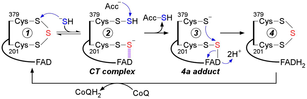
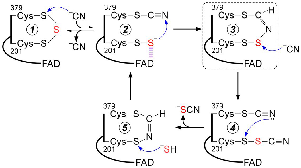
Sulfide quinone oxidoreductase (SQOR) catalyzes the first step in sulfide clearance, coupling H2S oxidation to coenzyme Q reduction. Recent structures of human SQOR revealed a sulfur atom bridging the SQOR active site cysteines in a trisulfide configuration. Here, we assessed the importance of this cofactor using kinetic, crystallographic, and computational modeling approaches. Cyanolysis of SQOR proceeds via formation of an intense charge transfer complex that subsequently decays to eliminate thiocyanate. We captured a disulfanyl-methanimido thioate intermediate in the SQOR crystal structure, revealing how cyanolysis leads to reversible loss of SQOR activity that is restored in the presence of sulfide. Computational modeling and MD simulations revealed an ∼105-fold rate enhancement for nucleophilic addition of sulfide into the trisulfide versus a disulfide cofactor. The cysteine trisulfide in SQOR is thus critical for activity and provides a significant catalytic advantage over a cysteine disulfide.


Conflict of interest statement

The authors declare no competing financial interest.

Figures

Figure 1.
Figure 1.
Postulated mechanism for sulfide oxidation catalyzed by human SQOR. Sulfide adds into the resting cysteine trisulfide (1) to generate a 379Cys-SSH persulfide and a 201Cys-SS persulfide, with the latter participating in a CT complex with FAD (2). Sulfur transfer to a small molecule acceptor proceeds through a putative 4a adduct (3) to generate the reduced enzyme (4). Electron transfer from FADH2 to CoQ regenerates the resting enzyme. The oxidized sulfur and bridging sulfur in the cysteine trisulfide are labeled in blue and red, respectively.
Figure 2.
Figure 2.
Cyanide-induced CT complex formation in SQOR. A, SQOR (10 μM, red line) in 100 mM potassium phosphate, pH 7.4 containing 0.03% DHPC, was mixed 1:1 (v/v) with KCN (4 mM) and monitored over 1.5 s at 4 °C for the formation of the cyanide-induced CT complex at 695 mn (blue line). B, Representative stopped flow kinetic trace for the reaction in (A) monitored at 695 mn. C, Dependence of the kobs at 4 °C for cyanide-induced CT complex formation on cyanide concentration. The data are representative of two independent experiments, with each data point obtained in triplicate. D, SQOR (5 μM, red line) was treated with KCN (5 mM) to form the CT complex (blue line), immediately followed by the addition of Na2S (200 μM) and incubated for 5 min at 20 °C, which led to CT complex decay and FAD reduction (black line). The data are representative of three independent experiments.
Figure 3.
Figure 3.
Cyanide-induced CT complex decay in SQOR. A, SQOR (25 μM) in Buffer A was treated with KCN (10 mM) to form the CT complex (red line), which was monitored over 43 min at 20 °C for the complete decay of the CT complex (blue line). B, Kinetic traces for the decay of the cyanide-induced CT complex in (A), monitored at 450 nm (open circles) and 695 nm (closed circles). C, SQOR (5 μM. solid black line) was treated with sodium sulfite (5 mM) and incubated for 1 min at 20 °C to form the CT complex (dashed black line). In tandem, SQOR (5 μM) pre-treated with KCN (10 mM) and desalted (solid red line) was then treated with sodium sulfite (5 mM). CT complex formation was not observed after incubation for 1 min at 20 °C (solid blue line). The data are representative of three independent experiments.
Figure 4.
Figure 4.
Regeneration of cyanide pre-treated SQOR by sulfide. A, Cyanide pre-treated SQOR (17 μM, red line) in Buffer A was rapidly mixed 1:1 (v/v) with Na2S (400 μM) and monitored over a period of 7 s at 4 °C for fonnation of the sulfide-induced CT complex (blue line). B, Comparison of the kinetic traces at 675 mn for cyanide pre-treated SQOR, as shown in A, versus native SQOR mixed with Na2S (400 μM) under the same conditions. C, SQOR (17 μM, solid black line) in Buffer A was treated with KCN (10 mM) to fonn the CT complex (dashed black line) and monitored over 40 min at 20 °C for the complete decay of the CT complex and desalted to remove excess cyanide (red line). Cyanide pre-treated SQOR was then incubated with Na2S (300 μM) for 1 h at 4 °C, which led to FAD reduction (blue trace). D, Cyanide pre-treated SQOR, pre-incubated with sulfide under the same conditions as (A) and desalted (5 μM, red line), was treated with sulfite (5 mM) and incubated for 1 min to fonn the sulfite-induced CT complex (blue line). The data are representative of three independent experiments.
Figure 5.
Figure 5.
Effect of bridging sulfur extraction on SQOR protein stability. SQOR (20 μM) in Buffer A was pre-treated with KCN (10 mM) for 45 min at 20 °C and desalted, followed by incubation with Na2S (300 μM) for 1 h at 4 °C and a second desalting. A final SQOR concentration of 5 μM was used for the thermal denaturation assays. The stability of native SQOR (black line) versus cyanide pre-treated SQOR before (red line) and after (blue line) incubation with sulfide was monitored by the increase in absorbance at 600 nm. The data are representative of three independent experiments.
Figure 6.
Figure 6.
Dithiol-mediated reduction of FAD in SQOR. A, SQOR (10 μM) in Buffer A was rapidly mixed 1:1 (v/v) with DTT (400 μM) and FAD reduction was monitored over 7 s at 4 °C. B, Proposed mechanism for the addition of DTT into the SQOR cysteine trisulfide, leading to FAD reduction. DTT adds into the cysteine trisulfide (1) at the solvent-accessible Cys-379 to generate a mixed disulfide and 201Cys-SS (2). An intramolecular thiol-disulfide exchange then regenerates the SQOR cysteine trisulfide, with electrons moving into FAD (3). C, SQOR (5 μM) in Buffer A (solid black line), was treated with DTT (200 μM), leading to FAD reduction (dashed black line), or β-mercaptoethanol (200 μM), leading to stable CT complex formation (solid gray line). FAD reduction was not observed in cyanide pre-treated SQOR (5 μM, solid red line) upon treatment with DTT (200 μM, solid blue line). D, SQOR (5 μM) under the same conditions as in (C) (solid black line), was treated with DHLA (200 μM), leading to FAD reduction (dashed black line), followed by re-oxidation by addition of CoQ1 (180 μM, solid gray line). FAD reduction was not observed in cyanide pre-treated SQOR (5 μM, solid red line) upon treatment with DHLA (200 μM, solid blue line). The data are representative of three independent experiments.
Figure 7.
Figure 7.
Structure and active site of SQOR-CoQ1 + cyanide. A, The overall structure of SQOR-CoQ1 + cyanide is shown with FAD, CoQ1, and cyanide in yellow, grey, and magenta spheres, respectively. The C-terminal membraneanchoring helices are highlighted in cyan. B, Electron density maps (2Fo-Fc) of the active site shown in mesh contoured at 1.0 σ. Cys-201, Cys-379, FAD, CoQ1, sulfur derived from the trisulfide, and cyanide are shown in stick display. C, Stereo image of the active site of SQOR-CoQ1 treated with cyanide. The electron densities (2Fo-Fc) are contoured at 1.0 σ. D, Stereo image of the active site in SQOR-CoQ1 treated with sulfide (PDB ID: 6O16) showing the resting trisulfide. Chain A is shown in tins figure.
Figure 8.
Figure 8.
MD simulations and computational modeling of SQOR. A, Active site architecture in representative structures corresponding to the most populated cluster from 600 ns MD simulations of SQOR in the trisulfide (left panel) or disulfide state (right panel). Condensed local softness for the most electrophilic Sγ atom between Cys-201 and Cys-379 is reported for each system in atomic units (a.u.). B, Sulfur-to-C4a FAD distances for SγC201–C4aFAD/Ssulfane–C4aFAD/SγC379–C4aFAD (trisulfide, left panel) and SγC201–C4aFAD/SγC379–C4aFAD (disulfide, right panel) monitored along the corresponding MD trajectories. C, Structure of the transition states (TS) located for the sulfide anion attack on the trisulfide (left panel) or disulfide (right panel) using a reduced model of the active site of SQOR at the IEFPCM-DFT level of theory in a dielectric of ε = 10.125. Data correspond to interatomic distances in Å and Gibbs free-energy associated barriers at 298 K in kcal mol−1.
Figure 9.
Figure 9.
Proposed mechanism for cyanolysis and cysteine trisulfide rebuilding in SQOR. Cyanide adds into the resting cysteine trisulfide (1) to generate a 379Cys-S-CN organic thiocyanate while the bridging sulfur is retained in the 201Cys-SS persulfide that participates in a CT complex with FAD (2). Conversion to the 379Cys N-(201Cys-disulfanyl)-methanimido thioate intennediate (3) leads to loss of the CT complex. Addition by a second cyanide at the sulfane sulfur of Cys-201 leads to intennediate (4), which can cyclize and eliminate thiocyanate (5), completing the cyanolysis reaction. Addition of sulfide to the Sγ of Cys-201 in the 379Cys N-(201Cys-sulfanyl)-methanimido thioate intennediate (5) regenerates the CT complex (2). Elimination of cyanide regenerates the resting trisulfide fonn of the enzyme. The bridging sulfur of the cysteine trisulfide is labeled in red. The dashed box highlights the intennediate observed in the crystal structure.

References

    1. Kimura H, Hydrogen sulfide: from brain to gut. Antioxid. Redox Signal 2010, 12 (9), 1111–23. - PubMed
    1. Kabil O; Banerjee R, Redox biochemistry of hydrogen sulfide. J. Biol. Chem 2010, 285 (29), 21903–7. - PMC - PubMed
    1. Filipovic MR; Zivanovic J; Alvarez B; Banerjee R, Chemical Biology of H2S Signaling through Persulfidation. Chem. Rev 2018, 118 (3), 1253–1337. - PMC - PubMed
    1. Singh S; Banerjee R, PLP-dependent H(2)S biogenesis. Biochim. Biophys. Acta 2011, 1814 (11), 1518–27. - PMC - PubMed
    1. Chiku T; Padovani D; Zhu W; Singh S; Vitvitsky V; Banerjee R, H2S biogenesis by human cystathionine gamma-lyase leads to the novel sulfur metabolites lanthionine and homolanthionine and is responsive to the grade of hyperhomocysteinemia. J. Biol. Chem 2009, 284 (17), 11601–12. - PMC - PubMed

Publication types

MeSH terms

Substances