

Retinoschisis associated with Kearns-Sayre syndrome
- PMID: 32787478
- PMCID: PMC8127726
- DOI: 10.1080/13816810.2020.1799416
Retinoschisis associated with Kearns-Sayre syndrome
Abstract
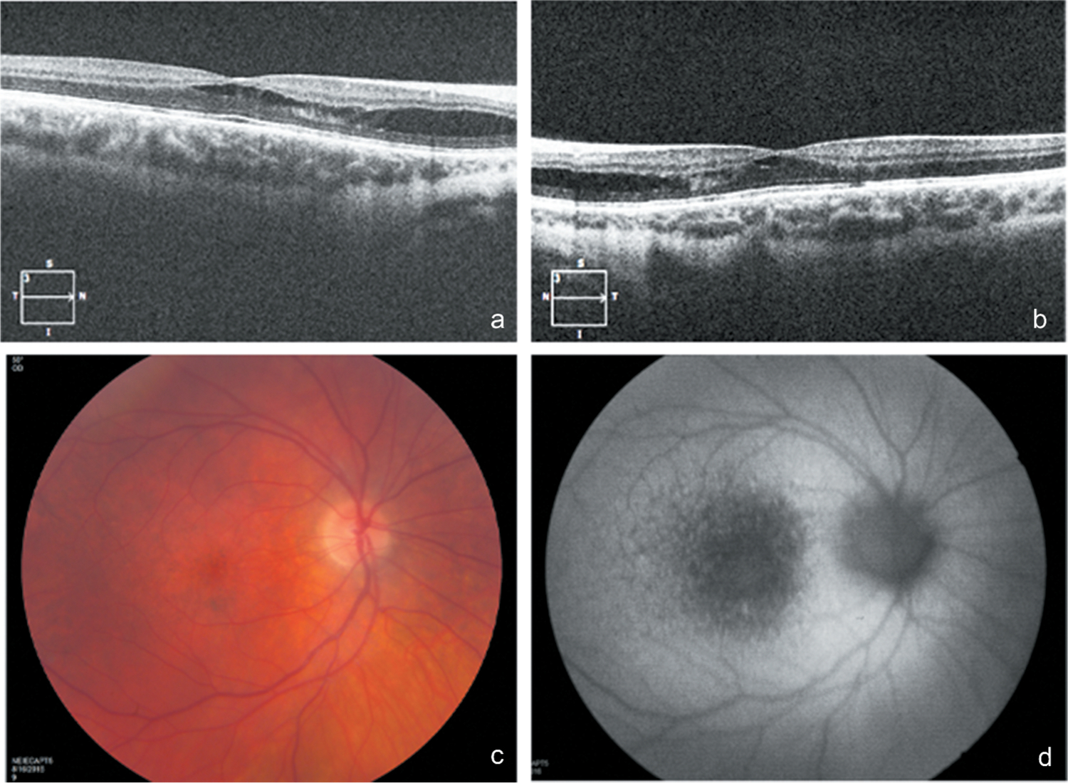
Background: Kearns-Sayre Syndrome (KSS) is characterized by pigmentary retinopathy, external ophthalmoplegia and heart block. We report on a now 24-year-old male with clinical retinoschisis and molecularly confirmed KSS.
Materials and methods: Physical and complete ophthalmic examination, molecular diagnosis.
Results: Over nine years of follow-up, the subject manifested progressive signs and symptoms of KSS, including external ophthalmoplegia/strabismus, ptosis, pigmentary retinopathy, corneal edema, Type I diabetes mellitus, gut dysmotility, sensorineural deafness and heart block. At age 21 he was incidentally found to have retinoschisis on optical coherence tomography that remained stable over three years follow-up. Sequencing of the RS1 gene revealed no pathogenic variants, effectively ruling out co-existing X-linked retinoschisis.
Conclusions: These findings suggest retinoschisis may be a rare manifestation of KSS. A trial of a carbonic anhydrase inhibitor was frustrated by coexisting corneal edema associated with the condition.
Keywords: Kearns-Sayre syndrome; mitochondria; retinoschisis.
Conflict of interest statement
Declaration of interest
The authors report no conflicts of interest. The authors alone are responsible for the content and writing of this article.
Figures
Comment in
-
Exclude hereditary and acquired differential disorders before attributing retinoschisis to Kears-Sayre syndrome.Ophthalmic Genet. 2021 Feb;42(1):99. doi: 10.1080/13816810.2020.1827444. Epub 2020 Sep 25. Ophthalmic Genet. 2021. PMID: 32975160 No abstract available.
-
Response to Finsterer's "Exclude hereditary and acquired differential disorders before attributing retinoschisis to Kears-Sayre syndrome".Ophthalmic Genet. 2021 Feb;42(1):100. doi: 10.1080/13816810.2020.1832295. Epub 2020 Nov 24. Ophthalmic Genet. 2021. PMID: 33233984 Free PMC article. No abstract available.
References
-
- Wallace DC, Lott MT, Brown MD, Kerstann K. Mitochondria and neuro-ophthalmologic diseases. In: The online metabolic and molecular bases of inherited diseases [Internet]. New York, NY: McGraw-Hill Education; 2019. ommbid.mhmedical.com/content.aspx?aid=1170091759
-
- Poulton J, Morten KJ, Marchington D, Weber K, Brown GK, Rotig A, Bindoff L. Duplications of mitochondrial DNA in Kearns-Sayre syndrome. Muscle Nerve Suppl. 1995;3:S154–8. - PubMed
Publication types
MeSH terms
Grants and funding
LinkOut - more resources
Full Text Sources