. 2020 Aug 12;24(1):495.
doi: 10.1186/s13054-020-03218-5.

Unspecific post-mortem findings despite multiorgan viral spread in COVID-19 patients

Affiliations

Unspecific post-mortem findings despite multiorgan viral spread in COVID-19 patients

Myriam Remmelink et al. Crit Care. .

Abstract

Background: Post-mortem studies can provide important information for understanding new diseases and small autopsy case series have already reported different findings in COVID-19 patients.

Methods: We evaluated whether some specific post-mortem features are observed in these patients and if these changes are related to the presence of the virus in different organs. Complete macroscopic and microscopic autopsies were performed on different organs in 17 COVID-19 non-survivors. Presence of SARS-CoV-2 was evaluated with immunohistochemistry (IHC) in lung samples and with real-time reverse-transcription polymerase chain reaction (RT-PCR) test in the lung and other organs.

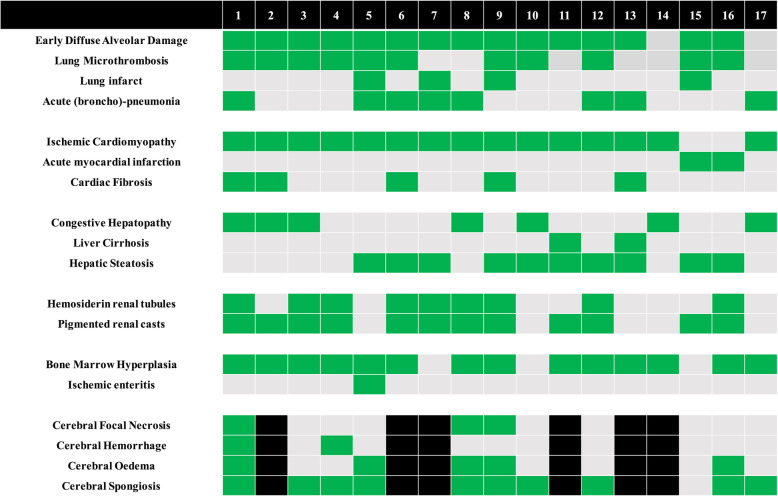
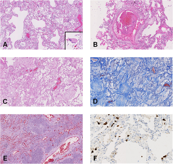
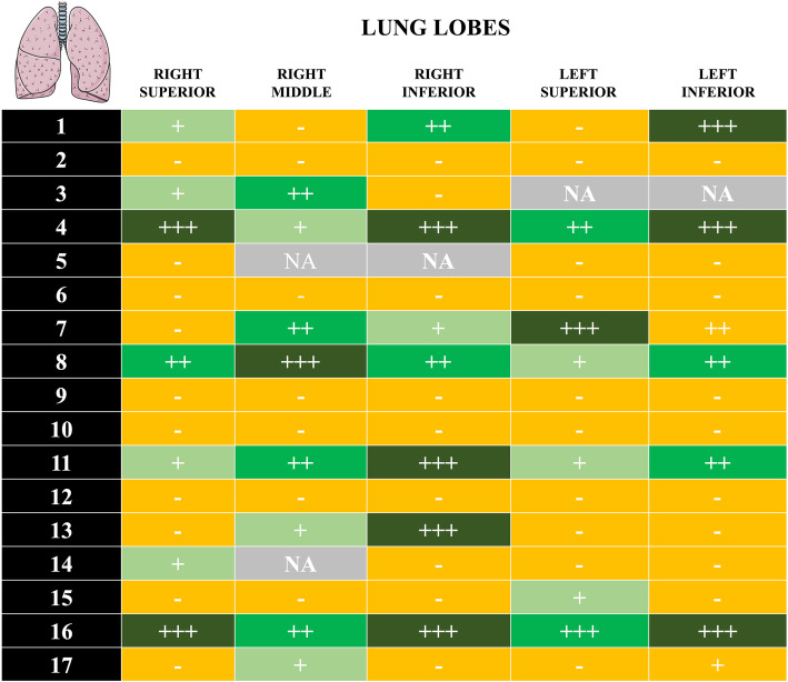
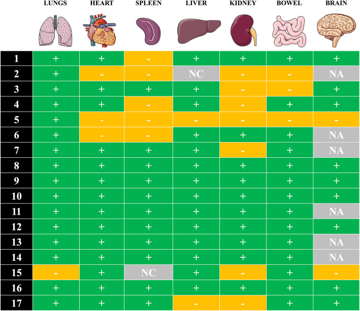
Results: Pulmonary findings revealed early-stage diffuse alveolar damage (DAD) in 15 out of 17 patients and microthrombi in small lung arteries in 11 patients. Late-stage DAD, atypical pneumocytes, and/or acute pneumonia were also observed. Four lung infarcts, two acute myocardial infarctions, and one ischemic enteritis were observed. There was no evidence of myocarditis, hepatitis, or encephalitis. Kidney evaluation revealed the presence of hemosiderin in tubules or pigmented casts in most patients. Spongiosis and vascular congestion were the most frequently encountered brain lesions. No specific SARS-CoV-2 lesions were observed in any organ. IHC revealed positive cells with a heterogeneous distribution in the lungs of 11 of the 17 (65%) patients; RT-PCR yielded a wide distribution of SARS-CoV-2 in different tissues, with 8 patients showing viral presence in all tested organs (i.e., lung, heart, spleen, liver, colon, kidney, and brain).

Conclusions: In conclusion, autopsies revealed a great heterogeneity of COVID-19-associated organ injury and the remarkable absence of any specific viral lesions, even when RT-PCR identified the presence of the virus in many organs.

Keywords: Autopsy; COVID-19; Immunohistochemistry; RT-PCR; SARS-CoV-2.


Conflict of interest statement

The authors declare that they have no competing interests.

Figures

Fig. 1
Fig. 1
Main histological findings. Green = finding present; gray = finding absent; black = unavailable
Fig. 2
Fig. 2
Pulmonary histological findings. a Early-stage diffuse alveolar damage (DAD): hyaline membrane (H&E, × 50 magnification) with a zoom on a giant cell (× 100 magnification). b Fibrin thrombi in a pulmonary artery (H&E, × 50 magnification). c Late-stage DAD: fibroblastic proliferation (H&E, × 50 magnification). d Late-stage DAD: fibroblastic proliferation (Trichrome staining, × 50 magnification). e Acute pneumonia (H&E, × 50 magnification). f Anti-SARS-CoV immunohistochemistry (IHC)-positive cells (× 200 magnification)
Fig. 3
Fig. 3
Detection of SARS-CoV-2 by immunohistochemistry (IHC) in FFPE post- mortem lung samples of 17 patients. Semi-quantitative evaluation: “−” negative result; “+” scattered positive cells (between 1 and < 5 positive cells/whole slide); “++” positive isolated cells (> 5 cells/whole slide, but no foci); “+++” foci of positive cells (more than 10 positive cells in one × 200 field). NA, not available
Fig. 4
Fig. 4
Molecular detection of SARS-Cov-2 RNA in post-mortem samples. Detection of SARS-CoV-2 by reverse transcription real-time polymerase chain reaction (RT-PCR) in FFPE post-mortem tissues of 17 patients. “+” positive result; “−” negative result; “NA” tissue not available; NC, non-informative test result (due to low-quality RNA)

References

    1. Guan WJ, Ni ZY, Hu Y, Liang WH, Ou CQ, He JX, et al. Clinical characteristics of coronavirus disease 2019 in China. N Engl J Med. 2020;382:1708–1720. doi: 10.1056/NEJMoa2002032. - DOI - PMC - PubMed
    1. Hoffmann M, Kleine-Weber H, Schroeder S, Krüger N, Herrler T, Erichsen S, et al. SARS-CoV-2 cell entry depends on ACE2 and TMPRSS2 and is blocked by a clinically proven protease inhibitor. Cell. 2020;181:1–10. doi: 10.1016/j.cell.2020.02.052. - DOI - PMC - PubMed
    1. Wichmann D, Sperhake JP, Lütgehetmann M, Steurer S, Edler C, Heinemann A, et al. Autopsy findings and venous thromboembolism in patients with covid-19: a prospective cohort study. Ann Intern Med. 2020. Epub ahead of print. 10.7326/M20-2003. - PMC - PubMed
    1. Varga Z, Flammer AJ, Steiger P, Haberecker M, Andermatt R, Zinkernagel AS, et al. Endothelial cell infection and endotheliitis in COVID-19. Lancet. 2020;395:1417–1418. doi: 10.1016/S0140-6736(20)30937-5. - DOI - PMC - PubMed
    1. Chen T, Wu D, Chen H, Yan W, Yang D, Chen G, et al. Clinical characteristics of 113 deceased patients with coronavirus disease 2019: retrospective study. BMJ. 2020. Epub ahead of print. 10.1136/bmj.m1091. - PMC - PubMed

Publication types

MeSH terms

LinkOut - more resources