. 2020 Oct 23;295(43):14578-14591.
doi: 10.1074/jbc.RA120.014345. Epub 2020 Aug 11.

Caspases from scleractinian coral show unique regulatory features

Affiliations

Caspases from scleractinian coral show unique regulatory features

Suman Shrestha et al. J Biol Chem. .

Abstract

Coral reefs are experiencing precipitous declines around the globe with coral diseases and temperature-induced bleaching being primary drivers of these declines. Regulation of apoptotic cell death is an important component in the coral stress response. Although cnidaria are known to contain complex apoptotic signaling pathways, similar to those in vertebrates, the mechanisms leading to cell death are largely unexplored. We identified and characterized two caspases each from Orbicella faveolata, a disease-sensitive reef-building coral, and Porites astreoides, a disease-resistant reef-building coral. The caspases are predicted homologs of the human executioner caspases-3 and -7, but OfCasp3a (Orbicella faveolata caspase-3a) and PaCasp7a (Porites astreoides caspase-7a), which we show to be DXXDases, contain an N-terminal caspase activation/recruitment domain (CARD) similar to human initiator/inflammatory caspases. OfCasp3b (Orbicella faveolata caspase-3b) and PaCasp3 (Porites astreoides caspase-3), which we show to be VXXDases, have short pro-domains, like human executioner caspases. Our biochemical analyses suggest a mechanism in coral which differs from that of humans, where the CARD-containing DXXDase is activated on death platforms but the protease does not directly activate the VXXDase. The first X-ray crystal structure of a coral caspase, of PaCasp7a determined at 1.57 Å resolution, reveals a conserved fold and an N-terminal peptide bound near the active site that may serve as a regulatory exosite. The binding pocket has been observed in initiator caspases of other species. These results suggest mechanisms for the evolution of substrate selection while maintaining common activation mechanisms of CARD-mediated dimerization.

Keywords: CARD-caspase; allosteric regulation; apoptosis; caspase; coral apoptosis; coral immunity; cysteine protease; functional divergence; substrate selection; substrate specificity.


Conflict of interest statement

Conflict of interest—The authors declare that they have no conflicts of interest with the contents of this article.

Figures

Figure 1.
Figure 1.
Domain organization and sequence comparison among caspases of O. faveolata and P. astreoides. A, domain organization of caspases in O. faveolata and P. astreoides. Processing site between large and small subunit, and after pro-domains are noted in biochemically characterized caspase. B, protein sequence identity (%) and similarity (%) among coral caspases.
Figure 2.
Figure 2.
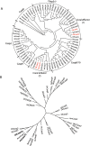
Coral caspase phylogenetic analysis. A, phylogenetic tree of cnidarian and vertebrate caspases: Orbicella faveolata (Of), Porites astreoides (Pa), Pocillophora damicornis (Pd), Stylophora pistillata (Sp), Nematostella vectensis (Nv), Exaiptasia pallida (Ep), Hydra vulgaris (Hv), Acropora digitophora (Ad), Homo sapiens (Hs), Mus musculus (Mm), Gallus gallus (Gg), Alligator mississippiensis (Am), Xenopus laevis (Xl), Danio rerio (Dr). Accession number of all used sequences are shown in Tables S1 and S2. B, phylogenetic analysis of CARD domains of caspases and CRADDs between cnidarians and vertebrates.
Figure 3.
Figure 3.
Biochemically characterized caspases of O. faveolata and P. astreoides aligned with human effector caspases. In the multiple sequence alignment, secondary structures (α-helices, β sheets, and loops) are indicated along with common position (CP) numbers among caspases. Gap positions, or sequences between common amino acid positions, are referred to as GP. Histidine (H) and cysteine (C), which forms a catalytic dyad, are colored in red and blue respectively. RYP motif insertions in OfCasp3a and PaCasp7a are colored in green.
Figure 4.
Figure 4.
Substrate preference determined by substrate-phage display. AD, amino acid preferences shown for substrate positions P5-P4-P3-P2-P1-P1′ for PaCasp7a (A), OfCasp3a (B), PaCasp3 (C) and OfCasp3b (D). Values of y-axes indicate number of phage sequences containing the specified amino acid (Count). Amino acids are shown on the x-axes in single letter code. Web logos are also shown in inset of respective graph for same results.
Figure 5.
Figure 5.
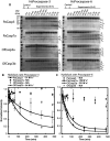
Cleavage kinetics of coral caspases using human procaspases-3 and -6 as a substrate. A, cleavage of full-length inactive HsCasp3 and HsCasp6 by coral caspases over time course. All cleaved products are labeled along with enzyme itself. (SL, large subunit of substrate; SS, small subunit of substrate; EL, large subunit of enzyme; ES, small subunit of enzyme; SL-P, large subunit with pro-domain cleaved; S, substrate; and E + S, enzyme and substrate). Bands with an asterisk (*) indicate only pro-domains were removed from full-length substrate. Molecular weight markers are the same for all gels. B and C, quantification of procaspase bands relative to the control (substrate without enzyme after 8 h incubation). Data were fit to a single exponential decay to calculate CF50 used to calculate hydrolysis rate of coral caspases (solid line). Procaspase-3 (B), procaspase-6 (C). Error bars represent S.D. from three different experiments.
Figure 6.
Figure 6.
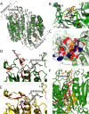
Structure of PaCasp7a. A, comparison of PaCasp7a (green) aligned with HsCasp3 (gray) (PDB ID: 2J30). B, PaCasp7a active site bound with inhibitor DEVD-CHO. Dashed lines show hydrogen bonding network to the P4 aspartate. C, surface map of active site residues in PaCasp7 within 5 Å of the inhibitor (yellow sticks). Neutral charges are gray, negative charges are red, and positive charges are blue. D, “out” orientation, of RYP residues in loop 1 in crystal structure of PaCasp7a. E, “in” orientation, of RYP residues in loop 1 in predicted model of PaCasp7a. Dashed lines show the hydrogen bonds formed by R and Y in In orientation. F, N-terminal peptide (orange) bound in hydrophobic pocket between helices 1 and 4. PaCasp7a residues that form the pocket are shown in yellow: Leu187 (CP-031), Ala190 (CP-034), Leu191 (CP-035), Phe330 (CP-177), Ala334 (CP-181), as well as Phe381 (CP-217) and Phe382 (CP-218) at the C terminus.
Figure 7.
Figure 7.
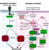
Proposed apoptotic pathways in coral compared with the apoptotic pathways in humans. All components in the pathways have homologs in O. faveolata and P. astreoides with the exception of BID (dotted box). A list of homologs is shown in Table S6. Dotted lines indicate that links have not yet been shown experimentally. Caspases in green background are initiators and those in red background are effectors. Pa refers to P. astreoides and Of refers to O. faveolata. The four caspases characterized here are shown in blue.

References

    1. Song Z., McCall K., and Steller H. (1997) DCP-1, a Drosophila cell death protease essential for development. Science 275, 536–540 10.1126/science.275.5299.536 - DOI - PubMed
    1. Dorstyn L., Mills K., Lazebnik Y., and Kumar S. (2004) The two cytochrome c species, DC3 and DC4, are not required for caspase activation and apoptosis in Drosophila cells. J. Cell Biol. 167, 405–410 10.1083/jcb.200408054 - DOI - PMC - PubMed
    1. Ellis H. M., and Horvitz H. R. (1986) Genetic control of programmed cell death in the nematode C. elegans. Cell 44, 817–829 10.1016/0092-8674(86)90004-8 - DOI - PubMed
    1. Schwartz H. T., and Horvitz H. R. (2007) The C. elegans protein CEH-30 protects male-specific neurons from apoptosis independently of the Bcl-2 homolog CED-9. Genes Dev. 21, 3181–3194 10.1101/gad.1607007 - DOI - PMC - PubMed
    1. Shaham S., and Horvitz H. R. (1996) Developing Caenorhabditis elegans neurons may contain both cell-death protective and killer activities. Genes Dev. 10, 578–591 10.1101/gad.10.5.578 - DOI - PubMed

Publication types