. 2020 Aug;8(2):e000896.
doi: 10.1136/jitc-2020-000896.

Efficient elimination of primary B-ALL cells in vitro and in vivo using a novel 4-1BB-based CAR targeting a membrane-distal CD22 epitope

Affiliations

Efficient elimination of primary B-ALL cells in vitro and in vivo using a novel 4-1BB-based CAR targeting a membrane-distal CD22 epitope

Talia Velasco-Hernandez et al. J Immunother Cancer. 2020 Aug.

Abstract

Background: There are few therapeutic options available for patients with B-cell acute lymphoblastic leukemia (B-ALL) relapsing as CD19- either after chemotherapy or CD19-targeted immunotherapies. CD22-chimeric antigen receptor (CAR) T cells represent an attractive addition to CD19-CAR T cell therapy because they will target both CD22+CD19- B-ALL relapses and CD19- preleukemic cells. However, the immune escape mechanisms from CD22-CAR T cells, and the potential contribution of the epitope binding of the anti-CD22 single-chain variable fragment (scFv) remain understudied.

Methods: Here, we have developed and comprehensively characterized a novel CD22-CAR (clone hCD22.7) targeting a membrane-distal CD22 epitope and tested its cytotoxic effects against B-ALL cells both in in vitro and in vivo assays.

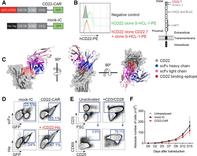
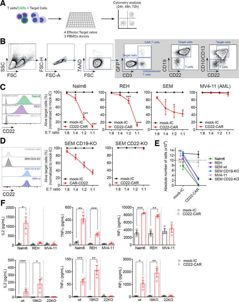
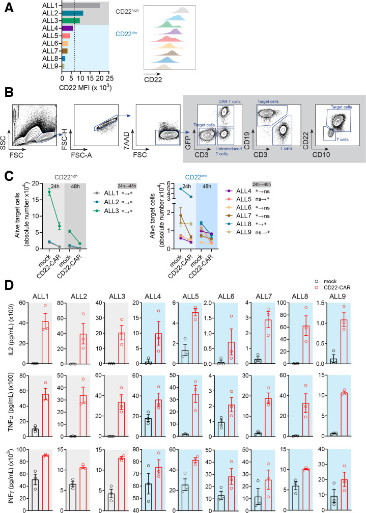
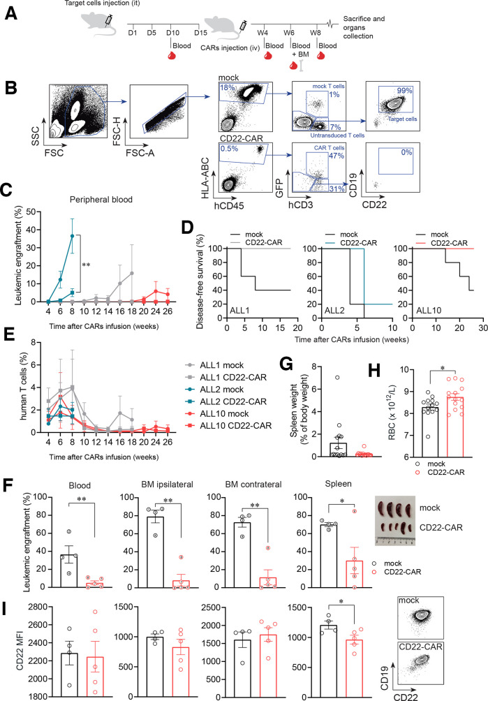
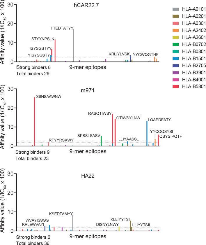
Results: Conformational epitope mapping, cross-blocking, and molecular docking assays revealed that the hCD22.7 scFv is a high-affinity binding antibody which specifically binds to the ESTKDGKVP sequence, located in the Ig-like V-type domain, the most distal domain of CD22. We observed efficient killing of B-ALL cells in vitro, although the kinetics were dependent on the level of CD22 expression. Importantly, we show an efficient in vivo control of patients with B-ALL derived xenografts with diverse aggressiveness, coupled to long-term hCD22.7-CAR T cell persistence. Remaining leukemic cells at sacrifice maintained full expression of CD22, ruling out CAR pressure-mediated antigen loss. Finally, the immunogenicity capacity of this hCD22.7-scFv was very similar to that of other CD22 scFv previously used in adoptive T cell therapy.

Conclusions: We report a novel, high-affinity hCD22.7 scFv which targets a membrane-distal epitope of CD22. 4-1BB-based hCD22.7-CAR T cells efficiently eliminate clinically relevant B- CD22high and CD22low ALL primary samples in vitro and in vivo. Our study supports the clinical translation of this hCD22.7-CAR as either single or tandem CD22-CD19-CAR for both naive and anti-CD19-resistant patients with B-ALL.

Keywords: T-lymphocytes; cell engineering; hematologic neoplasms; immunotherapy; receptors, chimeric antigen.


Conflict of interest statement

Competing interests: PM is co-founder of OneChain Immunotherapeutics (OCI), a IJC spin-off company focused on the development of CAR T-cell therapies. The patent of the membrane-distal CD22 scFv clone has been licenced to OCI.

Figures

Figure 1
Figure 1
Design, detection and expansion of hCD22.7-CAR T cells. (A) Scheme of the hCD22.7-CAR structure and the mock-IC. (B) Histogram showing the blocking of the binding of the anti-CD22 S-HCL-1 clone (PE signal) when CD22+ B cell line Raji cells are preincubated with the hCD22.7 clone (left panel). Right panel depicts the CD22 structure, indicating the binding site of hCD22.7 scFv (as well as m971, HA22 and BL22 scFvs). (C) Structural model of the CD22-hCD22.7–scFv complex. The structure is predicted based on the identified binding epitope of CD22 (ESTKDGKVP, in red) and the sequence of the heavy and light chains of the hCD22.7 scFv (blue and pink, respectively). (D) Representative CAR detection in primary T cells. CD22-CAR-transduced T cells are detected as GFP+ and are corecognized by an anti-human IgG F(ab')2 and an anti-His moAb (on incubation with rCD22-His). (E) Proper T cell activation determined by CD25 and CD69 staining after 48 hours exposure to anti-CD3/CD28 (PBMCs from n=3 healthy donors). (F) Robust expansion of activated T cells untransduced or transduced with either mock-IC or CD22-CAR (PBMCs from n=3 healthy donors). Plot shows mean±SEM (n=3 healthy donors). CAR, CD22-chimeric antigen receptor; FSC, forward scatter; His, histidine; mock-IC, mock-intracellular; PBMCs, peripheral blood mononuclear cells; rCD22-His, recombinant CD22-His; scFv, single-chain variable fragment; TM, transmembrane.
Figure 2
Figure 2
CD22-CAR T cells specifically target and eliminate B-ALL cell lines in vitro. (A) Experimental design of the in vitro cytotoxicity assays. (B) Flow cytometry gating strategy was used to analyze cytotoxicity. Alive target cells were identified as 7AADCD3GFPCD19+CD10+/CD13+ (CD33+ for MV4-11 cell line). CD22 was not used to avoid confounding detection of target cells due to potential antigen loss. Doublets were removed from the analysis. (C) Percentage of alive target cells after 48 hours incubation with CD22-CAR T cells at the indicated E:T ratios. Results are normalized respect to mock-IC data (PBMCs from n=3 healthy donors). CD22 expression in the different cell lines is shown previously in a histogram. (D) Specificity of CD22-CAR using CRISPR-Cas9 generated CD19-KO and CD22-KO SEM cells (PBMCs from n=3 healthy donors). CD22 expression in the different cell lines is shown previously in a histogram. (E) Absolute counts of alive target cells measured by FACS in 48 hours cytotoxicity assays at 1:1 E:T ratio (PBMCs from n=3 healthy donors). (F) ELISA showing robust secretion of proinflammatory cytokines by CD22-CAR T cells after 48 hours exposure to B-ALL and acute myeloid leukemia (AML) cell lines (upper panels) and wt, CD19-KO or CD22-KO SEM cell lines (bottom panels, PBMCs from n=3 healthy donors). Data are presented as mean±SEM (n=3 healthy donors). *p<0.05, **p<0.01, ***p<0.001. B-ALL, B-cell acute lymphoblastic leukemia; CAR, CD22-chimeric antigen receptor; E:T, effector:target ratio; FACS, fluorescence-activated cell sorting; FSC, forward scatter; FSC-A, forward scatter-area; FSC-H, forward scatter-high; mock-IC, mock intracellular; PBMCs, peripheral blood mononuclear cells; SSC, side scatter; KO, knock-out; MFI, median fluorescence intensity; wt, wild-type.
Figure 3
Figure 3
CD22-CAR T cells efficiently target and eliminate primary B-ALL cells in vitro although the expression level of CD22 impacts their efficacy. (A) MFI of CD22 in the different primary B-ALL samples used (n=9). The mean value for CD22 MFI was used to classify primary B-ALL samples as CD22high (ALL #1 to ALL #3) or CD22low (ALL #4 to ALL #9). (B) Flow cytometry gating strategy was used to analyze cytotoxicity. Alive target cells were identified as 7AADCD3GFPCD19+CD10+. CD22 was not used to avoid confounding detection of target cells due to potential antigen loss. Doublets were removed from the analysis. (C) Absolute counts of alive target CD22high and CD22low cells measured by FACS in 24 hours and 48 hours cytotoxicity assays at 1:1 E:T ratio. The statistical significance is indicated for each sample. A two-way ANOVA test (Sidak’s multiple comparison test) was used. (D) ELISA showing a similar robust production of IL2, TNFα, and IFNγ by CD22-CAR T cells after 48 hours exposure, among all B-ALL primary samples regardless the expression levels of CD22. Data are presented as mean±SEM (PBMCs from n=3 healthy donors). *p<0.05, **p<0.01. ANOVA, analysis of variance; B-ALL, B-cell acute lymphoblastic leukemia; CAR, CD22-chimeric antigen receptor; E:T, effector:target ratio; FACS, fluorescence-activated cell sorting; MFI, median fluorescence intensity; FSC, forward scatter; FSC-A, forward scatter-area; FSC-H, forward scatter-high; PBMCs, peripheral blood mononuclear cells; SSC, side scatter.
Figure 4
Figure 4
CAR T cells directed against a distal membrane CD22 epitope efficiently eliminate differentially aggressive PDX B-ALL blasts in vivo. (A) Experimental design of the in vivo experiments. (B) Gating strategy used to analyze FACS data from the in vivo experiments. Leukemic cells were identified as hCD45+hHLA-ABC+hCD3hCD19+hCD10+. CD22 was excluded from the identification to avoid confounding analysis due to potential antigen loss. CAR T cells were identified based on CD3 and GFP expression. Doublets were removed from the analysis. (C) Leukemic burden measured in PB over a 26 week follow-up period (n=4–5 mice/group; n=3 PDXs, CD22high (ALL#1 and ALL#2) and CD22low (ALL#10)). (D) Disease-free survival curves of mice transplanted with the three patient derived xenograft (PDXs). A mouse was considered ‘with disease’ when presenting >0.1% blasts in PB. (E) T cell persistence measured in PB of mice transplanted with the three PDXs. (F) Leukemic burden at sacrifice in PB, ipsilateral and contralateral BM and spleen (n=9 mice from the ALL#2 PDX). Each mouse is identified with the same number across the plots. Macroscopic images of these spleens are also shown. (G) Spleen weight (total animals, n=14) from mock-IC and CD22-CAR T cell-treated mice at sacrifice. (H) PB RBC counts in mock-IC and CD22-CAR T cell-treated mice at sacrifice (n=14). (I) CD22 MFI of persistent primary B-ALL in mock-IC-treated and CD22-CAR T cell-treated mice at sacrifice (ALL#2 mice, n=4–5) and representative FACS plots of contralateral BM samples showing the levels of CD22 of a mock-IC and a CD22-CAR T cell-treated mouse within the target cell population. Plot shows mean±SEM. *p<0.05, **p<0.01, ***p<0.001. B-ALL, B-cell acute lymphoblastic leukemia; BM, bone marrow; CAR, CD22-chimeric antigen receptor; D, day; FACS, fluorescence-activated cell sorting; PB, peripheral blood; it, intratibia; iv, intravenous; MFI, median fluorescence intensity; mock-IC, mock intracellular; RBC, red blood cell; W, week.
Figure 5
Figure 5
Immunogenic prediction of hCD22.7-scFv. The immunogenic potential of hCD22.7, m971 and HA22 CD22-scFvs was compared by in silico prediction analysis. CD22-scFvs were broken in 9-mer peptides, length for which human leukocyte antigen (HLA) molecules have a strong preference. Peptides are considered to bind MHC-I when a IC50 is <500 nM, being considered strong binders when IC50 <50 nM (affinity value >2, dotted line) and amino acid sequence is shown in the figure. Only the most representative HLA supertypes were analyzed. The predicted epitope-binding affinity was calculated as 1/IC50 100. Predicted epitopes are distributed along the x-axis according to their distribution in the protein. IC50, half-maximum inhibitory concentration; scFv, single-chain variable fragment.

References

    1. Greaves M. A causal mechanism for childhood acute lymphoblastic leukaemia. Nat Rev Cancer 2018;18:471–84. 10.1038/s41568-018-0015-6 - DOI - PMC - PubMed
    1. Smith MA, Altekruse SF, Adamson PC, et al. . Declining childhood and adolescent cancer mortality. Cancer 2014;120:2497–506. 10.1002/cncr.28748 - DOI - PMC - PubMed
    1. Pui C-H, Yang JJ, Hunger SP, et al. . Childhood acute lymphoblastic leukemia: progress through collaboration. J Clin Oncol 2015;33:2938–48. 10.1200/JCO.2014.59.1636 - DOI - PMC - PubMed
    1. Bhojwani D, Pui C-H. Relapsed childhood acute lymphoblastic leukaemia. Lancet Oncol 2013;14:e205–17. 10.1016/S1470-2045(12)70580-6 - DOI - PubMed
    1. Schrappe M, Hunger SP, Pui C-H, et al. . Outcomes after induction failure in childhood acute lymphoblastic leukemia. N Engl J Med 2012;366:1371–81. 10.1056/NEJMoa1110169 - DOI - PMC - PubMed

Publication types

MeSH terms