Review
. 2021:62:100033.
doi: 10.1194/jlr.TR120000954. Epub 2021 Feb 6.

Docosanoid signaling modulates corneal nerve regeneration: effect on tear secretion, wound healing, and neuropathic pain

Affiliations
Review

Docosanoid signaling modulates corneal nerve regeneration: effect on tear secretion, wound healing, and neuropathic pain

Thang L Pham et al. J Lipid Res. 2021.

Abstract

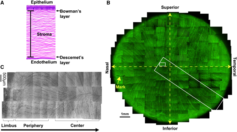
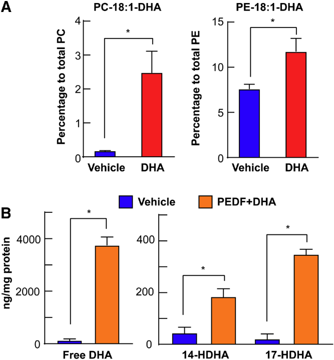
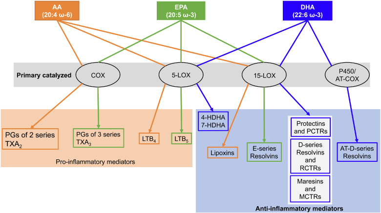
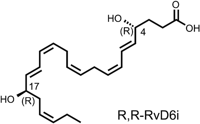
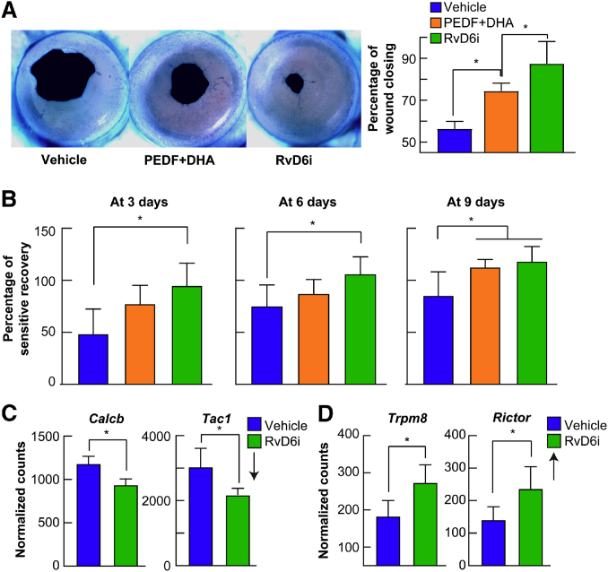
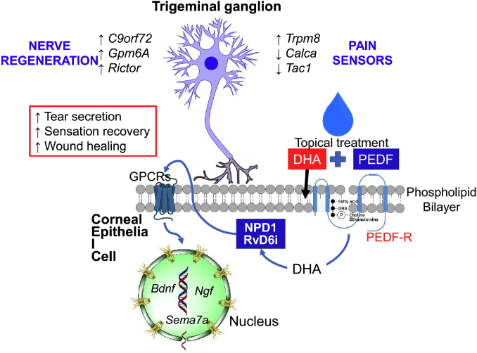
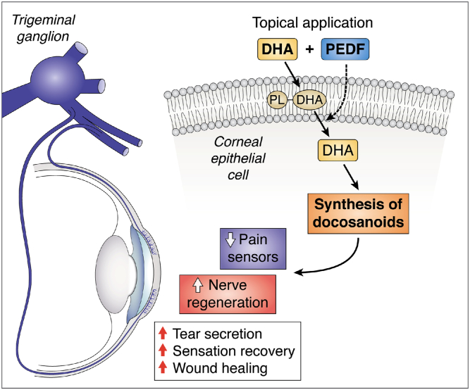
The cornea is densely innervated, mainly by sensory nerves of the ophthalmic branch of the trigeminal ganglia (TG). These nerves are important to maintain corneal homeostasis, and nerve damage can lead to a decrease in wound healing, an increase in corneal ulceration and dry eye disease (DED), and neuropathic pain. Pathologies, such as diabetes, aging, viral and bacterial infection, as well as prolonged use of contact lenses and surgeries to correct vision can produce nerve damage. There are no effective therapies to alleviate DED (a multifunctional disease) and several clinical trials using ω-3 supplementation show unclear and sometimes negative results. Using animal models of corneal nerve damage, we show that treating corneas with pigment epithelium-derived factor plus DHA increases nerve regeneration, wound healing, and tear secretion. The mechanism involves the activation of a calcium-independent phospholipase A2 that releases the incorporated DHA from phospholipids and enhances the synthesis of the docosanoids, neuroprotectin D1 (NPD1) and a new resolvin stereoisomer, resolvin D6i (RvD6i). NPD1 stimulates the synthesis of brain-derived neurotrophic factor, nerve growth factor, and semaphorin 7A. RvD6i treatment of injured corneas modulates gene expression in the TG resulting in enhanced neurogenesis, decreased neuropathic pain, and increased sensitivity. Taken together, these results represent a promising therapeutic option to reestablish the homeostasis of the cornea.

Keywords: cell signaling; docosahexaenoic acid; dry eye; gene expression; lipoxygenase; neuroprotectin D1; omega 3 fatty acids; phospholipase A2; pigment epithelium-derived factor; stereoisomer of resolvin D6.


Conflict of interest statement

Conflict of interest The authors declare that they have no conflicts of interest with the contents of this article.

Figures

Fig. 1
Fig. 1
Corneal structure and innervation. A: The anatomy of human cornea after hematoxylin and eosin histological stain. All five layers are shown: epithelium, Bowman’s layer, stroma, Descemet’s layer, and endothelium. B: Whole mount view of complete human corneal epithelial nerve network obtained from the left eye of a 45-year-old male donor. C: Detailed course of epithelial nerve bundles running from the periphery to the convergence at the center of the cornea (B and C are reproduced with permission from Elsevier, Ref. 5).
Fig. 2
Fig. 2
Incorporation of DHA into PC and PE after 1 h of DHA topical treatment to corneas of mice with damaged stromal nerves. A: Mouse corneas were injured and topically treated with DHA for 1 h and then lipids extracted and analyzed by LC-MS/MS (27). The proportion of PC and PE containing oleic acid (18:1) in the sn-1 position and DHA in the sn-2 position. PE was more enriched in DHA than PC. B: Release of DHA and synthesis of the monohydroxy-DHA derivatives after corneal injury and topical treatment with PEDF+DHA for 3 h. Corneal lipid profiles were analyzed by MS-based lipidomic analysis. ∗P < 0.05 with the t test statistical analysis to compare two groups at 95% of the confidence level.
Fig. 3
Fig. 3
Lipid mediators derived from the three most abundant essential fatty acids, AA, EPA, and DHA, esterified in the sn-2 position of the phospholipids. Depending on the primary catalyzing enzyme, COX-2, and 5- and 15-LOXs, there is synthesis of a variety of bioactive lipids involved in inflammation as well as in resolution of the inflammatory response. Mediators from AA are highlighted in orange, EPA in green, and DHA in blue.
Fig. 4
Fig. 4
Structure of the RvD6i. The new isomer was synthesized after topical stimulation of mouse injured corneas with PEDF+DHA and released in tears. It was analyzed by LC-MS/MS and showed at least six matched daughter ions with an RvD6 standard but with an earlier retention time (40). Posterior studies show that the peak retention time coincides with chemically synthetized R,R-RvD6i in a chiral column (unpublished observations).
Fig. 5
Fig. 5
RvD6i accelerates corneal wound healing and sensitivity. A: Representative images of mouse cornea wounded area stained with methylene blue after 20 h of an injury that damaged the epithelial and anterior stroma nerves. The animals received eye drops containing PEDF+DHA or RvD6i in similar concentrations three times per day. The images were taken with a dissecting microscope and quantified using Photoshop software (40). B: Recovery of cornea sensitivity at 3, 6, and 9 days after injury and treatment with PEDF+DHA or RvD6i (three times per day) using a noncontact aesthesiometer. RvD6i-treated mice recovered sensitivity sooner than PEDF+DHA-treated corneas. C: Expression of genes involved in inflammation and pain in the TG of RvD6i topically treated corneas. TG were obtained 12 days after cornea injury and treatment with RvD6i and analyzed by RNA sequencing (40). Calcb and Tac1 genes were downregulated while Trpm8 and Rictor genes were upregulated in the TG neurons by cornea treatment with RvD6i. ∗P < 0.05 with the t test statistical analysis to compare two groups at 95% of the confidence level.
Fig. 6
Fig. 6
Schematic model of signaling stimulated by the combination of PEDF+DHA. DHA is rapidly incorporated into membrane phospholipids from corneal epithelium and then released after stimulation by PEDF of the PEDF-R with calcium-independent phospholipase A2 (iPLA2ζ) activity. Free DHA is then the substrate for docosanoids such as NPD1 and the novel RvD6i. These docosanoids are then released into tears and, by autocrine stimulation, to an undefined GPRC receptor(s) that induces the gene and protein expression of neurotrophic factors NGF, BDNF, and semaphorin 7A (Sema7A) that are secreted into tears and enhance axon outgrowth. RvD6i stimulates corneal wound healing, corneal sensation and nerve recovery, and tear secretion. The mechanism involves changes in the TG transcriptome with activation of genes related to neurogenesis and modulation of genes implicated in neuropathic pain. Treatment with PEDF or DHA alone does not activate these pathways, and therefore, there was no increase in cornea nerve regeneration (19). Gpm6A, glycoprotein M6A; C9orf72, chromosome 9 open reading frame 72; Trpm8, transient receptor potential melastatin 8.
None

References

    1. DelMonte D.W., Kim T. Anatomy and physiology of the cornea. J. Cataract Refract. Surg. 2011;37:588–598. - PubMed
    1. Meek K.M., Knupp C. Corneal structure and transparency. Prog. Retin. Eye Res. 2015;49:1–16. - PMC - PubMed
    1. Hanna C., Bicknell D.S., O’Brien J.E. Cell turnover in the adult human eye. Arch. Ophthalmol. 1961;65:695–698. - PubMed
    1. Müller L.J., Marfurt C.F., Kruse F., Tervo T.M.T. Corneal nerves: structure, contents and function. Exp. Eye Res. 2003;76:521–542. - PubMed
    1. He J., Bazan N.G., Bazan H.E.P. Mapping the entire human corneal nerve architecture. Exp. Eye Res. 2010;91:513–523. - PMC - PubMed

Publication types

LinkOut - more resources